Comparative Metabolic Profiling in Pulp and Peel of Green and Red Pitayas (Hylocereus polyrhizus and Hylocereus undatus) Reveals Potential ...
←
→
Page content transcription
If your browser does not render page correctly, please read the page content below
Hindawi
BioMed Research International
Volume 2021, Article ID 6546170, 12 pages
https://doi.org/10.1155/2021/6546170

Research Article
Comparative Metabolic Profiling in Pulp and Peel of Green and
Red Pitayas (Hylocereus polyrhizus and Hylocereus undatus)
Reveals Potential Valorization in the Pharmaceutical and
Food Industries

 Xing’e Lin, Hongmao Gao, Zheli Ding, Rulin Zhan, Zhaoxi Zhou , and Jianhong Ming
 Haikou Experimental Station, Chinese Academy of Tropical Agricultural Sciences (CATAS), China

 Correspondence should be addressed to Zhaoxi Zhou; zhzx81@163.com and Jianhong Ming; jhming_326@163.com

 Received 25 May 2020; Revised 27 November 2020; Accepted 29 December 2020; Published 12 March 2021

 Academic Editor: Dorota Formanowicz

 Copyright © 2021 Xing’e Lin et al. This is an open access article distributed under the Creative Commons Attribution License,
 which permits unrestricted use, distribution, and reproduction in any medium, provided the original work is properly cited.

 Pitaya (Hylocereus genus) is a popular plant with exotic and nutritious fruit, which has widespread uses as a source of nutrients and
 raw materials in the pharmaceutical industry. However, the potential of pitaya peel as a natural source of bioactive compounds has
 not yet fully been explored. Recent advances in metabolomics have paved the way for understanding and evaluating the presence of
 diverse sets of metabolites in different plant parts. This study is aimed at exploring the diversity of primary and secondary
 metabolites in two commercial varieties of pitaya, i.e., green pitaya (Hylocereus undatus) and red pitaya (Hylocereus polyrhizus).
 A total of 433 metabolites were identified using a widely targeted metabolomic approach and classified into nine known diverse
 classes of metabolites, including flavonoids, amino acids and its derivatives, alkaloids, tannins, phenolic acids, organic acids,
 nucleotides and derivatives, lipids, and lignans. Red pitaya peel and pulp showed relatively high accumulation of metabolites viz.
 alkaloids, amino acids and its derivatives, and lipids. Differential metabolite landscape of pitaya fruit indicated the presence of
 key bioactive compounds, i.e., L-tyrosine, L-valine, DL-norvaline, tryptophan, γ-linolenic acid, and isorhamnetin 3-O-
 neohesperidoside. The findings in this study provide new insight into the broad spectrum of bioactive compounds of red and
 green pitaya, emphasizing the valorization of the biowaste pitaya peel as raw material for the pharmaceutical and food industries.

1. Introduction boxylated neohylocerenin [2]. These metabolites make the
 pitaya fruit an important diet source with high nutritional
Pitaya (Hylocereus genus) is an important exotic fruit native value, energy, and other health benefits like chemopreven-
to Central America and Mexico, usually cultivated in tropical tion [14, 15], anti-inflammatory [16], antimicrobial [17],
and subtropical habitats [1]. Pitaya species have several culti- antidiabetic, antioxidant properties [18–20], and prebiotic
vated types with different peel and pulp colors [2]. Classifica- effects [16, 21].
tion of pitayas in different species is mainly based on pulp Previous studies demonstrated the availability of second-
colors such as white (Hylocereus undatus), red (H. costaricen- ary metabolites in pitaya fruit pulp, i.e., antioxidants [2, 6,
sis and H. polyrhizus), and yellow (Selenicereus megalanthus) 22], polyphenolic compounds, organic acids, amino acids,
[3, 4]. H. undatus and H. polyrhizus are the most widely cul- and flavonoids [2, 23]. H. polyrhizus seed oil is also a rich
tivated species [5]. Besides its exotic appearance and striking source of fatty acids, i.e., linoleic acid, that is beneficial to
colors, pitaya fruit is well known for its nutritional properties human health [24, 25]. However, the concentration of these
and health benefits [2, 6–8]. Pitaya produces an array of metabolites varies in different fruit parts and is highly depen-
chemically and biologically diverse compounds, including dent on cultivars, environment, and management practices
betanin [9, 10], phyllocactin [11], betanidin [12], gluconic [26]. Usually, pitaya peel is discarded as biowaste during pro-
acid, tyrosine [13], tricarboxylated hylocerenin, and decar- cessing or direct consumption, approximately 22% of the
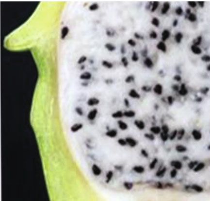
2 BioMed Research International

fruit weight [27]. Pitaya fruit peel contains soluble and insol- MS (Thermo, Ultimate 3000LC, Orbitrap Elite). The liquid
uble fiber, betalains, polyphenolic compounds, and other vol- phase conditions included (1) column: Hypergod C18
atile compounds [28–30]. It has been emphasized that the (100 × 4:6 mm 3 μm); (2) mobile phase: phase A = water +
concentration of metabolites tends to be significantly higher 0:1%formic acid and phase B = acetonitrile + 0:1%formic
in discarded fruit parts, i.e., peel and seed, than edible parts acid; (3) elution gradient: 0 min B = 5%in comparison and
[31]. However, not much work has been done to explore 2 min B = 5%, B was linearly increased to 95% in 12 min
the spectrum and concentration of bioactive metabolites in and maintained at 95% for 15 min, and B was reduced to
different parts of pitaya fruits, i.e., peel and seeds. The scenar- 5% and was balanced to 17 min; and (4) flow rate
ios of increasing yield potential and industrial demand for 0.3 mL/min: injection volume = 4 μL and automatic injector
natural bioactive compounds have raised prospects for temperature = 4° C. Whereas the MS conditions were as fol-
extending the utilization of this crop with the valorization lows: the positive electrospray ionization (ESI+) temperature
of neglected parts of the fruit, i.e., peel and seed as previously was 300°C, sheath gas flow rate was 45 arb, aux gas flow rate
demonstrated in citrus, apple, mango, pineapple, and pome- was 15 arb, sweep gas flow rate was 1 arb, spray voltage was
granate [31–33]. 3.0 kV, the capillary temperature was 350°C, and S-Lens RF
 The advance in the field of metabolomics has facilitated level was 30%. The ESI-conditions were the same as ESI+
the analysis and characterization of the metabolome in vari- except that the spray voltage was 3.2 kV, and the S-Lens RF
ous plant organs, subsequently helps in the detection of thou- level was 60%. The qualitative analysis of the material was
sands of bioactive metabolites [34–39]. The diversity of established on secondary spectrum information using the
metabolites accumulation and metabolic profiling of differ- public databases (KNApSAcK, METLIN, LipidMAps, and
ent fruit plants provides a promising approach to understand MassBank) of metabolites. The isotope signals were removed
and determine the commercial importance of the target fruit during the quantitative analysis of samples. Triple Q scans
plants [39]. were attained as multiple reaction monitoring (MRM) exper-
 In this study, we used the widely targeted metabolomic iments. Declustering potential (DP) and collision energy
approach to comparatively and comprehensively investigate (CE) for individual MRM transitions were done with further
the complex and distinct landscapes of metabolites in two DP and CE optimization [40]. A specific set of MRM transi-
main parts of pitaya fruits (peel and pulp) of the two major tions was monitored for each period according to the metab-
commercial varieties of pitaya viz. green pitaya (Hylocereus olites eluted within this period.
undatus) and red pitaya (H. polyrhizus). Our work provides
essential basic information for the valorization of pitaya fruit 2.5. Quality Control and Data Analysis. Quality control was
peel for its use as a functional food or a potential source for performed to check the reliability and reproducibility of the
raw material in the pharmaceutical industry. Further, it will data. Extracted samples were mixed and inserted into every
be helpful to dissect the biosynthetic pathways of important four samples, and changes were monitored. Data sets with
metabolites in pitaya fruit. the intensity of the metabolites from each sample, i.e., peel
 and pulp, were uploaded to the Analyst 1.6.1 software (AB
2. Materials and Methods SCIEX, Ontario, ON, Canada) for descriptive statistical anal-
 yses. The principal component analysis was performed using
2.1. Plant Materials. In this study, we used two commercial R package prcomp and visualized using ggbiplot.
varieties, i.e., green pitaya (Hylocereus undatus) and red
pitaya (H. polyrhizus) collected from Sanya fruit Island,
Dragon fruit planting base, Sanya, Hainan, China. Thirty
 3. Results
days after pollination, five fruits per tree were harvested from 3.1. Metabolome Profiling of the Pitaya Varieties. Two
three different trees of each variety, representing the three commercial pitaya varieties, green pitaya (Hylocereus
replications. Pulp and peel samples from each tree were undatus) and red-pitaya (H. polyrhizus), were used for meta-
mixed separately, and all samples were immediately placed bolomic analyses. These two commercial varieties possess
in liquid nitrogen and stored at –80°C until use. distinguished morphological appearances of peel and pulp
 (Figure 1(a)). Significant contrasting fruit colors suggested
2.2. Metabolomic Analyses. The sample preparation, extract
 the variable concentration of metabolites in peel and pulp tis-
analysis, metabolite identification, and quantification were
 sues. To support this hypothesis, in this study, we examined
performed at Wuhan Metware Biotechnology Co., Ltd.,
 the metabolome profile in the peel and pulp tissues of both
Wuhan, China, following their standard procedures.
 cultivars. Ultraperformance liquid chromatography and
2.3. Sample Preparation. All samples were ground to a fine tandem mass spectrometry (UPLC-MS/MS) techniques were
powder using a Grinding Mill at 65 Hz for 90 s. A total of employed and identified in total of 443 metabolites
50 mg of sample was weighed and extracted with 800 μL of (Table S1). Following the recommendations of international
methanol. The samples were vortexed for 30 s and centri- metabolomics society (http://metabolomicssociety.org/), the
fuged at 12000 rpm and 4°C for 15 min. 200 μL of superna- identified metabolites were categorized into first two levels
tant was transferred to the vial for LC-MS analysis. A (identified metabolites) and B (putatively annotated
 metabolites). Five random samples were selected, and their
2.4. Liquid Chromatography Coupled Mass Spectrometry (LC- graphs of mass scan data collected over time (TIC) have
MS). The data acquisition instrument system included LC- been presented in supplementary figures 1-5.The identified
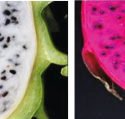
BioMed Research International 3

 (a)

 15

 10

 5
 PC3 (19.03%)
 0

 –5
 Total = 433
 –10 20
 34 Alkaloids 10
 0 )
 –10 .1%
 58 Amino acids & derivatives –15 –20 24
 2(
 96 Flavonoids –30 –20 –10 0 10 20 PC
 12 Lignans and coumarins PC1 (33.52%)
 63 Lipids
 31 Nucleotides & derivatives Green_pitaya_pulp Green_pitaya_peel
 Red_pitaya_peel Mix
 30 Organic acids
 Red_pitaya_pulp
 55 Phenolic acids
 3 Terpenoids
 51 Others
 (b) (c)

Figure 1: Metabolome landscape in peel and pulp tissue of red pitaya (Hylocereus polyrhizus) and green pitaya (Hylocereus undatus) varieties:
(a) pictorial description of green and red pitaya fruit; (b) distribution of identified metabolites classes; (c) principal component analysis with
3D visualization, comprising PC1 (33.52%), PC2 (24.1%), and PC3 (19.03%).

metabolites were further classified into nine known major ing among the fruit tissues rather than varieties, and all rep-
classes based on metabolites structure (Figure 1(b)). Among licates were grouped together, suggesting a high quality of
these 443 metabolites, 96 flavonoids, 63 lipids, 58 amino our metabolome profiling. Metabolome variance explained
acids and derivatives, 55 phenolic acids, 51 others, 34 by the first three PCA coordinates accumulatively higher as
alkaloids, 31 nucleotide and derivatives, 30 organic acids, 76.65%. First PCA coordinate (PC1) explained 33.52%, PC2
12 lignans, and coumarins, and 3 terpenoids were included explained 24.1%, and PC3 explained 19.03% variance of
(Figure 1(b)). These results indicate that flavonoids, lipids, metabolome data. These results indicated the high variance
amino acids and derivatives, and phenolic acids are in high of metabolite concentrations in pitaya fruit.
proportion in pitaya. Detailed information about the
identified landscape of metabolites, including molecular 3.2. Metabolite Landscape in Pitaya Fruit Peel from the
weights (Da), formula, compounds, major and minor Two Varieties. The metabolome landscape of pitaya fruit
classes, ionization, and KEGG maps, is described in Table S1. peel revealed a diverse array of metabolites. Top 20 metab-
 Principle component analysis (PCA) was performed to olites identified based on metabolites ion abundance were
summarize the descriptive assessment, help to understand N-benzylmethylene isomethylamine, choline, L-valine, DL-
the underlying characteristics and structure of the metabo- norvaline, tryptophan, D-(-)-valine, isorhamnetin 3-O-neo-
lome data. The 3D PCA plot in Figure 1(c) demonstrated hesperidoside, bioquercetin, γ-linolenic acid, stearic acid,
the four distinctive structures of metabolome data based on punicic acid, hexadecylsphingosine, 9,10-EODE, 9-hydroxy-
tissue and variety. Mix was used for quality control in the 10,12-octadecadienoic acid, 13-hydroxy-9,11-octadecadie-
analysis. PCA plot illustrated the closer grouping of red noic acid, 9S-hyroxy-10E,12E-octadecadienoic acid, 5 ′
pitaya peel to that green pitaya peel, similar to red and green -deoxy-5 ′ -(methylthio) adenosine, galactinol, chlorogenic
pitaya pulp samples. These results suggested the close group- acid, and protocatechuic acid-4-glucoside (Figure 2). Of these,
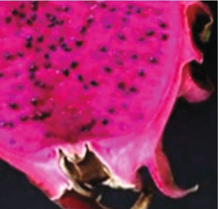
4 BioMed Research International

 8×10⁷ 8×10⁷

 6×10⁷ 6×10⁷

 Ion abundance
 Ion abundance

 4×10⁷ 4×10⁷

 2×10⁷
 2×10⁷

 0
 0
 N-Benzylmethylene isomethylamine
 Choline
 L-Valine
 DL-Norvaline
 Tryptophan
 D-(-)-Valine
 Isorhamnetin 3-O-neohesperidoside
 Bioquercetin
 γ-Linolenic acid
 Strearic acid
 Punicic acid

 9,10-EODE
 9-Hydroxy-10,12-octadecadienoic acid

 Protocatechuic acid-4-glucoside
 13-Hydroxy-9,11-octadecadienoic acid
 9S-Hyroxy-10E,12E-octadecadienoic acid
 5′-Deoxy-5′-(methylthio) adenosine
 Galactinol
 Chlorogenic acid
 Hexadecylsphingosine

 N-Benzylmethylene isomethylamine
 Sepermine
 Choline
 L-Valine
 DL-Norvaline
 Isorhamnetin 3-O-neohesperidoside
 γ-Linolenic acid

 Punicic acid
 9,10-EODE
 Strearic acid
 9-Hydroxy-10,12-octadecadienoic acid

 Protocatechuic acid-4-glucoside
 LysoPC (16:1)
 9S-Hyroxy-10E,12E-octadecadienoic acid

 2,5-Dihydroxy benzoic acid O-hexside
 Hexadecylsphingosine

 (Rs)-Mevalonic acid
 Galactinol
 D-Pantothenic acid
 Chlorogenic acid
 Alkaloids Nucleotide and derivatives
 Alkaloids Organic acid
 Amino acid and derivatives Phenolic acids
 Amino acid and derivatives Phenolic acids
 Flavonoids Others
 Flavonoids Others
 Lipids
 Lipids
Figure 2: Top 20 most abundant metabolites in pitaya fruit peel.
 Figure 3: Top 20 most abundant metabolites in fruit pulp.
choline (6:03E + 07), L-valine (5:99E + 07), γ-linolenic acid
(5:56E + 07), and N-benzylmethylene isomethylamine most abundant metabolites of nine major metabolite clas-
(4:90E + 07) were the most concentrated metabolites in fruit ses from each tissue separately. The top 10 highest abun-
peel (Table S2). These top 20 metabolites belong to seven dant metabolites from each class in fruit peel were
major classes, i.e., alkaloids, amino acid and derivatives, selected and compared between the two varieties
flavonoids, lipids, nucleotide and derivatives, phenolic acids, (Figure 4). In the present study, 34 metabolites belonged
and others. Lipids and amino acids and derivatives were in to the alkaloid class. Alkaloid is one of the most important
higher frequency compared to remaining metabolite classes. secondary metabolite classes which naturally occurred in
 plants [41]. The top 10 most accumulated metabolites
3.3. Metabolite Landscape in Pitaya Fruit Pulp from the identified in fruit peel tissues were N-benzylmethylene iso-
Two Varieties. Metabolome landscape was investigated in methylamine, choline, serotonin, spermine, trigonelline,
fruit pulp of green pitaya (Hylocereus undatus) and red gomphrenin I, 6-deoxyfagomine, dopamine hydrochloride,
pitaya (Hylocereus polyrhizus) varieties. The top 20 most amaranthin, and N-cis-feruloyltyramine (Figure 4(a)). N-
profuse metabolites identified in fruit pulp were N- Benzylmethylene isomethylamine metabolite showed the
benzylmethylene isomethylamine, spermine, choline, L- highest ion abundance among the top 10 alkaloids. N-
valine, DL-norvaline, isorhamnetin 3-O-neohesperidoside, Benzylmethylene isomethylamine and serotonin indicated
γ-linolenic acid, hexadecylsphingosine, punicic acid, 9,10- significantly higher ion abundance in red pitaya compared
EODE, stearic acid, 9-hydroxy-10,12-octadecadienoic acid, to green pitaya varieties. The top 10 abundant amino acids
LysoPC (16 : 1), 9S-hyroxy-10E,12E-octadecadienoic acid, and derivatives found in fruit peel were L-valine, DL-norva-
(Rs)-mevalonic acid, galactinol, D-pantothenic acid, chloro- line, tryptophan, D-(-)-valine, L-tyramine, L-2-chloropheny-
genic acid, protocatechuic acid-4-glucoside, and 2,5-dihydroxy lalanine, L-methionine, 2-aminoisobutyric acid, methionine
benzoic acid O-hexside (Figure 3). Of these metabolites, L- sulfoxide, and pipecolic acid (Figure 4(b)). L-Valine, DL-nor-
valine (7:33E + 07), γ-linolenic acid (7:21E + 07), and DL- valine, and tryptophan metabolites revealed the highest ion
norvaline (7:20E + 07) revealed the highest ion abundance abundance among the top 10 abundant amino acids and
(Table S3). These top 20 metabolites of fruit pulp also derivatives. L-Valine and DL-norvaline showed significantly
belonged to seven major classes, i.e., alkaloids, amino acid and higher ion accumulation in red pitaya peel than green
derivatives, flavonoids, lipids, organic acid, phenolic acids, and pitaya peel. Further, flavonoids were screened based on
others. While metabolites belonging to lipids, alkaloids, and the average value of ion abundance in green pitaya peel
phenolic acid classes had higher occurrences than other and red pitaya peel, and we selected the top 10 most abun-
metabolite classes. dant metabolites. These metabolites were isorhamnetin 3-
 O-neohesperidoside, bioquercetin, hyperin, rutin, isoquerci-
3.4. Comparative Analysis of Metabolome from the Pitaya trin, quercetin 3-O-glucoside (Isotrifoliin), spiraeoside,
Fruit Peel. For further investigation, comparative analyses kaempferol 3-O-rutinoside (Nicotiflorin), kaempferol 3-O-
between the two varieties were performed based on the robinobioside (Biorobin), and rhamnetin-O-glucoside-O-
Ion abundance Ion abundance

phenolic acids; (i) others.
 (Rs)-Mevalonic acid -Linolenic acid N-Benzylmethylene isomethylamine
 Sodium valproate Stearic acid Choline

 l-(+)-Tartaric acid Punicic acid Serotonin
 BioMed Research International

 Average
 4-Guanidinobutyric acid Hexadecylsphingosine Spermine

 9,10-EODE Trigonelline
 Aldehydo-D-galacturonate

 Red_Pitaya-Peel
 (a)

 (g)
 (d)
 Gomphrenin I

 Green_Pitaya-Peel
 Lipids
 D-Galacturonic acid (Gal A) 9-Hydroxy-10,12-octadecadienoic acid
 Alkaloids

 Organic acids
 13-Hydroxy-9,11-octadecadienoic acid 6-Deoxyfagomine
 6-Aminocaproic acid
 9S-Hyroxy-10E,12E-octadecadienoic acid Dopamine hydrochloride
 Anchoic acid
 Amaranthin
 Citraconic acid Myristic acid
 N-Cis-feruloyltyramine
 Methymalonic acid
 LysoPC (16:1)

 Chlorogenic acid Esculin (6,7-DihydroxyCoumarin-6-glucoside) L-Valine

 Protocatechuic acid-4-glucoside Syringaresional-aceglu DL-Norvaline

 2,5-Dihydroxy benzonic acid O-hexside Esculin gydrate Tryptophan
 7-Methoxycoumarin D-(-)-Valine
 Regaloside L
 (+)-Medioresinol-aceglu L-Tyramine
 Trihydroxycinnamoylquinic acid

 (e)
 (b)

 (h)
 Pinoresinol-acetylglucose L-2-Chlorophenylalanine
 1-O-[(E)-p-Cumaroyl]- -D-glcopyranose
 L-Methionine

 Phenolic acids
 Olivin diglucoside
 Phthalic anhydride
 Lignans & coumarins

 Pinoresinol diglucoside 2-Aminoisobutyric acid
 1-O-[(E)-Caffeoyl]- -D-glucopyranose
 Amino acid & its derivatives

 Syringaresinol-hex Methionine sulfoxide
 Coniferin
 Pinoresinol Pipecolic acid
 Echinacoside

 Galactinol 5′-Deoxy-5′-(methylthio) adenosine Isorhamnetin 3-O-neohesperidoside
 D-Pantothenic acid Guanosine Bioquercetin
 N-Hydroxy tryptamine Adenosine Hyperin
 D-(+)-Sucrose Adenine Rutin
 D-Glucose Guanine Isoquercitrin

 (i)
 (f)
 (c)

 Others
 D-(+)-Glucose Deoxyadenosine Quercetin 3-O-glucoside (isotrifoliin)
 Flavonoids

 Isomaltulose Cytidine Spiraeoside
 D-Glucoronic acid 2-(Dimethylamino) guanosine Kaempferol 3-O-rutinoside (nicotiflorin)
 Nucleotides & derivatives

 Phenethylamine 2-Deoxyribose 5-phosphate Kaempferol 3-O-robinobioside (biorobin)
 D-(+)-Trehaloseanhydrous Adenosine 5′-monophosphate Rhamnetin-O-glucoside-O-rhamnoside

(b) amino acid and derivatives; (c) flavonoids; (d) lipids; (e) lignans and coumarins; (f) nucleic acid and derivatives; (g) organic acids; (h)
Figure 4: Comparative analysis between red pitaya peel and green pitaya peel based on the top 10 metabolites from each class: (a) alkaloids;
 5
6

acids; (i) others.
 Ion abundance Ion abundance Ion abundance

 (Rs)-Mevalonic acid -Linolenic acid N-Benzylmethylene isomethylamine
 Sodium valproate Stearic acid Choline
 L-(+)-Tartaric acid Punicic acid Serotonin
 Spermine

 Average
 4-Guanidinobutyric acid Heaxadecylsphingosine
 9,10-EODE Trigonelline
 Aldehydo-D-galacturonate

 (a)

 (g)
 Gomphrenin l

 (d)
 Lipids
 9-Hydroxy-10,12-octadecadienoic acid

 Red_Pitaya-Pulp
 D-Galacturonic acid (Gal A)
 Alkaloids

 6-Deoxyfagomine

 Green_Pitaya-Pulp
 13-Hydroxy-9,11-octadecadienoic acid

 Organic acids
 6-Aminocaproic acid
 Dopamine hydrochloride
 Anchoic acid 9s-Hydroxy-10E,12E-octadecadienoic acid
 amaranthin
 Citraconic acid Myristic acid
 N-Cis-feruloyltyramine
 Methylmalonic acid LysoPC (16:1)

 Chlorogenic acid Esculin (6,7-Dihydroxycoumarin-6-glucoside) L-Valine
 Prorocatechuic acid-4-glucoside Syringgaresinol-aceGlu DL-Norvaline
 2,5-Dihydroxy benzonic acid O-hexside Esculin hydrate Tryptophan
 7-Methoxycoumarin D-(-)-Valine
 Regaloside L
 (+)-Medioresional-aceGlu L-Tyramine
 Coniferin

 (e)
 Pinoresinol-acetyglucose L-2-chlorophenylalanine
 (b)

 (h)
 Echinacoside
 Olovin diglucoside L-Methionine

 Phenolic acids
 3-O-p-Coumaroyl quinic acid
 Lignans & coumarins

 Pinoresionol diglucoside 2-Aminoisobutyric acid
 Ferulic acid
 Amino acid & its derivatives

 Syringaresinol-hex Methionine sulfoxide
 1-O-[(E)-Caffeoyl]- -D-glucopyranose Pinoresinol Pipecolic acid
 1-O-[(E)-p-Cumaroy]- -D-glucopyranose

 5’-Deoxy-5’- (methylthio) adenosine
 Galactinol Isorhamnetin 3-O-neohesperidoside
 Guanosine
 D-Pantothenic acid Bioquercetin
 Adenosine
 N-Hydroxy tryptamine Hyperin
 Adenine Isoquercitrin
 D-(+)-Sucrose
 Guanine Rutin
 D-Glucose
 Deoxyadenosine

 (i)
 (f)
 (c)

 Quercetin 3-O-glucoside (isotrifoliin)
 D-(+)-Glucose

 Others
 Flavonoids

 Cytidine Spiraeoside
 Isomaltulose
 2- (Diethylaino) guanosine Kaempferol 3-O-rutinoside (nicotiflorin)
 Nucleotides & derivatives

 D-Glucoronic acid
 2-Deoxyryibose 5-phosphate Kaempferol 3-O-robinoside (birobin)
 Phenethylamine
 Adenosine 5’-monophosphate Rhamnetin-O-glucoside-O-rhamnoside
 D-(+)-Trehaloseanhydrous

amino acid and derivatives; (c) flavonoids; (d) lipids; (e) lignans and coumarins; (f) nucleic acid and derivatives; (g) organic acids; (h) phenolic
Figure 5: Comparative analysis between red pitaya pulp and green pitaya pulp based on top 10 metabolites from each class: (a) alkaloids; (b)
 BioMed Research International
BioMed Research International 7

 6×10 8

 4×10 8

 2×10 8
 Ion abudance

 4×10 7

 2×10 7

 0
 Green_Pitaya-peel Green_Pitaya-pulp Red_Pitaya-peel Red_Pitaya-pulp

 Alkaloids Nucleotide and derivatives
 Amino acids & derivatives Organic acids and derivatives
 Flavanoids Phenolic acids
 Lignans and coumarins Terpenoids
 Lipids

Figure 6: Sum total of metabolite classes in different tissues, i.e., peel and pulp of green pitaya (Hylocereus undatus) and red pitaya
(Hylocereus polyrhizus) fruit.

rhamnoside (Figure 4(c)). These top 10 flavonoids revealed line, serotonin, spermine, trigonelline, gomphrenin I, 6-
significantly higher ion abundance in green pitaya peel deoxyfagomine, dopamine hydrochloride, amaranthin, and
compared to red pitaya peel. Lipid class was also evaluated N-cis-feruloyltyramine (Figure 5(a)). N-Benzylmethylene
for the top 10 most abundant metabolites based on ion isomethylamine, choline, and serotonin were the most
abundance average of red and green pitaya peels. The top abundant in pitaya fruit pulp. Choline metabolite demon-
10 most abundant metabolites were γ-linolenic acid, stearic strated higher ion accumulation in red pitaya pulp than
acid, punicic acid, hexadecylsphingosine, 9,10-EODE, 9- the green pitaya pulp. In contrast, serotonin showed higher
hydroxy-10,12-octadecadienoic acid, 13-hydroxy-9,11-octa- ion accumulation in green pitaya pulp compared to red
decadienoic acid, 9S-hyroxy-10E,12E-octadecadienoic acid, pitaya pulp. Amino acid and derivative classes were screened
myristic acid, and LysoPC (16 : 1) (Figure 4(d)). All metab- for the most abundant metabolites. Top 10 metabolites, L-
olites except LysoPC (16 : 1) displayed higher ion accumula- valine, DL-norvaline, tryptophan, D-(-)-valine, L-tyramine,
tion in red pitaya fruit compared to green pitaya fruit. L-2-chlorophenylalanine, L-methionine, 2-aminoisobutyric
Lignans and coumarins class were screened for the top 10 acid, methionine sulfoxide, and pipecolic acid, were selected
metabolites, and we identified esculin (6,7-dihydroxycou- (Figure 5(b)). The first two metabolites, L-valine and DL-
marin-6-glucoside), syringaresinol-aceGlu, esculin hydrate, norvaline, demonstrated higher ion accumulation compared
7-methoxycoumarin, (+)-medioresinol-aceGlu, pinoresinol- to the remaining metabolites. These metabolites also
acetylglucose, olivin diglucoside, pinoresinol diglucoside, syr- showed higher ion abundance in red pitaya pulp com-
ingaresinol-Hex, and pinoresinol metabolites (Figure 4(e)). pared to green pitaya pulp. Flavonoids are also among
The first three metabolites showed the highest concentrations. the important metabolite class. The top 10 abundant
While lignans and coumarins, organic acids (Figure 4(g)), metabolites from the flavonoid class were selected as iso-
phenolic acids (Figure 4(h)), and other (Figure 4(i)) class rhamnetin 3-O-neohesperidoside, bioquercetin, hyperin,
metabolites indicated similar behavior to flavonoid class as rutin, isoquercitrin, quercetin 3-O-glucoside (Isotrifoliin),
almost all top 10 metabolites exhibited significantly higher spiraeoside, kaempferol 3-O-rutinoside (Nicotiflorin),
abundance in green pitaya than the red pitaya varieties, while kaempferol 3-O-robinobioside (Biorobin), and rhamnetin-
nucleotides and derivatives displayed the opposite pattern O-glucoside-O-rhamnoside (Figure 5(c)). Isorhamnetin 3-
(Figure 4(f)). O-neohesperidoside metabolite revealed higher ion accu-
 mulation among the flavonoid class and also revealed
3.5. Comparative Analysis of Metabolome from Pitaya Fruit higher abundance in red pitaya pulp compared to green
Pulp. To understand the important metabolites from each pitaya pulp. Lipid and nucleotides and derivative classes
class in fruit pulp, the top 10 abundant metabolites were were screened for the top 10 metabolites, as shown in
selected and compared between red pitaya pulp and green Figures 5(d) and 5(f), respectively. Metabolites from both
pitaya pulp. Firstly, metabolites of the alkaloid class were of these classes demonstrated higher ion abundance in
evaluated for the top 10 most abundant metabolites. These red pitaya pulp compared to green pitaya pulp. Further,
metabolites were N-benzylmethylene isomethylamine, cho- metabolites from lignans and coumarins (Figure 5(e))
8 BioMed Research International

and phenolic acids (Figure 4(h)) indicated contrasting with the highest abundance in red pitaya peel. This study
behavior. Few metabolites showed higher ion abundance also indicated the highest concentration of L- and D-
in green pitaya pulp, while few demonstrated higher ion amino acids like L-valine, DL-norvaline, D-(-)-valine, L-
accumulation in red pitaya pulp. Besides, organic acids tyramine, L-2-chlorophenylalanine, and L-methionine in
(Figure 4(g)) and other (Figure 4(i)) class metabolites the peel and pulp tissues of pitaya fruit.
demonstrated significantly higher ion abundance in red Pitaya fruit, with its nutritional benefits and culinary
pitaya pulp than green pitaya pulp. applications, is an excellent natural source for use as a raw
 material in the cosmetics and pharmaceutical industry [48,
3.6. Comparative Analysis of Metabolome between Fruit 49]. Large scale metabolic profiling of pitaya peel and pulp
Pulp and Peel. Ion abundance of major classes in peel tissues indicated a diverse array of metabolites from different
and pulp tissues was compared among the two commer- major metabolite classes. The top 10 most abundant metabo-
cial cultivars, as shown in Figure 6. Comparative analysis lites are conserved in peel and pulp tissues, while their abun-
revealed that peel tissues of both cultivars have higher dance varied in green pitaya and red pitaya cultivars. This
metabolite abundance compared to the pulp tissues. More- metabolic profiling between the two cultivars suggests a dif-
over, red pitaya varieties demonstrated higher ion abundance ferential pattern of metabolite abundance, as it is evident that
in peel and pulp compared to green pitaya varieties. Overall, the metabolic landscape varies according to the variety,
metabolite results indicated that few metabolite classes, i.e., underlying tissues, and environment [50–53]. Metabolome
amino acid and derivatives, alkaloids, lipids, organic acid landscapes of peel and pulp tissues showed that alkaloids,
and derivatives, and phenolic acids, were most abundant in amino acid and derivatives, lipids, organic acid and deriv-
peel and pulp tissues. In contrast, lipids demonstrated higher atives, and phenolic acids have the highest concentrations.
accumulation in red pitaya (Hylocereus polyrhizus) fruits. Alkaloid class has not only important role in human diet
Furthermore, flavonoids exhibited tissue-specific patterns of but also is famous for its pharmaceutical properties like
ion abundance as this class of metabolites was enriched in antioxidative [54], antibacterial [55, 56], antiparasitic [57],
peel tissues compared to pulp tissues. N-Benzylmethylene insecticidal [58], anticorrosive [59], and antiplasmodial
isomethylamine, choline, L-valine, DL-norvaline, isorham- [59]. N-Benzylmethylene isomethylamine, spermine, and
netin 3-O-neohesperidoside, γ-linolenic acid, punicic acid, choline were the most abundant alkaloids estimated in this
stearic acid, 9-hydroxy-10,12-octadecadienoic acid, LysoPC study which have potential pharmaceutical applications [60,
(16 : 1), 9S-hyroxy-10E,12E-octadecadienoic acid, galactinol, 61]. Protein extricates from plants are the essential raw mate-
chlorogenic acid, and protocatechuic acid-4-glucoside rial in the cosmetic and pharmaceutical industries [62, 63].
showed higher abundance in both peel and pulp tissues Peel extracts of pitaya fruit unveiled a noticeable abundance
(Figures 2 and 3, Table S2 and 3). However, tryptophan, D- of amino acids, including L-valine, DL-norvaline, and trypto-
(-)-valine, bioquercetin, hexadecylsphingosine, 9,10-EODE, phan. These amino acids are well-known for their beneficial
13-hydroxy-9,11-octadecadienoic acid, and 5 ′ -deoxy-5 ′ characteristics in the human body like insulin secretion, mus-
-(methylthio) adenosine showed higher abundance in peel cle strengthening, mammary health, mucin production, and
tissues. several other metabolic functions [64–67]. Among the lipid
 class, γ-linolenic acid is the highest concentrated metabolite
4. Discussion which is important for several beneficial human health effects
 like atopic eczema, platelet aggregation, and immune func-
It has been documented that discarded fractions of fruits tion [68–70].
contain the highest accumulation of bioactive compounds Besides, isorhamnetin 3-O-neohesperidoside metabolite
[26, 42]. However, comparative metabolic profiling of pitaya exhibited the highest abundances among flavonoid class in
peel and pulp in different cultivars is lacking. Such a study peel tissue. This metabolite is widely employed in clinical
will provide insight into the importance of discarded parts, practices, i.e., scrofula, abscesses, abdominal pain, and many
which in turn will help valorize the pitaya crop for its nutri- other chronic diseases [71, 72], and also acts as antioxidant
tional and industrial utilization. Keeping this in view, the [73], antimicrobial [74], and antiatherogenic [75]. Therapeu-
present study was performed to evaluate the metabolic pro- tic effects of hexadecylsphingosine with a sphingolipid base,
files of pitaya peel and pulp tissues. Previous studies revealed one of the most abundant lipid metabolite found in this
that red pitaya cultivars are rich in amino acids like L- and D- study, during intestinal cancer have been documented [76].
amino acids [43]. A recent increase in synthetic coloration in Furthermore, another lipid metabolite, punicic acid, abun-
the food industry is becoming a significant concern for scien- dantly present in green pitaya pulp, has been reported in
tists due to its adverse effects on human health [44]. Recently, many studies for its therapeutic effects during different
albedo parts of pitaya fruit were taken to form a coloring chronic diseases in human viz. obesity, diabetics, inflamma-
powder as a natural food additive [44], in addition to previ- tion, and metabolic syndromes [77–79]. Pomegranate seed
ous utilization of pitaya as a natural colorant for the food oil is considered as the primary natural source of punicic acid
industry [45, 46]. Betalain, a secondary metabolite, is derived [77, 80]. Stearic acid from lipids was also found to be one of
for L-tyrosine with its known use as natural food colorant the highly abundant metabolites in pulp tissues of red pitaya
[47]. Although L-tyrosine was not among the most abundant pulp. Previous reports suggested an active role of stearic acid
metabolites in our study, we identified a significant abun- in lowering cholesterol levels in humans [81, 82]. Phenolics
dance of L-tyrosine in both red and green pitaya (Table S1), are natural compounds involved in potential health benefits,
BioMed Research International 9

as they possess cardioprotective, anti-inflammatory, antial- Supplementary Materials
lergic, anticarcinogenic, antiarthritic, antioxidant, and anti-
microbial activities [83]. D-Pantothenic acid was found Supplementary 1. Table S1: metabolome landscape in peel
among the most abundant phenolic acids in this study. Disul- and pulp tissues of green pitaya and red pitaya varieties.
fide pantothenic acid is considered as the most active form of Table S2: top 20 most abundant metabolites in pitaya fruit
vitamin B5 [84]. Another phenolic acid, chlorogenic acid peel. Table S3: top 20 most abundant metabolites in pitaya
(with high abundance in green pitaya peel and pulp tissue), fruit pulp.
is considered as helpful bioactive compound against neuro- Supplementary 2. Figure S1: graph of mass scan data collected
degenerative conditions beside its widespread use as an anti- over time (TIC) of MRM from sample 20-Meta2 (9-hydroxy-
oxidant [85–87]. Presented results and previously reported 10,12-octadecadienoic acid). Figure S2: graph of mass scan
statistics suggested that fruit peel, especially red pitaya peel, data collected over time (TIC) of MRM from sample 26-
is a rich source of metabolites with potential applications in Meta 26 (trigonelline). Figure S3: graph of mass scan data
pharmaceutics as a natural raw material. collected over time (TIC) of MRM from sample 30-Meta30
 (baicalein). Figure S4: graph of mass scan data collected over
5. Conclusions time (TIC) of MRM from sample 37-Meta37 (tangeretin).
 Figure S5: graph of mass scan data collected over time
Metabolome landscape of pitaya fruit provides insight into (TIC) of MRM from sample 43-Meta43 (isoquercitrin).
the natural variations between peel and pulp tissues of green
pitaya (Hylocereus undatus) and red pitaya (Hylocereus References
polyrhizus) varieties. An array of 433 metabolites was identi-
fied in pitaya fruit, which includes nine known diverse [1] Y. Mizrahi, A. Nerd, and P. S. Nobel, “Cacti as crops,” Horti-
metabolite classes. Results suggested that both tissues of cultural Reviews, vol. 18, pp. 291–320, 1997.
pitaya fruit are abundant in important metabolite classes like [2] D. H. Suh, S. Lee, D. Y. Heo et al., “Metabolite profiling of red
amino acid and derivatives and lipids. Our study also sheds and white pitayas (Hylocereus polyrhizus and Hylocereus
light on the importance of pitaya fruit peel, which is usually undatus) for comparing betalain biosynthesis and antioxidant
discarded as a waste product. Peel and pulp of red pitaya have activity,” Journal of Agricultural and Food Chemistry, vol. 62,
 no. 34, pp. 8764–8771, 2014.
a high abundance of major classes; therefore, may have more
health benefits and pharmaceutical importance compared to [3] A. Nerd, Y. Sitrit, R. A. Kaushik, and Y. Mizrahi, “High
green pitaya. Further exploitation and understanding of summer temperatures inhibit flowering in vine pitaya crops
 (Hylocereus spp.),” Scientia Horticulturae, vol. 96, no. 1-4,
physio-chemical properties of pitaya peel metabolome can
 pp. 343–350, 2002.
pave the way for a better valorization of pitaya fruit as a
 [4] T. Hoa, C. Clark, B. Waddell, and A. Woolf, “Postharvest qual-
raw material in the food and pharmaceutical industries.
 ity of Dragon fruit (Hylocereus undatus) following disinfesting
 hot air treatments,” Postharvest Biology and Technology,
Data Availability vol. 41, no. 1, pp. 62–69, 2006.
 [5] F. Le Bellec, F. Vaillant, and E. Imbert, “Pitahaya (Hylocer-
All data used in this work could be found inside the text and eusspp.): a new fruit crop, a market with a future,” Fruits,
in the supplementary tables. vol. 61, no. 4, pp. 237–250, 2006.
 [6] H. Kim, H. K. Choi, J. Y. Moon, Y. S. Kim, A. Mosaddik, and
Conflicts of Interest S. K. Cho, “Comparative antioxidant and antiproliferative
 activities of red and white pitayas and their correlation with
The authors declare no conflict of interest. flavonoid and polyphenol content,” Journal of Food Science,
 vol. 76, no. 1, pp. C38–C45, 2011.
 [7] S. Li, X. Liu, J. Wu, Z. Chen, N. Shu, and Z. Zhu, “The devel-
Authors’ Contributions opment of pitaya,” Science and Technology of Food Industry,
 vol. 7, pp. 88–90, 2003.
XL, ZZ, and JM designed the study. XL, HG, ZD, RZ, and ZZ
performed the material sampling, data analysis, and interpre- [8] X. Chen and B. He, “Oxygen free radical scavening activity and
 anti-lipid peroxidation of tea polyphenol,” Zhong Yao Cai,
tation. XL drafted the manuscript. ZZ and JM revised the
 vol. 21, no. 3, pp. 141–144, 1998.
manuscript. All authors have read and approved the final
 [9] H. Sekiguchi, Y. Ozeki, and N. Sasaki, “In vitro synthesis of
version of this manuscript.
 betaxanthins using recombinant DOPA 4, 5-dioxygenase and
 evaluation of their radical-scavenging activities,” Journal of
Acknowledgments Agricultural and Food Chemistry, vol. 58, no. 23, pp. 12504–
 12509, 2010.
This work was funded by the Hainan key research and devel- [10] U. Steiner, W. Schliemann, H. Böhm, and D. Strack, “Tyrosi-
opment plan (ZDYF2018057, ZDYF2019102), the Develop- nase involved in betalain biosynthesis of higher plants,”
ment and demonstration of pitaya’s winter fruit regulation Planta, vol. 208, no. 1, pp. 114–124, 1999.
technology (ZDYF2018057), and the Introduction and dem- [11] Y. Cai, M. Sun, and H. Corke, “Antioxidant activity of beta-
onstration cultivation of new varieties of golden pitaya lains from plants of the Amaranthaceae,” Journal of Agricul-
(ZDYF2019102). tural and Food Chemistry, vol. 51, no. 8, pp. 2288–2294, 2003.
10 BioMed Research International

[12] K. Herbach, F. Stintzing, and R. Carle, “Impact of thermal [29] H. Luo, Y. Cai, Z. Peng, T. Liu, and S. Yang, “Chemical compo-
 treatment on color and pigment pattern of red beet (Beta vul- sition and in vitroevaluation of the cytotoxic and antioxidant
 garis L.) preparations,” Journal of Food Science, vol. 69, no. 6, activities of supercritical carbon dioxide extracts of pitaya
 pp. C491–C498, 2004. (dragon fruit) peel,” Chemistry Central Journal, vol. 8, no. 1,
[13] Q. Wu, H. Gao, Z. Zhang et al., “Deciphering the metabolic p. 1, 2014.
 pathways of pitaya peel after postharvest red light irradiation,” [30] S. M. Lira, A. P. Dionísio, M. O. Holanda et al., “Metabolic
 Metabolites, vol. 10, no. 3, p. 108, 2020. profile of pitaya (Hylocereus polyrhizus (FAC Weber) Britton
[14] L.-c. Wu, H.-W. Hsu, Y.-C. Chen, C.-C. Chiu, Y.-I. Lin, and & Rose) by UPLC-QTOF-MSE and assessment of its toxicity
 J.-A. A. Ho, “Antioxidant and antiproliferative activities of and anxiolytic-like effect in adult zebrafish,” Food Research
 red pitaya,” Food Chemistry, vol. 95, no. 2, pp. 319–327, International, vol. 127, article 108701, 2020.
 2006. [31] J. Villacís-Chiriboga, K. Elst, J. Van Camp, E. Vera, and
[15] U. Chavan and R. Amarowicz, “Effect of various solvent sys- J. Ruales, “Valorization of byproducts from tropical fruits:
 tems on extraction of phenolics, tannins and sugars from extraction methodologies, applications, environmental, and
 beach pea (Lathyrus maritimus L.),” International Food economic assessment: a review (part 1: general overview of
 Research Journal, vol. 20, no. 3, pp. 1139–1144, 2013. the byproducts, traditional biorefinery practices, and possible
 applications),” Comprehensive Reviews in Food Science and
[16] J. C. Choo, R. Y. Koh, and A. P. K. Ling, “Medicinal properties
 Food Safety, vol. 19, no. 2, pp. 405–447, 2020.
 of pitaya: a review,” Spatula DD, vol. 6, no. 2, pp. 69–76, 2016.
 [32] N. A. Sagar, S. Pareek, S. Sharma, E. M. Yahia, and M. G. Lobo,
[17] M. Nurmahani, A. Osman, A. A. Hamid, F. M. Ghazali, and
 “Fruit and vegetable waste: bioactive compounds, their extrac-
 M. P. Dek, “Antibacterial property of Hylocereus polyrhizus
 tion, and possible utilization,” Comprehensive Reviews in Food
 and Hylocereus undatus peel extracts,” International Food
 Science and Food Safety, vol. 17, no. 3, pp. 512–531, 2018.
 Research Journal, vol. 19, no. 1, p. 77, 2012.
 [33] M. Chandrasekaran, Valorization of Food Processing By-Prod-
[18] F. C. Stintzing, A. Schieber, and R. Carle, “Betacyanins in fruits
 ucts, CRC press, 2012.
 from red-purple pitaya, Hylocereus polyrhizus (Weber) Brit-
 ton & Rose,” Food Chemistry, vol. 77, no. 1, pp. 101–106, 2002. [34] Y. Tikunov, A. Lommen, C. R. De Vos et al., “A novel
 approach for nontargeted data analysis for metabolomics.
[19] P. Cos, T. D. Bruyne, N. Hermans, S. Apers, D. V. Berghe, and
 Large-scale profiling of tomato fruit volatiles,” Plant Physiol-
 A. Vlietinck, “Proanthocyanidins in health care: current and
 ogy, vol. 139, no. 3, pp. 1125–1137, 2005.
 new trends,” Current Medicinal Chemistry, vol. 11, no. 10,
 pp. 1345–1359, 2004. [35] U. Vrhovsek, D. Masuero, M. Gasperotti et al., “A versatile
 targeted metabolomics method for the rapid quantification
[20] S. Wybraniec and Y. Mizrahi, “Fruit flesh betacyanin pigments of multiple classes of phenolics in fruits and beverages,”
 in Hylocereus cacti,” Journal of Agricultural and Food Chemis- Journal of Agricultural and Food Chemistry, vol. 60,
 try, vol. 50, no. 21, pp. 6086–6089, 2002. no. 36, pp. 8831–8840, 2012.
[21] G. R. Gibson, H. M. Probert, J. Van Loo, R. A. Rastall, and [36] E. Pujos-Guillot, J. Hubert, J.-F. Martin et al., “Mass
 M. B. Roberfroid, “Dietary modulation of the human colonic spectrometry-based metabolomics for the discovery of bio-
 microbiota: updating the concept of prebiotics,” Nutrition markers of fruit and vegetable intake: citrus fruit as a case
 Research Reviews, vol. 17, no. 2, pp. 259–275, 2004. study,” Journal of Proteome Research, vol. 12, no. 4,
[22] Y. Wu, J. Xu, Y. He et al., “Metabolic profiling of pitaya (Hylo- pp. 1645–1659, 2013.
 cereus polyrhizus) during fruit development and maturation,” [37] L. Vaclavik, A. Schreiber, O. Lacina, T. Cajka, and J. Hajslova,
 Molecules, vol. 24, no. 6, article 1114, 2019. “Liquid chromatography–mass spectrometry-based metabolo-
[23] P. Esquivel, F. C. Stintzing, and R. Carle, “Comparison of mics for authenticity assessment of fruit juices,” Metabolomics,
 morphological and chemical fruit traits from different pitaya vol. 8, no. 5, pp. 793–803, 2012.
 genotypes (Hylocereus sp.) grown in Costa Rica,” Journal of [38] A. Moing, A. Aharoni, B. Biais et al., “Extensive metabolic
 Applied Botany and Food Quality, vol. 81, no. 1, p. 7, 2007. cross-talk in melon fruit revealed by spatial and developmental
[24] M. ed Stems and B. Roots, Edible Medicinal and Non- combinatorial metabolomics,” New Phytologist, vol. 190, no. 3,
 Medicinal Plants, Springer, 2016. pp. 683–696, 2011.
[25] W. Liaotrakoon, N. De Clercq, V. Van Hoed, D. Van de Walle, [39] D. Cuthbertson, P. K. Andrews, J. P. Reganold, N. M. Davies,
 B. Lewille, and K. Dewettinck, “Impact of thermal treatment and B. M. Lange, “Utility of metabolomics toward assessing
 on physicochemical, antioxidative and rheological properties the metabolic basis of quality traits in apple fruit with an
 of white-flesh and red-flesh dragon fruit (Hylocereus spp.) emphasis on antioxidants,” Journal of Agricultural and Food
 purees,” Food and Bioprocess Technology, vol. 6, no. 2, Chemistry, vol. 60, no. 35, pp. 8552–8560, 2012.
 pp. 416–430, 2013. [40] W. Chen, L. Gong, Z. Guo et al., “A novel integrated method
[26] B. Yang, Y. Jiang, J. Shi, F. Chen, and M. Ashraf, “Extraction for large-scale detection, identification, and quantification of
 and pharmacological properties of bioactive compounds from widely targeted metabolites: application in the study of rice
 longan (Dimocarpus longan_ Lour.) fruit – a review,” Food metabolomics,” Molecular Plant, vol. 6, no. 6, pp. 1769–1780,
 Research International, vol. 44, no. 7, pp. 1837–1842, 2011. 2013.
[27] M. I. Khan and P. Giridhar, “Plant betalains: chemistry and [41] J. Kurek, “Introductory chapter: alkaloids-their importance in
 biochemistry,” Phytochemistry, vol. 117, pp. 267–295, 2015. nature and for human life,” in Alkaloids-Their Importance in
[28] F. Fathordoobady, H. Mirhosseini, J. Selamat, and M. Y. A. Nature and Human Life, IntechOpen, 2019.
 Manap, “Effect of solvent type and ratio on betacyanins and [42] J. Sun, J. Shi, Y. Jiang, S. J. Xue, and X. Wei, “Identification of
 antioxidant activity of extracts from Hylocereus polyrhizus_ two polyphenolic compounds with antioxidant activities in
 flesh and peel by supercritical fluid extraction and solvent longan pericarp tissues,” Journal of Agricultural and Food
 extraction,” Food Chemistry, vol. 202, pp. 70–80, 2016. Chemistry, vol. 55, no. 14, pp. 5864–5868, 2007.
BioMed Research International 11

[43] M.-n. Cheng, Z.-j. Huang, Q.-z. Hua et al., “The WRKY [58] Y. Ge, P. Liu, R. Yang et al., “Insecticidal constituents and
 transcription factor HpWRKY44 regulates CytP450-like1 activity of alkaloids from Cynanchum mongolicum,” Mole-
 expression in red pitaya fruit (Hylocereus polyrhizus),” Hor- cules, vol. 20, no. 9, pp. 17483–17492, 2015.
 ticulture Research, vol. 4, no. 1, pp. 1–9, 2017. [59] M. Frédérich, M.-J. Jacquier, P. Thépenier et al., “Antiplas-
[44] N. Moshfeghi, O. Mahdavi, F. Shahhosseini, S. Malekifar, modial activity of alkaloids from various Strychnos species,”
 and S. K. Taghizadeh, “Introducing a new natural product Journal of Natural Products, vol. 65, no. 10, pp. 1381–1386,
 from dragon fruit into the market,” International Journal 2002.
 of Recent Research and Applied Studies, vol. 15, no. 2, [60] A. E. Pegg, “The function of spermine,” IUBMB Life, vol. 66,
 pp. 269–272, 2013. no. 1, pp. 8–18, 2014.
[45] B. Jamilah, C. Shu, M. Kharidah, M. Dzulkily, and [61] A. Sarada and B. Ramasastri, “A simple thin layer chromato-
 A. Noranizan, “Physico-chemical characteristics of red pitaya graphic identification of choline in human semen,” Journal of
 (Hylocereus polyrhizus) peel,” International Food Research the Forensic Science Society, vol. 23, no. 3, pp. 233–236, 1983.
 Journal, vol. 18, no. 1, 2011. [62] D. D. Kitts and K. Weiler, “Bioactive proteins and peptides
[46] P. Esquivel and Y. Araya, “Características del fruto de la pita- from food sources. Applications of bioprocesses used in isola-
 haya (Hylocereus sp.) y su potencial de uso en la industria ali- tion and recovery,” Current Pharmaceutical Design, vol. 9,
 mentaria,” Revista Venezolana de Ciencia y Tecnología de no. 16, pp. 1309–1323, 2003.
 Alimentos, vol. 3, no. 1, pp. 113–129, 2012. [63] K. Sitthiya, L. Devkota, M. B. Sadiq, and A. K. Anal, “Extrac-
[47] N. Fadzliana, S. Rogayah, N. Shaharuddin, and O. Janna, tion and characterization of proteins from banana (Musa
 “Addition of L-tyrosine to improve betalain production in Sapientum L) flower and evaluation of antimicrobial activi-
 red pitaya callus,” Pertanika Journal of Tropical Agricultural ties,” Journal of Food Science and Technology, vol. 55, no. 2,
 Science, vol. 40, no. 4, 2017. pp. 658–666, 2018.
[48] M. C. Jeronimo, J. V. C. Orsine, and M. R. C. G. Novaes, [64] G. Chevrier, P. Mitchell, M.-S. Beaudoin, and A. Marette,
 “Nutritional pharmacological and toxicological characteristics “Impact of dietary proteins on energy balance, insulin sensitiv-
 of pitaya (Hylocereus undatus): a review of the literature,” ity and glucose homeostasis: from proteins to peptides to
 African Journal of Pharmacy and Pharmacology, vol. 11, amino acids,” in The Molecular Nutrition of Amino Acids
 no. 27, pp. 300–304, 2017. and Proteins, pp. 241–264, Elsevier, 2016.
[49] D. Molina, J. Cruz, and C. Quinto, “Producción y expertación [65] J. M. M. Sanz, A. Norte, E. S. García, and I. Sospedra,
 de la pitahaya hacia el mercado europeo,” in Monografia (Espe- “Branched chain amino acids and sports nutrition and energy
 cializacion en Finanzas), p. 115, Facultad de Economía y homeostasis,” in Sustained Energy for Enhanced Human Func-
 Negocios, Guayaquil, 2009. tions and Activity, pp. 351–362, Elsevier, 2017.
[50] M. M. Wall, “Ascorbic acid, vitamin A, and mineral composi- [66] S. Zhang, X. Zeng, M. Ren, X. Mao, and S. Qiao, “Novel meta-
 tion of banana (Musa sp.) and papaya (Carica papaya) culti- bolic and physiological functions of branched chain amino
 vars grown in Hawaii,” Journal of Food Composition and acids: a review,” Journal of Animal Science and Biotechnology,
 Analysis, vol. 19, no. 5, pp. 434–445, 2006. vol. 8, no. 1, p. 10, 2017.
[51] L. A. Mattos, E. P. Amorim, V. B. de Oliveira Amorim, K. de [67] J. Kałużna-Czaplińska, P. Gątarek, S. Chirumbolo, M. S. Char-
 Oliveira Cohen, C. A. da Silva Ledo, and S. de Oliveira e Silva, trand, and G. Bjørklund, “How important is tryptophan in
 “Agronomical and molecular characterization of banana human health?,” Critical Reviews in Food Science and Nutri-
 germplasm,” Pesquisa Agropecuária Brasileira, vol. 45, no. 2, tion, vol. 59, no. 1, pp. 72–88, 2019.
 pp. 146–154, 2010. [68] D. Horrobin, “Nutritional and medical importance of gamma-
[52] A. Pereira and M. Maraschin, “Banana (Musa spp) from peel linolenic acid,” Progress in Lipid Research, vol. 31, no. 2,
 to pulp: ethnopharmacology, source of bioactive compounds pp. 163–194, 1992.
 and its relevance for human health,” Journal of Ethnopharma- [69] J.-L. Richard, C. Martin, M. Maille, F. Mendy, B. Delplanque,
 cology, vol. 160, pp. 149–163, 2015. and B. Jacotot, “Effects of dietary intake of gamma-linolenic
[53] C. Vilela, S. A. Santos, J. J. Villaverde et al., “Lipophilic phyto- acid on blood lipids and phospholipid fatty acids in healthy
 chemicals from banana fruits of several Musa species,” Food human subjects,” Journal of Clinical Biochemistry and Nutri-
 Chemistry, vol. 162, pp. 247–252, 2014. tion, vol. 8, no. 1, pp. 75–84, 1990.
[54] D. Karou, A. Savadogo, A. Canini et al., “Antibacterial activity [70] M. A. Schirmer and S. D. Phinney, “γ-Linolenate reduces
 of alkaloids from Sida acuta,” African Journal of Biotechnology, weight regain in formerly obese humans,” The Journal of
 vol. 5, no. 2, pp. 195–200, 2006. Nutrition, vol. 137, no. 6, pp. 1430–1435, 2007.
[55] H. Zhang, C.-R. Zhang, Y.-S. Han, M. A. Wainberg, and J.- [71] A. C. Fruet, L. N. Seito, V. L. M. Rall, and L. C. Di Stasi, “Die-
 M. Yue, “New Securinega alkaloids with anti-HIV activity tary intervention with narrow-leaved cattail rhizome flour
 from Flueggea virosa,” RSC Advances, vol. 5, no. 129, (Typha angustifolia L.) prevents intestinal inflammation in
 pp. 107045–107053, 2015. the trinitrobenzenesulphonic acid model of rat colitis,” BMC
[56] C. Onyema, C. Ofor, V. Okudo, and A. Ogbuagu, “Phytochem- Complementary and Alternative Medicine, vol. 12, no. 1,
 ical and antimicrobial analysis of banana pseudo stem (Musa p. 62, 2012.
 acuminata),” Journal of Pharmaceutical Research Interna- [72] L.-y. Du, M. Zhao, J.-h. Tao et al., “The metabolic profiling of
 tional, vol. 10, no. 1, pp. 1–9, 2016. isorhamnetin-3-O-neohesperidoside produced by human
[57] L. S. Fernandez, M. L. Sykes, K. T. Andrews, and V. M. Avery, intestinal flora employing UPLC-Q-TOF/MS,” Journal of
 “Antiparasitic activity of alkaloids from plant species of Papua Chromatographic Science, vol. 55, no. 3, pp. 243–250, 2017.
 New Guinea and Australia,” International Journal of Antimi- [73] S.-J. Liu, P.-D. Chen, G.-L. Dai et al., “Analysis of isorhamne-
 crobial Agents, vol. 36, no. 3, pp. 275–279, 2010. tin-3-O-neohesperidoside in rat plasma by liquid
12 BioMed Research International

 chromatography/electrospray ionization tandem mass spec-
 trometry and its application to pharmacokinetic studies,” Chi-
 nese Journal of Natural Medicines, vol. 11, no. 5, pp. 572–576,
 2013.
[74] B. Dar, S. Lone, W. Shah, and K. Bhat, “LC–MS guided isola-
 tion of bioactive principles from Iris hookeriana and bioeva-
 luation of isolates for antimicrobial and antioxidant
 activities,” Drug Research, vol. 66, no. 8, pp. 427–431, 2016.
[75] X. Wang, R. Zhang, L. Gu et al., “Cell-based screening iden-
 tifies the active ingredients from Traditional Chinese Medi-
 cine formula Shixiao San as the inhibitors of atherosclerotic
 endothelial dysfunction,” PLoS One, vol. 10, no. 2, article
 e0116601, 2015.
[76] A. H. Merrill Jr., “Sphingolipid and glycosphingolipid meta-
 bolic pathways in the era of sphingolipidomics,” Chemical
 Reviews, vol. 111, no. 10, pp. 6387–6422, 2011.
[77] M. A. Shabbir, M. R. Khan, M. Saeed, I. Pasha, A. A. Khalil,
 and N. Siraj, “Punicic acid: a striking health substance to com-
 bat metabolic syndromes in humans,” Lipids in Health and
 Disease, vol. 16, no. 1, p. 99, 2017.
[78] A. A. Hennessy, P. R. Ross, G. F. Fitzgerald, and C. Stanton,
 “Sources and bioactive properties of conjugated dietary fatty
 acids,” Lipids, vol. 51, no. 4, pp. 377–397, 2016.
[79] W. N. Liu and K. N. Leung, “The immunomodulatory activity
 of jacaric acid, a conjugated linolenic acid isomer, on murine
 peritoneal macrophages,” PLoS One, vol. 10, no. 12, article
 e0143684, 2015.
[80] E. P. Lansky and R. A. Newman, “Punica granatum (pome-
 granate) and its potential for prevention and treatment of
 inflammation and cancer,” Journal of Ethnopharmacology,
 vol. 109, no. 2, pp. 177–206, 2007.
[81] A. Bonanome and S. M. Grundy, “Effect of dietary stearic acid
 on plasma cholesterol and lipoprotein levels,” New England
 Journal of Medicine, vol. 318, no. 19, pp. 1244–1248, 1988.
[82] P. L. Zock and M. B. Katan, “Hydrogenation alternatives:
 effects of trans fatty acids and stearic acid versus linoleic acid
 on serum lipids and lipoproteins in humans,” Journal of Lipid
 Research, vol. 33, no. 3, pp. 399–410, 1992.
[83] D. J. Bhuyan and A. Basu, “Phenolic compounds potential
 health benefits and toxicity,” in Utilisation of Bioactive Com-
 pounds from Agricultural and Food Production Waste,
 pp. 27–59, CRC Press, 2017.
[84] A. Sampedro, J. Rodriguez-Granger, J. Ceballos, and L. Aliaga,
 “Pantothenic acid: an overview focused on medical aspects,”
 European Scientific Journal, vol. 11, no. 21, 2015.
[85] E. Heitman and D. K. Ingram, “Cognitive and neuroprotective
 effects of chlorogenic acid,” Nutritional Neuroscience, vol. 20,
 no. 1, pp. 32–39, 2017.
[86] R. Niggeweg, A. J. Michael, and C. Martin, “Engineering plants
 with increased levels of the antioxidant chlorogenic acid,”
 Nature Biotechnology, vol. 22, no. 6, pp. 746–754, 2004.
[87] M. Plazas, I. Andujar, S. Vilanova et al., “Breeding for chloro-
 genic acid content in eggplant: interest and prospects,” Notu-
 lae Botanicae Horti Agrobotanici Cluj-Napoca, vol. 41, no. 1,
 pp. 26–35, 2013.
You can also read