Comprehensive health assessment and blood analyte reference intervals of gopher tortoises (Gopherus polyphemus) in southeastern FL, USA
←
→
Page content transcription
If your browser does not render page correctly, please read the page content below
Volume 9 • 2021                  10.1093/conphys/coab015

                                                                                                                              Research article

                                                                                                                                                             Downloaded from https://academic.oup.com/conphys/article/9/1/coab015/6190176 by Florida Atlantic University, jperrault@marinelife.org on 31 March 2021
Comprehensive health assessment and blood
analyte reference intervals of gopher tortoises
(Gopherus polyphemus) in southeastern FL, USA
Annie Page-Karjian1, *, Kathleen Rafferty1 , Clerson Xavier1 , Nicole I. Stacy2 , Jon A. Moore1,3 , Sarah E. Hirsch4 ,
Samantha Clark4 , Charles A. Manire4 and Justin R. Perrault4

1 Harbor Branch Oceanographic Institute, Florida Atlantic University, Fort Pierce, FL 34946, USA
2 University of Florida College of Veterinary Medicine, Gainesville, FL 32609, USA
3 Wilkes Honors College, Florida Atlantic University, Jupiter, FL 33458, USA
4 Loggerhead Marinelife Center, Juno Beach, FL 33408, USA

*Corresponding author. Harbor Branch Oceanographic Institute, Florida Atlantic University, Fort Pierce, FL 34946, USA. Tel: 772-206-1163. Email:
cpagekarjian@fau.edu

..........................................................................................................................................................

The gopher tortoise (Gopherus polyphemus), a keystone species, is declining throughout its geographic range. Lack of
knowledge with respect to the potential infectious diseases present within wild populations creates a dilemma for wildlife
biologists, conservationists and public policy makers. The objective of this study was to conduct a health assessment of two
previously unstudied gopher tortoise aggregations located at two sites in southeastern FL. Samples were collected from
91 tortoises (48 adults, 35 juveniles, 8 hatchlings) captured at Florida Atlantic University’s Harbor Branch Oceanographic
Institute, in Fort Pierce, FL, USA in 2019, and Loggerhead Park in Juno Beach, FL, USA, during 2018–2019. Samples of blood,
nasal swabs and oral/cloacal swabs were analyzed for hematology, plasma protein electrophoretic profiles and infectious
disease testing including Mycoplasma spp. serology and polymerase chain reaction (PCR) assays for Ranavirus, Herpesvirus and
Anaplasma spp. Hematological and plasma protein electrophoresis reference intervals are presented for adult and juvenile
tortoises from both sites combined. Clinical signs consistent with upper respiratory tract disease (URTD) were observed in
18/91 (20%) tortoises, and antibodies to Mycoplasma agassizii were detected in 33/77 (42.9%) tortoises. Adult tortoises were
significantly more likely than juveniles to have URTD clinical signs, and statistically significant, positive relationships were
observed between the presence of antibodies to Mycoplasma spp. and carapace length, packed cell volume and plasma
globulin concentrations. Anaplasma spp. inclusions were observed in 8/82 (10%) tortoises, but PCR detected Anaplasma sp.
in 21/83 (25%) tortoises. Herpesvirus and Ranavirus were not detected in any blood or swab samples. This work contributes
important baseline information on the health of gopher tortoises toward the southern end of the species’ range.

Key words: Anaplasma, epidemiology, Herpesvirus, Mycoplasma, Ranavirus, upper respiratory tract infection
Editor: Steven Cooke
Received 22 November 2020; Revised 20 January 2021; Editorial Decision 27 February 2021; Accepted 1 March 2021
Cite as: Page-Karjian A, Rafferty K, Xavier C, Stacy NI, Moore JA, Hirsch SE, Clark S, Manire CA, Perrault JR (2021) Comprehensive health
assessment and blood analyte reference intervals of gopher tortoises (Gopherus polyphemus) in southeastern FL, USA. Conserv Physiol 9(1): coab015;
doi:10.1093/conphys/coab015.

..........................................................................................................................................................

..........................................................................................................................................................
© The Author(s) 2021. Published by Oxford University Press and the Society for Experimental Biology.
This is an Open Access article distributed under the terms of the Creative Commons Attribution License (http://creativecommons.org/licenses/            1
by/4.0/), which permits unrestricted reuse, distribution, and reproduction in any medium, provided the original work is properly cited.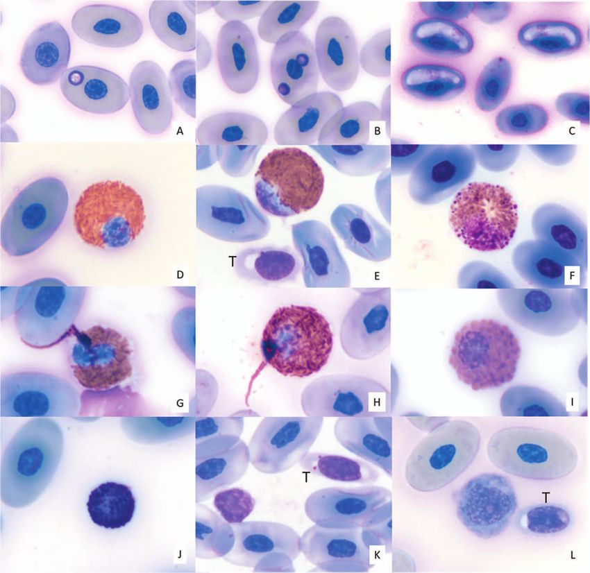
Research article                                                                                           Conservation Physiology • Volume 9 2021
..........................................................................................................................................................

Introduction                                                                       A number of other pathogens are known to cause (e.g.
                                                                               Herpesvirus) or potentially cause (e.g. Ranavirus, Helicobac-
                                                                               ter sp.) similar clinical signs to URTD (Jacobson, 1994;
The gopher tortoise (Gopherus polyphemus) is declining                         Pettan-Brewer et al., 1996; Westhouse et al., 1996; Origgi
throughout its range due to habitat loss and fragmentation,                    and Jacobson, 2000; Origgi et al., 2004; Johnson, 2006;

                                                                                                                                                             Downloaded from https://academic.oup.com/conphys/article/9/1/coab015/6190176 by Florida Atlantic University, jperrault@marinelife.org on 31 March 2021
human interaction including vehicular collision, predation by                  Wellehan et al., 2016). For example, Ranavirus has been
domestic animals and disease (Auffenberg and Franz, 1982;                      associated with nasal and ocular discharge, conjunctivitis
Diemer-Barish et al., 2010; Smith et al., 2006). Gopher tor-                   and subcutaneous edema in tortoise species, including gopher
toises are federally listed in the western portion of their range,             tortoises (Westhouse et al., 1996; Johnson et al., 2008). In
state-listed in FL and currently a candidate for federal listing               several other species of tortoise, Herpesvirus infections can
in the eastern portion of their range (U.S. Fish & Wildlife                    result in necrotizing stomatitis, glossitis, tracheitis, laryngitis
Service, 2011). The inherent impacts of infectious diseases on                 and rhinitis (Jacobson et al., 1985, Drury et al., 1998, Muro
wildlife conservation and biodiversity are evident; however,                   et al., 1998, Johnson et al., 2005). However, because diag-
until recently, these impacts were not often considered. Lack                  nostic tests are not readily available, little is known about
of knowledge with respect to the potential infectious diseases                 the importance and prevalence of these microorganisms in
present within wild populations, the impact of disease status                  wild tortoises. Other pathogens reported in gopher tortoises
on relocation or reproduction of species and disease impacts                   include intestinal parasites such as pinworms, ascarids, flukes
to long-term population viability create a major dilemma for                   and protozoans such as Cryptosporidium spp., a zoonotic
wildlife biologists, conservationists and public policy makers.                pathogen (McGuire et al., 2013, Huffman, 2017); various
This is especially critical for a keystone species such as the                 hemoparasites including Anaplasma spp., which has been
gopher tortoise (Eisenberg, 1983).                                             associated with anemia, and hemogregarines, which are typ-
                                                                               ically considered an incidental finding (Cooney et al., 2016,
    Most disease researches in wild gopher tortoises have                      2019, Raskin et al., 2020), and ectoparasites including ticks
focused on upper respiratory tract disease (URTD) caused                       (e.g. Amblyomma tuberculatum; Ennen and Qualls, 2011).
by Mycoplasma agassizii and Mycoplasma testudineum                             In cases of an immunocompromised and/or stressed host,
(McLaughlin, 1997; Smith et al., 1998; Brown et al., 1999;                     nutritional imbalance, reduced body condition and/or pres-
Berish et al., 2000; McLaughlin et al., 2000; Wendland,                        ence of co-infections, these pathogens may cause chronic
2007). These contagious bacteria, transmitted via direct                       disease, which can lead to reduced reproductive capacity,
contact between tortoises (McLaughlin, 1997), can infect                       abnormal growth and development, increased susceptibility
the respiratory tract of tortoises (Brown et al., 2001)                        to secondary infections and, in some cases, a reduced life span
and cause mild to severe nasal and ocular discharge,                           (U.S. Fish & Wildlife Service, 2019). Identifying the impacts of
conjunctivitis and swelling of the eyes and nares (Jacobson et                 such diseases can be a difficult task. The full effect of chronic
al., 1991; Schumacher et al., 1997; McGuire et al., 2014a).                    disease on a long-lived species such as the gopher tortoise may
Mycoplasmosis is perhaps the most important chronic                            take months to years to manifest in a population. Therefore, it
infectious disease of wild and captive tortoises in North                      is important that populations are monitored using standard-
America and Europe (Jacobson et al., 2014). Diagnostic                         ized techniques so that any changes associated with health
tests specifically validated for gopher tortoise URTD are                      problems may be detected over time (Wendland et al., 2009).
available, including polymerase chain reaction (PCR) to
detect Mycoplasma spp. DNA in nasal sections (Brown                               Despite seemingly healthy adult tortoise populations in
et al., 1995) and an enzyme-linked immunosorbent assay                         many areas of south FL, low fecundity has been observed in
(ELISA) that detects antibodies to M. agassizii 6–8 weeks                      several fragmented habitats, demonstrated by a lack of nests
post-exposure (Schumacher et al., 1993; Wendland et al.,                       within active burrows and a lack of juveniles and sub-adults
2007). Although it is considered a primary cause of disease,                   (Zeiger and Frazier, 2012). This top-heavy demographic struc-
serological tests have indicated that exposure to Mycoplasma                   ture raises concern about population sustainability. Assess-
spp. is widely distributed within the gopher tortoise’s range                  ment of fecund tortoise populations living in fragmented
and exposed animals are not always clinically ill (Jacobson et                 habitats will generate data to help explain health and fecun-
al., 2014). Exposed (i.e. antibody-positive) gopher tortoises                  dity differences observed in geographically and ecologically
have been found in MS (Smith et al., 1998), GA (McGuire                        similar sites. The objective of this study was to conduct a
et al., 2014a, 2014b), AL (Goessling et al., 2019) and                         comprehensive health assessment of two previously unstudied
throughout much of FL (Beyer, 1993; Epperson, 1997;                            gopher tortoise aggregations in southeastern FL.
Smith et al., 1998; Berish et al., 2000; McCoy et al., 2007;
Wendland 2007). Previous investigations have demonstrated
antibody prevalence of 30% and 22% in FL tortoises (Berish                     Materials and methods
et al., 2000; Wendland, 2007), and a more recent study
demonstrated URTD prevalence in FL gopher tortoises
                                                                               Study sites
ranged from 0% to 78%, depending on site (Diemer-Barish                        To obtain estimated population sizes, we conducted surveys
et al., 2010).                                                                 over the 17.3-acre area of Loggerhead Park, Juno Beach,

..........................................................................................................................................................
2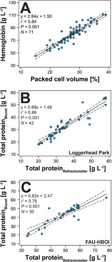
Conservation Physiology • Volume 9 2021 Research article
..........................................................................................................................................................
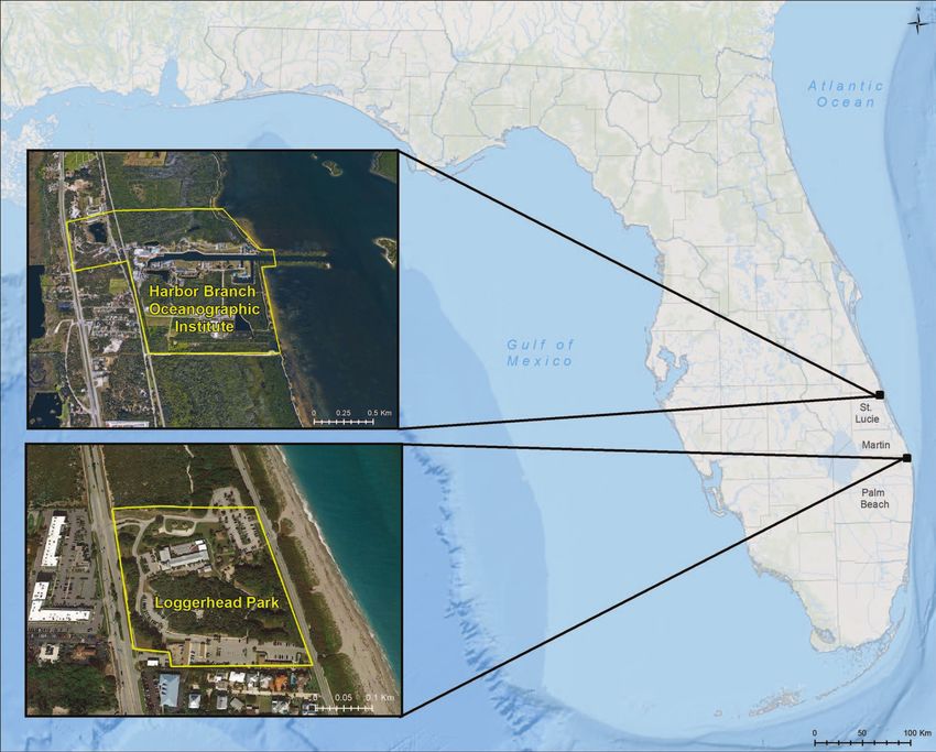
FL (26.8847◦ N, −80.0563◦ W), during March 2018–August 235 mm SCL considered sexually mature adults (Moore
2019, and over the 144-acre campus of Florida Atlantic et al., 2009). All captured adult tortoises were permanently
University’s Harbor Branch Oceanographic Institute (HBOI; and uniquely marked using a triangular file or Dremel tool to
27.5360◦ N, −80.3614◦ W) (Fig. 1) during May–August 2019. notch one or a combination of the eight rearmost marginal
The 17.3-acre Loggerhead Park consists of ∼6.1 acres of scutes, following the Florida Fish and Wildlife Commission
Downloaded from https://academic.oup.com/conphys/article/9/1/coab015/6190176 by Florida Atlantic University, jperrault@marinelife.org on 31 March 2021
marginally to moderately suitable habitat including sandy (FWC)’s gopher tortoise marking guidelines (FWC, 2008).
pine-scrub, oak maritime forest and sandhills (Ashton et al., Prior to notching, the shell was swabbed with povidone iodine
2008). This area is bounded by highly developed commercial and care was taken to avoid injury to the limbs.
parking lot areas on the north and south sides; by a 4-lane
highway with very heavy traffic on the western side, across
from which lie commercially developed paved sites; and by a
Sample collection
2-lane road on the eastern side, across from which is a sandy Venous blood samples (0.5–4 mL;Research article Conservation Physiology • Volume 9 2021
..........................................................................................................................................................
Downloaded from https://academic.oup.com/conphys/article/9/1/coab015/6190176 by Florida Atlantic University, jperrault@marinelife.org on 31 March 2021
Figure 1: Map depicting sampling sites of two gopher tortoise aggregations in southeastern FL, USA.
Hematology and plasma protein Hemoglobin concentration was analyzed in ∼20 μL of
electrophoresis previously frozen, thawed whole blood using a HemoCue®
Hb 201+ photometer (HemoCue® , Inc., Lake Forest, CA,
Blood films were stained using Wright–Giemsa stain USA) with HemoCue® Hb 201 microcuvettes, which has been
(Harleco® , EMD Millipore, Billerica, MA, USA). Light validated for use in birds and used in sea turtles (Velguth et
microscopy was used to conduct complete blood cell counts al., 2010, Harter et al., 2015, Stacy et al., 2019, Page-Karjian
and evaluation of any hemoparasites by one evaluator. et al., 2020) and has a measuring range of 0–256 g l−1 .
Evaluation of blood films included white blood cell (WBC) Frozen plasma aliquots (0.5–1.0 mL) were shipped overnight
estimate (Weiss, 1984) and differential (including mature on dry ice to the University of Miami Avian & Wildlife
heterophils, immature heterophils, lymphocytes, monocytes, Laboratory (UMAW), where they were analyzed for protein
eosinophils and basophils) based on 200 WBC counts, and fractions using the SPIFE 3000 system (Helena Laboratories
morphological evaluation of red blood cells (RBCs), WBCs Inc., Beaumont, TX, USA) and accompanying gels (Dickey
and thrombocytes. Immature heterophils were quantified as et al., 2014), with protein fraction delimits placed using the
a separate WBC category in addition to mature heterophils following conventions: pre-albumin, albumin and alpha-1,
(Stacy et al., 2017), and immature RBCs were counted alpha-2, beta and gamma globulins; total globulins were cal-
as number of immature RBC per 100 mature RBCs. The culated. Total protein was quantified using the Biuret method
heterophil:lymphocyte ratio was calculated using numbers of (TP-B) at UMAW, and the albumin:globulin (A:G) ratio was
mature and immature heterophils combined. calculated for each sample.
..........................................................................................................................................................
4Conservation Physiology • Volume 9 2021 Research article
..........................................................................................................................................................
Parasite and pathogen analyses Statistical analyses
Frozen plasma aliquots were shipped overnight on dry ice Measures of central tendency and range were calculated for
to the University of Florida College of Veterinary Medicine, SCL (mm) and body mass (kg) in juvenile and adult tortoises
Mycoplasma Research Laboratory where they were analyzed from each sampling site. Parametric methods for sample sizes
Downloaded from https://academic.oup.com/conphys/article/9/1/coab015/6190176 by Florida Atlantic University, jperrault@marinelife.org on 31 March 2021
for antibodies to Mycoplasma agassizii and M. testudineum ≥20 but 32), of reference intervals. Mean and standard deviation were
negative (Research article Conservation Physiology • Volume 9 2021
..........................................................................................................................................................
Institutional Animal Care and Use Committee under protocol was absent (N = 1), minimal (N = 2) or mild (N = 1); and
#A17–11. anisocytosis (erythrocytes unequal in size) was absent (N = 1),
minimal (N = 1) or mild (N = 2). One of the tortoises had
two ticks removed at physical examination, while the other
three had no ticks observed. Results of morphological
Downloaded from https://academic.oup.com/conphys/article/9/1/coab015/6190176 by Florida Atlantic University, jperrault@marinelife.org on 31 March 2021
Results evaluation of RBCs, WBCs and thrombocytes are shown in
Overall, 91 tortoises were captured and evaluated for this Table 3. Figure 2 depicts erythrocytes containing inclusions
study, including 57 at Loggerhead Park and 34 at HBOI, suggestive of Anaplasma spp. (A, B) as well as hemogregarine
representing three age classes (Table 1). The HBOI campus gametocytes (C), and Fig. 2D–J shows various examples of
had significantly fewer juvenile tortoises (including hatch- WBCs observed in gopher tortoises in this study. Linear
lings) and more adults—nearly 3 adults for every juvenile regression analysis revealed very strong positive relationships
compared to Loggerhead Park (approximately 1.2 juveniles between PCV and plasma hemoglobin concentration (Fig. 3a)
for every adult) (P = 0.003). Physical examination revealed and between TP-R and TP-B for Loggerhead Park tortoises
that 18/91 (19.8%) of the tortoises had clinical signs consis- (Fig. 3b) and HBOI tortoises (Fig. 3c). Additionally, the
tent with URTD, including nasal discharge (N = 10), asymmet- hemoglobin concentration is about three times the PCV (PCV
rical nares (N = 6), wheezing (N = 5), palpebral/conjunctival ∗ 2.9) using the SI unit (g l−1 ), or a third of the PCV (PCV ∗
swelling (N = 3) and ocular discharge (N = 2). Additionally, 0.29) using the conventional unit (g dl−1 ).
12/91 (13.2%) tortoises had some other form of physical PCV, immature heterophil counts, plasma concentrations
abnormality noted during physical examination, including of hemoglobin, total protein, albumin, alpha-2 globulin, beta
limb (N = 1), eye (N = 1) and shell abnormalities (N = 3), or globulin, gamma globulin, total globulins and A:G were sig-
extra scutes (N = 7). The five tortoises sampled at Loggerhead nificantly correlated to SCL, a proxy for age (Tuberville et al.,
Park that tested ‘suspect’ for antibodies to M. testudineum 2011) (Table 4). Examples of protein electrophoretograms for
were also positive for antibodies to M. agassizii; however, hatchling, juvenile and adult gopher tortoises from this study
none of these five tortoises exhibited clinical signs of URTD are shown in Fig. 4, demonstrating the progression toward
at the time of sampling. All sampled ticks were identified as increased plasma proteins as animals mature, especially with
gopher tortoise ticks (A. tuberculatum). regards to beta globulin and gamma globulin.
All plasma samples had hemolysis scores of either 0 or
A significantly positive relationship (using power regres-
1+, and no lipemia was documented. Mann–Whitney U-
sion) was observed between SCL and body mass in all tor-
tests revealed significant differences between the jugular
toises, including hatchlings, juveniles and adults (R2 = 0.97,
and brachial vein blood sampling sites in adult tortoises
P < 0.01, N = 90). Fisher’s exact tests revealed that adult tor-
[samples from brachial veins had higher median immature
toises were significantly more likely to have intraerythrocytic
heterophils (U = 127.5, N = 44, P = 0.026), gamma globulins
hemogregarine gametocytes (P = 0.001) and more likely to
(U = 84.5, N = 44, P = 0.001) and total globulins (U = 137.5,
have ticks (P = 0.002) than juvenile tortoises. There were no
N = 44, P = 0.049) and lower median A:G ratio (U = 130.5,
significant differences between age classes for PCR results for
N = 44, P = 0.032) and pre-albumin (U = 126.5, N = 44,
Ranavirus, Herpesvirus or Anaplasma spp., or for polychro-
P = 0.025) than samples from jugular veins] and juveniles
masia, anisocytosis or intraerythrocytic inclusions suggestive
[samples from brachial veins had lower median heterophils
of Anaplasma spp. infection (all P > 0.05).
(U = 46.5, N = 32, P = 0.033), alpha-1 globulins (U = 36.5,
N = 29, P = 0.041) and beta globulins (U = 34.5, N = 29, Fisher’s exact tests showed that adult tortoises were signifi-
P = 0.032)]. These differences, however, did not consistently cantly more likely to have clinical signs of URTD compared to
indicate that either sampling site influenced clinical pathology juveniles (P = 0.002). Tortoise sex was not significantly related
results via lymph dilution. Two samples suspected to to Mycoplasma spp. serology results (all P > 0.05). Logistic
have slight lymph dilution based on blood color during regression models (Table 5) revealed statistically significant,
sampling also had low PCVs (10% and 12%) and were not positive relationships between the presence of antibodies to
included in hematological and plasma biochemical analyses. Mycoplasma spp. and SCL, PCV and plasma concentrations
Hematological reference intervals for juveniles and adults of albumin, alpha-2 globulin, beta globulin, and gamma glob-
are presented in Table 2. There were four juvenile tortoises, ulin. Total WBC estimates and heterophil and lymphocyte
two at HBOI and two at Loggerhead Park, that had PCV counts were not significantly related to Mycoplasma spp.
values less than 20%, ranging from 15%–19%. None of serology results (all P > 0.05).
these samples were suspected of lymph dilution, which
can alter blood analyte values (Gottdenker and Jacobson, Tortoises with ticks were significantly more likely to
1995). Of those four tortoises, none had clinical signs have intraerythrocytic hemogregarine gametocytes noted on
or URTD, none had hemoparasites or intraerythrocytic examination of blood films (P < 0.001). Logistic regression
inclusions observed on blood films and all tested negative showed a significant association between PCV and presence
for antibodies to Mycoplasma spp. and for Anaplasma of intraerythrocytic inclusions suggestive of Anaplasma spp.,
spp. via qPCR. Polychromasia (erythrocyte color variation) but not between PCV and qPCR assay results (Table 6). All
..........................................................................................................................................................
6Conservation Physiology • Volume 9 2021 Research article
..........................................................................................................................................................
Table 1: Descriptive statistics resulting from physical examination and pathogen surveys of two gopher tortoise (G. polyphemus) aggregations in
southeastern FL, USA
Loggerhead Park N = 57 HBOI Campus N = 34
Downloaded from https://academic.oup.com/conphys/article/9/1/coab015/6190176 by Florida Atlantic University, jperrault@marinelife.org on 31 March 2021
Physical examination
Age class
Adults 23 (40%)∗ 25 (73.5%)∗
Juveniles 28 (49%)∗ 7 (20.6%)∗
Hatchlings 6 (11%)∗ 2 (5.9%)∗
Sex
Males 15 (26%) 8 (24%)
Females 10 (18%) 17 (50%)
Unknown 32 (56%) 9 (26%)
Pathogen survey
A. tuberculanum ticks 1 (2%)∗ 20 (59%)∗
Intraerythrocytic hemogregarine gametocytes 0/49 (0%)∗ 10/33 (30%)∗
M. agassizii ELISA ‘positive’ (titers > 32) 24/47 (51%) 19/30 (63%)
M. agassizii ELISA ‘suspect’ (titers = 32) 6/47 (13%) 5/30 (17%)
M. testudineum ELISA ‘positive’ (titers > 32) 0/47 (0%) 0/30 (0%)
M. testudineum ELISA ‘suspect’ (titers = 32) 5/47 (11%) 0/30 (0%)
Ranavirus qPCR 0/53 (0%) 0/29 (0%)
Herpesvirus cPCR 0/53 (0%) 0/29 (0%)
Intraerythrocytic inclusions suggestive of Anaplasma spp. 7/49 (14%) 1/33 (3%)
Anaplasma spp. qPCR 12/53 (23%) 9/30 (30%)
∗ Statistically significant differences between sampling sites.
of the tortoises with intraerythrocytic inclusions suggestive of and Jacobson, 1982, Rosenberg et al., 2018) and were not
Anaplasma spp. also had polychromasia, and all but one also indicative of active clinical disease. The plasma protein elec-
had mild to moderate anisocytosis. Cohen’s Kappa coefficient trophoretic profiles were generally higher than those reported
indicated a slight level of diagnostic agreement between by Rosenberg et al. (2018) for a healthy captive group of
Anaplasma-like inclusions viewed during microscopic gopher tortoises. Because consistent methodologies were used
evaluation of blood films and the results of qPCR assays for both studies, these differences are likely real and may
targeting Anaplasma spp. DNA (κ = 0.14, SE of κ = 0.06, 95% indicate a higher degree of antigenic stimulation in the wild
CI = 0.02–0.26). Fisher’s exact tests showed no statistically gopher tortoises sampled in this study. This is consistent
significant relationships between presence of A. tuberculanum with the fact that free-ranging animals typically have higher
ticks and Anaplasma spp. infection (diagnosed via either internal and external parasite burdens and are likely exposed
identification of intraerythrocytic inclusions suggestive of to pathogens more frequently than captive animals, which are
Anaplasma spp., or via qPCR), or between Anaplasma spp. often regularly treated with parasiticides and also receive sup-
infection and Mycoplasma spp. serology results (all were portive care, including anti-microbials, when sick (Jacobson et
P > 0.05). al., 1998). There were four juvenile tortoises in this study with
PCV values less than 20% (range: 15%–19%). Two of these
samples were collected from the brachial vein and two were
collected from the jugular vein. While PCV values ranging
Discussion from 14% to 34% are considered ‘normal’ for healthy gopher
Hematology and plasma biochemistry tortoises (Hernandez et al., 2011, Rosenberg et al., 2018, this
study), PCV values less than 20% are on the low end of the
parameters reference intervals calculated for tortoises in this study. There
The hematology data presented here fall within previously were no data, however, to suggest that these tortoises were
determined reference intervals for gopher tortoises (Taylor unhealthy.
..........................................................................................................................................................
78
Table 2: Reference intervals with 90% confidence interval for upper and lower limits for PCV (N = 35 for juveniles; N = 48 for adults), hemoglobin (N = 26 for juveniles; N = 44 for
Research article
adults), WBC count with differentials (N = 36 for juveniles; N = 44 for adults) and plasma protein electrophoresis (N = 27 for juveniles; N = 44 for adults) in standard international units
for juvenile and adult gopher tortoises (G. polyphemus) from southeastern FL, USA. Mean hematological data are provided for six hatchling gopher tortoises
Juveniles Adults Hatchlings
RI LRL 90% CI URL 90% CI RI LRL 90% CI URL 90% CI Mean ± SD
Hematology
PCV (%) 16–34 14–19 32–36 21–37 19–23 36–39 20 ± 9
Hemoglobin (g L−1 ) 46–99 38–53 91–107 59–110 53–64 105–116 11 ± 9
3
tWBC (x10 μ1−1 ) 4.31–17.31 2.72–5.90 15.72–18.90 5.07–21.13 4.33–5.94 18.05–24.73 9.85 ± 5.17
Total heterophils (×103 μ1−1 ) 0.78–6.59 0–1.56 5.81–7.37 1.39–8.88a 1.13–1.70 7.23–10.90 3.22 ± 2.48
Lymphocytes (×103 μ1−1 ) 1.61–8.26a 1.32–1.97 6.76–10.09 1.24–6.57 0.65–1.83 5.99–7.16 3.30 ± 2.20
3 a
Monocytes (×10 μ1−1 ) 0–1.22 0–0.15 1.07–1.36 0.12–1.86 0.09–0.16 1.37–2.53 0.76 ± 0.39
3 a a
Eosinophils (×10 μ1−1 ) 0.21–4.66 0.15–0.31 3.18–6.84 0.29–5.20 0.21–0.40 3.76–7.18 0.90 ± 0.64
Basophils (×103 μ1−1 ) 0.17–4.22a 0.12–0.26 2.86–6.24 0.19–3.36a 0.14–0.26 2.45–4.62 1.53 ± 1.14
a a
Heterophil:lymphocyte 0.25–3.56 0.17–0.35 2.49–5.10 0.32–2.88 0.25–0.41 2.26–3.67 0.98 ± 0.89
Plasma proteins
Total protein (g L−1 ) 15.8–37.0 12.8–18.8 34.0–40.1 27.2–60.2 23.6–30.9 56.6–63.9 –
Albumin:globulin 0.35–1.27 0.22–0.48 1.14–1.40 0.23–0.74a 0.20–0.26 0.65–0.84 –
a
Pre-albumin (g L−1 ) 2.9–9.7 1.9–3.8 8.8–10.7 4.1–9.5 3.8–4.5 8.7–10.5 –
a
Albumin (g L−1 ) 2.9–7.3 2.3–3.6 6.6–7.9 3.6–9.9 3.2–4.0 8.8–11.1 –
Alpha-1 globulins (g L−1 ) 0.5–2.3 0.3–0.8 2.1–2.6 0.7–2.4 0.6–0.9 2.2–2.5 –
Alpha-2 globulins (g L−1 ) 1.1–4.9 0.6–1.6 4.4–5.5 2.5–7.5 1.9–3.0 7.0–8.1 –
Beta globulins (g L−1 ) 2.6–12.5 1.2–4.0 11.1–13.9 8.1–29.6 5.7–10.4 27.2–31.9 –
Gamma globulins (g L−1 ) 1.1–4.8 0.5–1.6 4.3–5.3 2.6–8.5 1.9–3.2 7.8–9.1 –
Total globulins (g L−1 ) 7.5–22.3 5.4–9.6 20.2–24.3 16.2–46.0 12.9–19.4 42.7–49.3 –
a
Reference intervals were calculated using logarithmic transformations, as original data were non-normal.
Abbreviations: CI, confidence interval; LRL, lower reference limit; RI, reference interval; URL, upper reference limit; SD, standard deviation.
..........................................................................................................................................................
Conservation Physiology • Volume 9 2021
..........................................................................................................................................................
Downloaded from https://academic.oup.com/conphys/article/9/1/coab015/6190176 by Florida Atlantic University, jperrault@marinelife.org on 31 March 2021Conservation Physiology • Volume 9 2021 Research article
..........................................................................................................................................................
Table 3: Morphological evaluation of RBCs, WBCs and thrombocytes for two gopher tortoise (G. polyphemus) aggregations in southeastern FL,
USA. For immature RBC/100 mature RBC and hemogregarines/100 RBC, mean ± standard deviation are reported, with the range parenthetically
Loggerhead Park HBOI Campus
Downloaded from https://academic.oup.com/conphys/article/9/1/coab015/6190176 by Florida Atlantic University, jperrault@marinelife.org on 31 March 2021
Thrombocytes Adequate: 100% (49/49) Adequate: 100% (33/33)
Polychromasia∗ Absent: 31% (15/49)∗ Absent: 9% (3/33)∗
Minimal: 33% (16/49) Minimal: 36% (12/33)
Mild: 29% (14/49) Mild: 55% (18/33)
Moderate: 8% (4/49)
Anisocytosis∗ Minimal: 55% (27/49)∗ Absent: 6% (2/33)
Mild: 37% (18/49)∗ Minimal: 21% (7/33)∗
Moderate: 8% (4/49) Mild: 73% (24/33)∗
Immature RBC/100 3.8 ± 5.7 (0–27) 2.8 ± 1.8 (0–7)
mature RBC
Erythrocyte morphology NSCF: 96% (47/49) NSCF: 97% (32/33)
Rare early stage precursors: 2% (1/49) Rare early stage precursors: 3%
Occasional variably sized clear RBC vacuoles of unknown (1/33)
significance; one to multiple per RBC: 2% (1/49)
RBC inclusions 0: 86% (42/49) 0: 97% (32/33)
suggestive of AnaplasmaResearch article Conservation Physiology • Volume 9 2021
..........................................................................................................................................................
Downloaded from https://academic.oup.com/conphys/article/9/1/coab015/6190176 by Florida Atlantic University, jperrault@marinelife.org on 31 March 2021
Figure 2: Composite of photomicrographs of blood films from gopher tortoises (G. polyphemus) in this study. (A) Erythrocyte with granular
inclusion most consistent with Anaplasma spp. (confirmed by PCR); (B) Erythrocyte with two granular inclusions most consistent with
Anaplasma spp. (confirmed by PCR); (C) Erythrocytes with hemogregarine gametocytes; (D) Mature heterophil; (E) Immature heterophil and
thrombocyte (T); (F) Immature heterophil with primary granules; (G, H) Heterophils with ‘whip-like’ projections; (I) Eosinophil; (J) Basophil;
(K) Small lymphocyte and thrombocyte (T); (L) Monocyte and thrombocyte (T). ×100 objective. Wright–Giemsa stain.
carapace length and antibody prevalence, with juvenile turtles documented to result in factors increasing opportunities for
less likely to have antibodies to Mycoplasma spp. (Beyer, social interactions, including higher incidences of shared
1993, Wendland, 2007) except in populations undergoing burrows, greater home range overlap and increased mating
epizootic events (Wendland, 2010). Interestingly, in this attempts (Guyer et al., 2012). In contrast, although the total
study we detected antibodies to M. agassizii in 8 juveniles prevalence of tortoises with antibodies to M. agassizii was
sampled at Loggerhead Park, representing 29% of the higher at HBOI, only 1 juvenile tortoise (14%) sampled
juveniles sampled at that site. This result, along with the at HBOI had antibodies to M. agassizii. No tortoises at
detection of antibodies to M. agassizii in 70% of the adults either sampling site were confirmed to have antibodies
sampled at Loggerhead Park, suggests that this tortoise to M. testudineum, although 11% of tortoises sampled at
aggregation may be undergoing an M. agassizii epizootic. Loggerhead Park had ‘suspect positive’ titers. Antibodies to
This finding may be related to density-dependent factors, M. testudineum in gopher tortoises have typically been shown
since increased gopher tortoise population density has been in tortoises captured in more northern latitudes, including the
..........................................................................................................................................................
10Conservation Physiology • Volume 9 2021 Research article
..........................................................................................................................................................
Table 4: Significant Spearman correlations between SCL (a proxy for age) and measured blood analytes for two gopher tortoise (G. polyphemus)
aggregations in southeastern FL, USA. Blood values were combined for the two aggregations
Analyte rs P N
Downloaded from https://academic.oup.com/conphys/article/9/1/coab015/6190176 by Florida Atlantic University, jperrault@marinelife.org on 31 March 2021
PCV 0.38Research article Conservation Physiology • Volume 9 2021
..........................................................................................................................................................
Table 6: Results of logistic regression analysis to examine the relationships between PCV and Anaplasma spp. diagnostics, including
presence/absence of intraerythrocytic inclusions and qPCR results, for both sampling sites combined. ∗ Statistically significant associations
Data for explanatory variables Model fit
Downloaded from https://academic.oup.com/conphys/article/9/1/coab015/6190176 by Florida Atlantic University, jperrault@marinelife.org on 31 March 2021
Variable Coefficient SE P Odds ratio 95% CI χ2 P df
Intraerythrocytic inclusions suggestive of −0.17 0.09 0.048∗ 0.84 0.71–1.00 4.35 0.037 1
Anaplasma spp. vs. PCV
Anaplasma spp. qPCR results vs. PCV 0.07 0.06 0.230 1.07 0.96–1.20 1.48 0.224 1
29%, respectively). This, along with the lack of a significant it is not possible to speciate them based on morphological
correlation between presence of antibodies to Mycoplasma characteristics alone, and molecular characterization was not
spp. and clinical signs of URTD, may be explained by the fact performed in this study (Hernandez et al., 2011).
that detection of circulating antibodies is typically associated
with past or chronic infection (Jacobson et al., 1995), while Upon hematological examination, variably-sized (2–5 μm),
clinical signs of URTD may signify current mycoplasmal round to oval, basophilic, stippled, intracytoplasmic inclu-
infections in tortoises that have not had time to seroconvert sions were observed within erythrocytes of 7 tortoises (14%)
(Diemer-Barish et al., 2010). In these instances, infection with captured at Loggerhead Park, and in 1 (3%) tortoise captured
another mycoplasmal species or other respiratory pathogens at HBOI. These inclusions were consistent with previous
must also be considered (Wendland, 2007). The presence descriptions of Anaplasma spp., a bacterial hemoparasite
of a statistically significant relationship between antibodies associated with anemia and an emerging pathogen in gopher
to M. agassizii and PCV and plasma alpha-1 globulin, beta tortoises (Crosby et al., 2016, Wellehan et al., 2016, Raskin et
globulin and gamma globulin concentrations is intriguing, al., 2020). Although Anaplasma infections in other species are
particularly since the beta and gamma protein fractions known to be transmitted by ticks (Vanstreels et al., 2018), in
contain antibodies. Both PCV and total globulins were also this study statistically significant relationships were not found
positively correlated to tortoise size; therefore, size and/or between the presence of A. tuberculanum ticks and intraery-
hydration status may be a confounding variable in these throcytic inclusions, nor between ticks and blood samples that
results. While the effects of mycoplasmosis on the long-term were positive for Anaplasma spp. via qPCR. Ticks were not
health and viability of gopher tortoise populations is not well tested for Anaplasma spp. using qPCR in this study; future
understood, it seems likely that physiological stress associated studies should include directly testing ticks for this emerg-
with extrinsic stressors including human impacts on tortoises ing pathogen. Anaplasmosis in previously reported gopher
and their habitats and population density are related to both tortoise cases has been associated with anemia that was
overt morbidity and mortality, as well as seroconversion that attributed to hemolytic disease (Raskin et al., 2020). Here,
is detectable via molecular assays (Jacobson et al., 2014). statistical analysis revealed a significant, negative association
between PCV and presence of intraerythrocytic inclusions
None of the tortoises tested positive for Ranavirus or suggestive of Anaplasma spp. Additionally, all tortoises with
Herpesvirus via PCR; this represents important baseline data, Anaplasma-like inclusions also had polychromasia, an indica-
since these viruses are thought to be emerging pathogens tion of increased release of erythrocytes from hematopoietic
of other tortoise and turtle species (Johnson et al., 2005, tissues, and all but one also had anisocytosis, characterized
2008, 2010, Jacobson et al., 2012). Adult tortoises were by erythrocytes of unequal size. None of these tortoises had
significantly more likely than juvenile tortoises to have both abnormally colored plasma samples that would be indicative
ticks and intraerythrocytic hemogregarine gametocytes, and of hemolysis. Both polychromasia and anisocytosis can be
tortoises with ticks were significantly more likely to have associated with regenerative anemia in reptiles; thus, this
intraerythrocytic hemogregarine gametocytes. These results observation could indicate underlying hemolysis and contin-
are noteworthy because hemogregarines in tortoises are ued erythrocyte regeneration in Anaplasma spp.-infected tor-
thought to be transmitted by ticks (Cook et al., 2009). toises (Jackson, 2007, Stacy et al., 2011). The mean ± SD PCV
Differences observed between sampling sites also support this for tortoises with Anaplasma-like intraerythrocytic inclusions
hypothesis, since ticks were found on 59% and hemoparasites was 23 ± 7, while the mean ± SD PCV for tortoises without
were identified in 30% of the tortoises sampled at HBOI, these inclusions was 28 ± 5; however, only one of the tortoises
while only a single tick was found, and no hemoparasites with Anaplasma-like intraerythrocytic inclusions was clini-
were identified in tortoises sampled at Loggerhead Park. cally anemic based on previously published reference intervals
These hemoprotozoans were considered an incidental finding (Rosenberg et al., 2018). Moreover, lymph dilution of the
in these cases (Stacy et al., 2017); in general, the clinical blood sample from this tortoise cannot be excluded since TP-
significance of hemoparasite infections is related to the level R and TP-B were also low (Conservation Physiology • Volume 9 2021 Research article
..........................................................................................................................................................
heterophil projections are considered artifact or associated
with inflammation (non-specific) (Stacy et al., 2017). Given
consistent sample handling and processing times at both sites,
artifact is less likely. An association with the presence of a
pathogen such as Anaplasma spp., or other pathogens, and
Downloaded from https://academic.oup.com/conphys/article/9/1/coab015/6190176 by Florida Atlantic University, jperrault@marinelife.org on 31 March 2021
an immune response is plausible. There was only a slight level
of diagnostic agreement between Anaplasma-like inclusions
viewed during microscopic evaluation of blood films and
the results of qPCR assays targeting Anaplasma spp. DNA.
This result was driven by the number of negative agreements
between the two diagnostic techniques (N = 49). While PCR
is likely a more sensitive method for diagnosing this blood
parasite, infection confirmation is most reliable when the two
diagnostic methods are applied in tandem, since the number
of organisms may be too low for PCR detection or be missed
by blood film review, respectively. Because there were no
tortoises that had both the inclusions and a positive qPCR
result, it is difficult to make conclusive statements about the
presence and significance of Anaplasmosis in these gopher
tortoise aggregations.
Conclusions
This work contributes important baseline health informa-
tion on gopher tortoises toward the southern end of the
species’ range. Because the gopher tortoise is one of the most
commonly translocated species in North America (Tuberville
et al., 2011; Cozad et al., 2020), it is important to understand
pathogen distributions within their populations (McGuire
et al., 2014b). This study highlights the importance of con-
tinued health surveillance of gopher tortoise populations, as
we detected an emerging pathogen (Anaplasma spp.), docu-
mented the absence of two other emerging pathogens (Her-
pesvirus, Ranavirus) and provided evidence for a potential
M. agassizii epizootic within the tortoise aggregation inhabit-
ing Loggerhead Park. Long-term studies of these and other
populations of management concern will help us to better
understand the consequences of disease and various stressors
on important variables including behavior and reproductive
potential (McGuire et al., 2014a, 2014b). Thus, further health
assessments and pathogen surveillance in the gopher tortoises
of southeastern FL are warranted.
Figure 3: Linear regression analysis revealed strong positive
relationships between (A) PCV and plasma hemoglobin
concentration and between plasma total protein by refractometer Funding
and total protein by biuret method for both (B) Loggerhead Park and
(C) HBOI gopher tortoises (G. polyphemus). Total protein relationships This work was supported by grants from Association of Rep-
by refractometer were analyzed separately for the two aggregations tile and Amphibian Veterinarians, Chicago Herpetological
as different brands of refractometer were used. Society, Wildlife Disease Association Challenge in association
with experiment.com; generous donations from the Albert E.
and Birdie W. Einstein Fund, Bonnie Simes; and various
icant relationship between PCV and qPCR assay results. donors to our crowdfunding efforts to fund this project. Sum-
Interestingly, the Loggerhead Park tortoise aggregation with mer internship funds were provided by the Link Foundation
higher frequency of Anaplasma spp. had heterophil pro- [to K.R.] and by the James Pomponi Memorial Scholarship
jections, which were absent in tortoises at HBOI. These Fund [to C.X.].
..........................................................................................................................................................
13Research article Conservation Physiology • Volume 9 2021
..........................................................................................................................................................
Downloaded from https://academic.oup.com/conphys/article/9/1/coab015/6190176 by Florida Atlantic University, jperrault@marinelife.org on 31 March 2021
Figure 4: Representative plasma protein electrophoretograms of (A) hatchling, (B) juvenile, (C) adult female and (D) adult male gopher
tortoises (G. polyphemus) from southeastern FL showing the fractions of interest: pre-albumin, albumin, alpha-1 globulins, alpha-2 globulins,
beta globulins and gamma globulins. An adult male tortoise that was seropositive for M. agassizii (E) had notably larger fractions of alpha-2
globulin, beta globulin and gamma globulin compared (F) to an adult male tortoise that was seronegative for M. agassizii. All data were
determined in non-hemolyzed plasma samples. By convention, no units are reported on the y-axis (Gicking et al., 2004).
Acknowledgements collection; Debra Miller for sharing Ranavirus qPCR tar-
get sequences; and the ZooMed Diagnostic Laboratory, the
Mycoplasma Research Laboratory at the University of Florida
We thank Adrienne McCracken, Nicole Montgomery, Jennifer and the Infectious Diseases Laboratory at the University of
Reilly and Ashley Sabater for their assistance with sample Georgia for sample analysis.
..........................................................................................................................................................
14Conservation Physiology • Volume 9 2021 Research article
..........................................................................................................................................................
Cooney BT, Elhassani D, Frazier E, Caruso J (2016) A comparative sur-
References vey of Gopherus polyphemus hemoparasites in four different South
Florida habitats. J Immunol 196 (1S): 216.5.
Allender MC, Bunick D, Mitchell MA (2013) Development and validation
of TaqMan quantitative PCR for detection of frog virus 3-like virus Cozad RA, Hernandez SM, Norton TM, Tuberville TD, Stacy NI, Stedman
Downloaded from https://academic.oup.com/conphys/article/9/1/coab015/6190176 by Florida Atlantic University, jperrault@marinelife.org on 31 March 2021
in eastern box turtles (Terrapene carolina carolina). J Virol Methods NL, Aresco MJ (2020) Epidemiological investigation of a mortality
188(1–2): 121–125. event in a translocated gopher tortoise (Gopherus polyphemus) pop-
ulation in northwest Florida. Front Vet Sci 7: 120.
Ashton KG, Engelhardt BM, Branciforte BS (2008) Gopher tortoise
(Gopherus polyphemus) abundance and distribution after prescribed Crosby FL, Peltierra L, Weeden AL, Wellehan JFX, Brown MB, Lundgren
fire reintroduction to Florida scrub and sandhill at Archbold Biologi- AM, Barbet AF (2016) Novel Anaplasma species in the environmen-
cal Station. J Herpetol 42: 523–529. tally threatened Florida gopher tortoise. Proceedings of the 28th Meet-
ing of the American Society for Rickettsiology.
Auffenberg W, Franz R (1982) The status and distribution of the gopher
tortoise (Gopherus polyphemus). In RB Bury, ed, North American Tor- Dickey M, Cray C, Norton T, Murray M, Barysauskas C, Arheart K, Nelson
toises: Conservation and Ecology. Wildlife Research Report No. 12, U.S. S, Rodriguez M (2014) Assessment of hemoglobin binding protein in
Fish and Wildlife Service, Washington, pp. 95–126. loggerhead sea turtles (Caretta caretta) undergoing rehabilitation. J
Zoo Wildl Med 45(3): 700–703.
Berish JE, Medica PA (2014) Home range and movements of North Amer-
ican tortoises. In DC Rostal, ED McCoy, HR Mushinsky, eds, Biology and Diemer-Barish JE, Wendland LD, Kiltie RA, Garrison EP, Gates CA (2010)
Conservation of North American Tortoises. Johns Hopkins University Effects of mycoplasmal upper respiratory tract disease on morbidity
Press, USA, pp. 96–182. and mortality of gopher tortoises in northern and central Florida. J
Wildl Dis 46(3): 695–705.
Berish JED, Wendland LD, Gates CA (2000) Distribution and prevalence
of upper respiratory tract disease in gopher tortoises in Florida. J Dodd CK. 1995. Disarticulation of turtle shells in North-Central Florida—
Herpetol 34(1): 5–12. how long does a shell remain in the woods. Am Midl Nat 134(2):
378–387.
Beyer SM (1993) Habitat relations of juvenile gopher tortoises and a pre-
liminary report of upper respiratory tract disease (URTD) in gopher Drury SEN, Gough RE, McArthur S, Jessop M (1998) Detection of Her-
tortoises. MS Thesis, Iowa State University, Ames, Iowa. pesvirus-like and papilloma-like particles associated with diseases of
tortoises. Vet Rec 143 (23): 639.
Brown MB, Brown DR, Klein PA, McLaughlin GS, Schumacher IM, Jacob-
son ER, Adams HP, Tully JG (2001) Mycoplasma agassizii sp. nov., Eisenberg J (1983) The gopher tortoise as a keystone species. In
isolated from the upper respiratory tract of the desert tortoise RJ Bryant, R Franz, eds, The Gopher Tortoise: A Keystone Species,
(Gopherus agassizii) and the gopher tortoise (Gopherus polyphemus). pp. 1–4. Florida State Museum, GainesvilleProceedings of the 4th
Int J Syst Evol Microbiol 51(2): 413–418. Annual Meeting of the Gopher Tortoise Council
Brown DR, Crenshaw BC, McLaughlin GS, Schumacher IM, McKenna Ennen J, Qualls C (2011) Distribution and habitat utilization of the
CE, P. A. Klein, Jacobson ER, Brown MB (1995) Taxonomic analysis gopher tortoise tick (Amblyomma tuberculatum) in Southern Missis-
of the tortoise mycoplasmas Mycoplasma agassizii and Mycoplasma sippi. J Parasitol 97(2): 202–206.
testudinis by 16S rRNA gene sequence comparison. Int J Syst Bacteriol Epperson DM (1997) Gopher tortoise (Gopherus polyphemus) popula-
45(2): 348–350. tions: Activity patterns, upper respiratory tract disease, and man-
Brown MB, McLaughlin GS, Klein PA, Crenshaw BC, Schumacher IM, agement on a military installation in northeast Florida. MS Thesis.
Brown DR, Jacobson ER (1999) Upper respiratory tract disease in the University of Florida, Gainesville, Florida.
gopher tortoise is caused by Mycoplasma agassizii. J Clin Microbiol Eubanks JO, Michener WK, Guyer C (2003) Patterns of movement and
37(7): 2262–2269. burrow use in a population of gopher tortoise (Gopherus polyphe-
mus). Herpetologica 59(3): 311–321.
Campbell TW (2006) Clinical pathology of reptiles. In DR Mader, ed,
Reptile Medicine and Surgery, EdEd 2. W.B. Saunders, Philadelphia, pp. Fleming KA, Perrault JR, Stacy NI, Coppenrath CM, Gainsbury AM (2020)
453–470. Heat, health and hatchlings: associations of in situ nest tempera-
tures with morphological and physiological characteristics of log-
Cook CA, Smit NJ, Davies AJ (2009) A redescription of Haemogrega-
gerhead sea turtle hatchlings from Florida. Cons Physiol 8(1): 1–17.
rina fitzsimonsi Dias, 1953 and some comments on Haemgregarina
doi:10.1093/conphys/coaa046.
parvula Dias, 1953 (Adeleorina: Haemogregarinidae) from southern
African tortoises (Cryptodira: Testudinidae), with new host data and Florida Fish and Wildlife Conservation Commission (FWC) (2008) Gopher
distribution records. Folia Parasit 56: 173–179. tortoise permitting guidelines. Tallahassee, Florida.
Cooney BT, Elhassani D, Bari A, Huffman J, Frazier E (2019) Prevalence Friedrichs KR, Harr KE, Freeman KP, Szladovits B, Walton RM, Barnhart KF,
and levels of parasitemia of Hepatozoon sp. (Apicomplexa: Adeleo- Blanco-Chavez J (2012) ASVCP reference interval guidelines: deter-
rina) in four gopher tortoise (Gopherus polyphemus) populations of mination of de novo reference intervals in veterinary species and
south Florida, USA. J Wildl Dis 55(3): 654–657. other related topics. Vet Clin Pathol 41: 441–453.
..........................................................................................................................................................
15Research article Conservation Physiology • Volume 9 2021
..........................................................................................................................................................
Gicking JC, Foley AM, Harr KE, Raskin RE, Jacobson E (2004) Plasma Agassizii, in the Colorado desert of Southern California. J Zoo Wildl
protein electrophoresis of the Atlantic loggerhead sea turtle, Caretta Med 25(1): 68–81.
caretta. J Herp Med Surg 14(3): 13–18.
Johnson AJ (2006) Iridovirus infections of captive and free-ranging che-
Goessling JM, Guyer C, Godwin JC, Hermann SM, Sandmeier FC, Smith lonians in the United States. Ph.D. Dissertation. Veterinary Sciences,
Downloaded from https://academic.oup.com/conphys/article/9/1/coab015/6190176 by Florida Atlantic University, jperrault@marinelife.org on 31 March 2021
LL, Mendonça (2019) Upper respiratory tract disease and associ- University of Florida, Gainesville, FL.
ated diagnostic tests of mycoplasmosis in Alabama populations
Johnson AJ, Pessier AP, Wellehan JFX, Brown R, Jacobson ER (2005)
of gopher tortoises, Gopherus polyphemus. PLoS One 14(4): 1–12.
Identification of a novel Herpesvirus from a California desert tortoise
doi:10.1371/journal.pone.0214845
(Gopherus agassizii). Vet Microbiol 111 (1–2): 107–116.
Gottdenker NL, Jacobson ER (1995) Effect of venipuncture sites on
hematologic and clinical biochemical values in desert tortoises Johnson AJ, Pessier AP, Wellehan JFX, Childress A, Norton TM, Stedman
(Gopherus agassizii). Am J Vet Res 56(1): 19–21. NL, Bloom DC, Belzer W, Titus VR, Wagner R, Brooks JW, Spratt J,
Jacobson ER (2008) Ranavirus infection of free-ranging and cap-
Guyer C, Johnson VM, Hermann SM (2012) Effects of population den- tive box turtles and tortoises in the United States. J Wildl Dis 44(4):
sity on patterns of movement and behavior of gopher tortoises 851–863.
(Gopherus polyphemus). Herpetol Monogr 26: 122–134.
Johnson AJ, Wendland L, Norton TM, Blezer B, Jacobson ER (2010) Devel-
Harter TS, Reichert M, Brauner CJ, Milsom WK (2015) Validation of the opment and use of an indirect enzyme-linked immunosorbent assay
i-STAT and HemoCue systems for the analysis of blood parameters for detection of iridovirus exposure in gopher tortoise (Gopherus
in the bar-headed goose, Anser indicus. Conserv Physiol 3(1): 1–9. polyphemus) and eastern box turtles (Terrapene carolina carolina). Vet
doi:10.1093/conphys/cov021. Microbiol 142: 160–167.
Hernandez SM, Tuberville TD, Frank P, Stahl SJ, McBride MM, Buhlmann Keirans JE, Litwak TR (1989) Pictorial key to the adults of hard ticks,
KA, Divers SJ (2011) Health and reproductive assessment of a free- family Ixodidae (Ixodida:Ixodoidea), East of the Mississippi River. J
ranging gopher tortoise (Gopherus polyphemus) population follow- Med Entomol 26(5): 435–448.
ing translocation. J Herpetol Med Surg 20(2–3): 84–93.
McCoy ED, Mushinsky HR, Lindzey J (2007) Conservation strategies and
Huffman JN (2017) A survey of Gopherus polyphemus intestinal parasites emergent diseases: The case of upper respiratory tract disease in the
in South Florida. Florida Atlantic University, Boca Raton, M.S. Thesis. gopher tortoise. Chel Cons Biol 6(2): 170–176.
Jacobson ER, Behler JL, Jarchow JL (1998) Health assessment of chelo- McGuire JL, Miller EA, Norton TM, Raphael BL, Spratt JS, Yabsley
nians and release into the wild. In ME Fowler, RE Miller, eds, Zoo and MJ (2013) Intestinal parasites of the gopher tortoise (Gopherus
Wild Animal Medicine, Current Therapy 4. W.B. Saunders Co, Philadel- polyphemus) from eight populations in Georgia. Parasitol Res 112(12):
phia. 4205–4210.
Jackson ML (2007) Chapter 1: Erythrocytes. In ML Jackson, ed, Veterinary McGuire JL, Smith LL, Guyer C, Lockhart JM, Lee GW, Yabsley MJ (2014a)
Clinical Pathology: An Introduction. Blackwell Publishing, Ames, Iowa. Surveillance for upper respiratory tract disease and Mycoplasma in
Jacobson ER, Berry KH, Wellehan JFX Jr, Origgi F, Childress AL, Braun J, free-ranging gopher tortoises (Gopherus polyphemus) in Georgia,
Schrenzel M, Yee J, Rideout B (2012) Serologic and molecular evi- USA. J Wildl Dis 50(4): 733–744.
dence for testudinid Herpesvirus 2 infection in wild Agassiz’s desert McGuire JL, Smith LL, Guyer C, Yabsley MJ (2014b) Effects of Mycoplas-
tortoises, Gopherus agassizii. J Wildl Dis 48: 747–757. mal upper-respiratory-tract disease on movement and thermoregu-
Jacobson ER, Brown MB, Wendland LD, Brown DR, Klein PA, Christopher latory behavior of gopher tortoises (Gopherus polyphemus) in Geor-
MM, Berry KH (2014) Mycoplasmosis and upper respiratory tract gia, USA. J Wildl Dis 50(4): 745–756.
disease of tortoises: a review and update. Vet J 201(3): 257–264.
McLaughlin GS (1997) Upper respiratory tract disease in
Jacobson ER, Brown MB, Schumacher IM, Collins BR, Harris RK, Klein PA gopher tortoises, Gopherus polyphemus: pathology, immune
(1995) Mycoplasmosis and the desert tortoise (Gopherus agassizii) in responses, transmission, and implications for conservation and
Las Vegas Valley, Nevada. Chel Cons Biol 1: 279–284. management. PhD Dissertation, University of Florida, Gainesville,
Florida.
Jacobson ER, Clubb S, Gaskin JM, Gardiner C (1985) Herpesvirus-
like infection in Argentine tortoises. J Am Vet Med Assoc 187(11): McLaughlin GS, Jacobson ER, Brown DR, McKenna CE, Schumacher IM,
1227–1229. Adams HP, Brown MB, Klein PA (2000) Pathology of upper respiratory
tract disease of gopher tortoises in Florida. J Wildl Dis 36(2): 272–283.
Jacobson ER, Gaskin JM, Brown MB, Harris RK, Gardiner CH, Lapointe
JL, Adams HP, Reggiardo C (1991) Chronic upper respiratory tract McRae WA, Landers JL, Cleveland GD (1981) Sexual dimorphism in the
disease of free-ranging desert tortoises (Xerobates agassizii). J Wildl gopher tortoise (Gopherus polyphemus). Herpetologica 37(1): 46–52.
Dis 27(2): 296–316.
Moore JA, Strattan M, Szabo V (2009) Evidence for year-round reproduc-
Jacobson ER, Wronski TJ, Schumacher J, Reggiardo C, Berry KH (1994) tion in the gopher tortoise (Gopherus polyphemus) in southeastern
Cutaneous dyskeratosis in free-ranging desert tortoises, Gopherus- Florida. Bull Peabody Mus Nat Hist 50(2): 387–392.
..........................................................................................................................................................
16You can also read