Pediatric lupus nephritis - Nefrite lúpica em pediatria - SciELO
←
→
Page content transcription
If your browser does not render page correctly, please read the page content below
Review Articles | Artigos de Revisão

                                             Pediatric lupus nephritis
                                             Nefrite lúpica em pediatria

      Authors                                Abstract                                        Resumo
      Sergio Veloso Brant Pinheiro1
                                             Involvement of the kidneys by lupus ne-         A nefrite lúpica (NL) é caracterizada pelo aco-
      Raphael Figuiredo Dias1                phritis (LN) is one of the most severe          metimento dos rins no contexto das diversas
      Rafaela Cabral Gonçalves               clinical manifestations seen in individu-       manifestações clínicas do Lupus Eritematoso
      Fabiano1
                                             als with systemic lupus erythematosus           Sistêmico (LES), e representa uma das ma-
      Stanley de Almeida Araujo1
                                             (SLE). LN is more frequent and severe           nifestações clínicas mais graves da doença.
      Ana Cristina Simões e Silva1
                                             in pediatric patients and has been asso-        A NL é mais frequente e mais grave nos pa-
                                             ciated with higher morbidity and mor-           cientes pediátricos, em comparação com os
                                             tality rates. This narrative review aimed       adultos, e causa maiores taxas de morbidade
      1
       Universidade Federal de Minas
      Gerais, Hospital das Clínicas,
                                             to describe the general aspects of LN           e mortalidade. O objetivo desta revisão nar-
      Unidade de Nefrologia Pediátrica,      and its particularities when affecting          rativa foi descrever os aspectos gerais da NL
      Belo Horizonte, MG, Brasil.            children and adolescents, while focus-          e suas particularidades em crianças e adoles-
                                             ing on the disease’s etiopathogenesis,          centes, com foco em sua etiopatogênese, nas
                                             clinical manifestations, renal tissue al-       manifestações clínicas, nas alterações histopa-
                                             terations, and treatment options.               tológicas renais e na abordagem terapêutica.

                                             Keywords: Lupus Nephritis; Pediatrics;          Palavras-chave: Nefrite Lúpica; Pediatria;
                                             Autoimmunity; Antibodies, Antinuclear.          Autoimunidade; Anticorpos Antinucleares.

                                             Introduction                                    higher morbidity and mortality rates.5,6
                                                                                             This review aimed to describe the general
                                             Systemic lupus erythematosus (SLE) is a
                                                                                             and particular features of LN in children
                                             chronic inflammatory condition that af-
                                                                                             and adolescents and to shed light on the
                                             fects numerous organs such as the skin,
                                                                                             disease’s etiopathogenesis, clinical mani-
                                             joints, lungs, heart, kidneys, and nervous
                                                                                             festations, histopathology, and treatment.
                                             system.1 Its etiology is multifactorial and
                                             includes genetic and environmental fac-
                                                                                             Epidemiology
                                             tors. The involved pathophysiological
                                             mechanisms include decreased immune             SLE preferentially affects non-Caucasian
                                             tolerance, production of antibodies, de-        young women.7,8 Patients aged 18 years
                                             position of immune complexes on target          or younger account for up to 20% of the
                                             tissues, and activation of the complement       cases.9 The prevalence of SLE in children
                                             system.2-4                                      and adolescents (juvenile SLE) varies as a
                                                 Involvement of the kidneys by lupus ne-     function of the ethnicity and age range of
                                             phritis (LN) is one of the most severe clini-   the individuals enrolled in different stud-
                                             cal manifestations observed in individuals      ies.9 Juvenile SLE is a rare disease, with an
      Submitted on: 04/24/2018.              with systemic lupus erythematosus (SLE).        incidence of 0.3-0.9/100,000 children per
      Approved on: 09/05/2018.
                                             LN is more frequent and severe in pediat-       year and a prevalence of 3.3-8.8/100,000
                                             ric patients and has been associated with       children.10
      Correspondence to:
      Ana Cristina Simões e Silva.
      E-mail: acssilva@hotmail.com

      DOI: 10.1590/2175-8239-JBN-2018-0097

252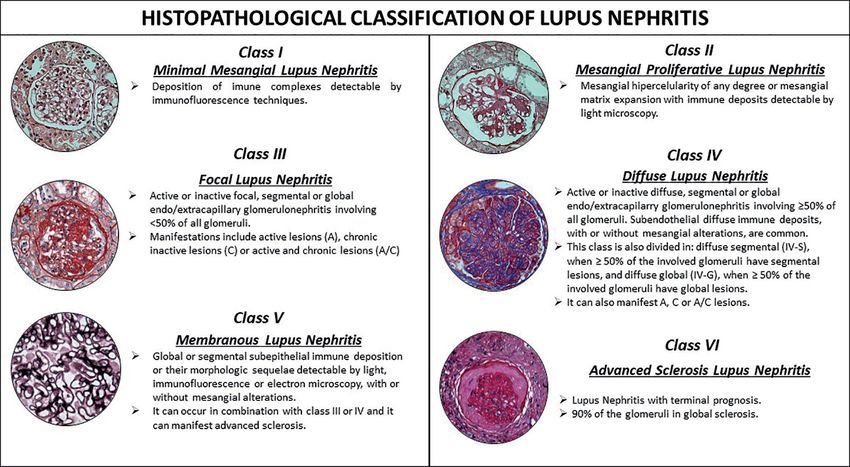
Pediatric lupus nephritis

    Neonatal SLE is a rare condition that equally af-      to neutrophil apoptosis (NETosis) - rarely persist
fects individuals of both sexes. It is usually associ-     for long enough to be processed by antigen-present-
ated with maternal SLE and other autoimmune dis-           ing cells.22,25 The clearance of dead cells and genetic
eases.11,12 Multicenter studies performed in Brazil and    material is impaired in SLE on account of apoptosis
the USA suggested that SLE in infants is usually asso-     and NETosis defects, which expose self-antigens to
ciated with complement deficiencies.13,14 Female chil-     the immune system.22,25 Some genetic defects of the
dren and adolescents develop SLE more commonly,            complement system may introduce flaws in opsoniza-
possibly due to the hormonal changes of puberty.15         tion and thus impair the clearance of self-antigens.22
The predominance of SLE in female pediatric patients       Nuclear self-antigen internalization and recognition
increases gradually with age to the values observed in     by toll-like receptors (TLR 2 and 9 in particular) pro-
adults.16-19                                               motes the conversion of dendritic cells into antigen-
    Although similar to the manifestations observed in     presenting cells, and consequently the activation of
adults with SLE, the clinical events present in juvenile   autoreactive T cells.22-25 By their turn, autoreactive T
SLE are usually more severe and involve multiple or-       cells amplify the immune response by increasing the
gans.1,5,6,20,21 Renal involvement occurs in 50-75% of     production of T and B cells in the bone marrow and
pediatric patients with SLE and more than 90% de-          lymphoid organs.22-25 Active B cells may differentiate
velop LN within two years of diagnosis.1 Individuals       into plasma B cells or memory B cells.22-25 Active B cells
aged 10-13 years are preferentially involved and pres-     continuously exposed to nuclear self-antigens produce
ent an incidence of 0,72/100,000 per year.1,20 The risk    large quantities of autoantibodies, which then react
of patients with juvenile LN developing LN is higher       with nuclear self-antigens to form circulating immune
among Asians, African Americans, and Hispanics.21          complexes (CIC).22-25 CIC are not adequately cleared
    The 5-year renal survival of children with LN has      and deposit in various tissues.22-25 A few physiological
improved markedly in recent decades, and currently         phenomena protect self-DNA against identification
ranges from 77% to 93%.21 However, when com-               by the immune system.26 Impaired clearance of dead
pared to healthy children, the mortality rate seen in      cells and genetic material has been associated with
pediatric individuals with LN is 19 times greater.21       loss of discrimination between self-genetic and viral
The prognosis of children with LN and end-stage            material by the immune system.26
renal disease is particularly somber. Mortality rates          Renal involvement in SLE derives from the deposi-
within the first five years of renal replacement therapy   tion of CIC in renal tissue or from the formation of IC
may reach 22%, mainly on account of cardiopulmo-           in situ (Figure 1).23-25 The deposition of IC in renal tis-
nary complications.21                                      sue activates the classical complement, macrophage,
                                                           and neutrophil pathways from the binding of phago-
Etiopathogenesis                                           cyte surface Fc receptors and immunoglobulin com-
The pathogenesis of SLE involves a complex interac-        plexes.23,25 Complement system protein C1q binds to
tion between genetic susceptibility and environmen-        the Fc region of IgG (IgG1 and IgG3 in particular) or
tal factors, which result primarily in loss of immune      IgM present in IC deposits to promote neutrophil ac-
tolerance and onset of chronic autoimmunity.22-25          tivation.25 The activation of the classical complement
Genetic susceptibility stems from genetic mutations        pathway leads to the formation of chemoattractant
that may predispose patients to developing SLE.22-25       complement system proteins (C3a and C5a), which
Environmental factors induce epigenetic alterations -      also induce neutrophil recruitment.23-25 Local neu-
variations in gene expression caused by DNA meth-          trophil activation and recruitment trigger the release
ylation and histone modification and/or non-coding         of reactive oxygen species (ROS), the production of
RNA - that may trigger the onset of SLE in genetically     proinflammatory cytokines, and the amplification
predisposed individuals. Epigenetic changes may be         of immune and inflammatory response in renal tis-
caused by factors such as viral infection, sun expo-       sues.23-25 Proinflammatory and profibrotic cytokines
sure, hormonal alterations, nutrition, physical and        [mainly interleukin-4 (IL-4), transforming growth
mental stress, and medication.22-25                        factor-beta (TGF-beta), tumor necrosis factor (TNF),
    Loss of immune tolerance is the initial trigger for    and interferon gamma (IFN-gamma)] induce different
SLE.22-25 Immune tolerance is not lost under normal        grades of podocyte injury, proliferation of mesangial,
conditions, since nuclear self-antigens - subsequently     endothelial, and parietal epithelial cells, increased

                                                                          Braz. J. Nephrol. (J. Bras. Nefrol.) 2019;41(2):252-265   253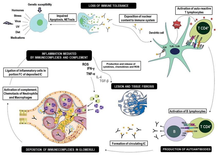
Pediatric lupus nephritis

      Figure 1. Schematic representation of the pathogenesis of lupus nephritis.

      extracellular matrix synthesis and deposition, and re-                       reflect the severity of the disease. Additionally, clinical
      nal impairment.23-25                                                         findings do not predict the clinical development or the
                                                                                   prognosis of patients with the disease. Therefore, kidney
      Clinical       manifestations                                                biopsy becomes an essential measure at assessing tissue
      The glomerulus is the most severely affected structure                       involvement, categorizing LN, and choosing the course
      in the nephrons of individuals with LN.21 Altered ultra-                     of therapy.5,21
      filtration membrane permeability is a common finding
      - often associated with proteinuria of varying degrees
                                                                                   Complementary         workup

      and local inflammation - linked to glomerular hematuria                      Diagnosis
      and decreased glomerular filtration.21 Glomerular inju-                      Early detection of LN is of the essence, since renal in-
      ries may be focal or diffuse.21 Therefore, the presenta-                     volvement may decrease the 10-year survival by 88%.28
      tion and clinical development of LN in pediatric patients                    According to the guidelines established by the Systemic
      vary considerably - from benign, slow-progressing cases                      Lupus International Collaborating Clinics (SLICC) in
      to rapidly progressing disease.21 Patients may present                       2012, LN may occur in patients diagnosed with SLE or
      with asymptomatic hematuria, mild proteinuria, ne-                           LN alone.29 The diagnosis of SLE requires patients to
      phrotic syndrome, acute nephrotic syndrome, rapidly                          present at least four of the criteria defined by the SLICC,
      progressive glomerulonephritis, acute or chronic kid-                        including one clinical and one immunological not nec-
      ney injury.1,2,5,21,27 In some cases the interstitium and                    essarily occurring simultaneously (Table 1).29 Renal
      renal tubules may be compromised, thus impairing the                         involvement in patients with SLE is defined by the fol-
      mechanisms of urine concentration and electrolyte reab-                      lowing: 24-hour urinary protein ≥ 500 mg (or urine pro-
      sorption.2,5,21,27 Despite the large number of clinical mani-                tein to creatinine ratio ≥ 0.5) OR red blood cell casts in
      festations, the signs and symptoms of LN do not always                       urine. A possibly ideal additional criterion is renal biopsy

254   Braz. J. Nephrol. (J. Bras. Nefrol.) 2019;41(2):252-265
Pediatric lupus nephritis
Table 1 Clinical and immunologic criteria used in the classification of the Systemic Lupus International
Collaborating Clinics (SLICC)
Clinical criteria
1. Acute cutaneous lupus, including:
Lupus malar rash (do not count if malar discoid)
Bullous lupus
Toxic epidermal necrolysis variant of SLE
Maculopapular lupus rash
Photosensitive lupus rash
In the absence of dermatomyositis
OR subacute cutaneous lupus (nonindurated psoriasiform and/or annular polycyclic lesions that resolve without
scarring, although occasionally with post-inflammatory dyspigmentation or telangiectasias)
2. Chronic cutaneous lupus, including:
Classical discoid rash
Localized (above the neck)
Generalized (above and below the neck)
Hypertrophic (verrucous) lupus
Lupus panniculitis (Profundis)
Mucosal lupus
Lupus erythematosus tumidus
Chilblains lupus
Discoid lupus/lichen planus overlap
3. Oral ulcers
Palate
Buccal
Tongue
OR nasal ulcers
In the absence of other causes such as vasculitis, Behçet's disease, infection (herpesvirus), inflammatory bowel
disease, reactive arthritis, and acidic foods
4. Non-scarring alopecia (diffuse thinning or hair fragility with visible broken hairs)
In the absence of other causes such as alopecia areata, drugs, iron deficiency, and androgenic alopecia.
5. Synovitis involving two or more joints, characterized by swelling or effusion
OR tenderness in two or more joints and at least 30 minutes of morning stiffness
6. Serositis
Typical pleurisy for more than one day
OR pleural effusions
OR pleural rub
Typical pericardial pain (pain with recumbency improved by sitting forward) for more than one day
OR pericardial effusion
OR pericardial rub
OR pericarditis by electrocardiography
In the absence of other causes such as infection, uremia, and Dressler’s pericarditis
7. Renal
Urine protein-to-creatinine ratio (or 24-hour urine protein) equal to or greater than 500 mg protein/24 hours OU red blood
cell casts
8. Neurologic
Braz. J. Nephrol. (J. Bras. Nefrol.) 2019;41(2):252-265 255Pediatric lupus nephritis
Continued Table 1.
Seizures
Psychosis
Mononeuritis multiplex
In the absence of other known causes such as primary vasculitis
Myelitis
Peripheral or cranial neuropathy
In the absence of other known causes such as primary vasculitis, infection, and diabetes mellitus
Acute confusional state
In the absence of other causes, including toxic/metabolic, uremia, drugs
9. Hemolytic anemia
10. Leukopenia (< 4000/mm3 at least once)
In the absence of other known causes such as Felty’s syndrome, drugs, and portal hypertension
OR lymphopenia (< 1000/mm3 at least once)
In the absence of other known causes such as corticosteroids, drugs, and infection
11. Thrombocytopenia (< 100,000/mm3 at least once)
In the absence of other known causes such as drugs, portal hypertension, thrombotic thrombocytopenic purpura
Immunologic criteria
1. ANA level above laboratory reference range
2. Anti-dsDNA antibody level above laboratory reference range (or 2-fold the reference range if tested by ELISA)
3. Anti-Sm: the presence of antibody to Sm nuclear antigen
4. Antiphospholipid antibody positivity, as determined by:
Positive test for lupus anticoagulant
False-positive test result for rapid plasma reagin
Moderate titer anticardiolipin level (IgA, IgG, or IgM)
Positive test result for anti-2-glycoprotein I (IgA, IgG, or IgM)
5. Low complement
Low C3
Low C4
Low CH50
6. Direct Coombs’ test in the absence of hemolytic anemia
Source: Petri M et al., 2012.29
Notes: The criteria are cumulative and do not have to be present simultaneously.
Anti-dsDNA: anti-double stranded DNA; ELISA: enzyme-linked immunosorbent assay; ANA: antinuclear antibodies.
showing immune-complex-mediated nephritis with com- tubulointerstitial nephritis, ascending tubulointersti-
plement deposition associated with varying degrees of tial infection, opportunistic renal infection, and drug-
cell injury.30 Renal biopsy must be ordered whenever LN induced nephrotoxicity.31
is suspected.30 In order to be diagnosed with LN alone,
patients must have renal biopsy findings consistent with Serum biomarkers
LN along with high levels of antinuclear antibodies Autoantibodies
(ANA) and/or increased circulating levels of anti-double The main antinuclear antibodies related to SLE are anti-
stranded DNA (anti-dsDNA) antibodies.29 dsDNA antibodies, ribonucleic protein (anti-Smith or
Patients with SLE may present with numerous renal anti-Sm and anti-RNP) antibodies, and RNA polymerase
disorders not linked to LN, such as thrombotic micro- antibodies.21,27,29,30,32 Elevated ANA and anti-dsDNA
angiopathy, amyloidosis, immune-complex-mediated
256 Braz. J. Nephrol. (J. Bras. Nefrol.) 2019;41(2):252-265Pediatric lupus nephritis
antibody levels have been incorporated in the diagnostic studies are required to determine the role of these markers
criteria set out by the SLICC (Table 1). Other immuno- in predicting the activity of LN or renal function decline.
logical criteria include: increased anti-Sm antibody lev-
els; high antiphospholipid antibody levels (positive lupus Urinary biomarkers
anticoagulant test, false-positive rapid plasma reagin Urinary sediment (white and red blood cells)
test, moderate to high anticardiolipin antibody levels, Hematuria, red blood cell casts, and leukocyturia are ge-
and positive anti-β2 glycoprotein I antibody testing); de- nerally suggestive of active glomerulonephritis in infection-
creased complement levels (C3, C4, CH50); and positive -free individuals.21,29,30,32,38 The combination of hematuria
direct Coombs test in the absence of hemolytic anemia.29 and red blood cell casts is one of the diagnostic criteria for
Although autoantibodies are required in the diagnosis of LN.21,32,36 Recent studies indicated that glomerular hema-
SLE, their role in monitoring LN is unclear. Recent stud- turia may be associated with progression of renal disease.39
ies showed that LN may recur without prior increases in
anti-dsDNA antibody levels.21,32 Proteinuria
Proteinuria is one of the diagnostic criteria for LN,
Complement system proteins
although its absence does not rule out active LN.21,32,36
Complement system protein levels decrease in response Although lacking in specificity, urinary protein va-
to the activation of the classical complement pathway by lues above 1g/day may indicate severe renal involve-
IC deposited locally.21,25,33 Decreased plasma levels of C3 ment.21,32,36,40 On the other hand, some studies sug-
and C4 have been traditionally associated with disease gested that significant drops in urinary protein after
activity, particularly in proliferative LN.21,32 However, three or six months of therapy were associated with
these proteins are generally not very sensitive or specific increased long-term renal survival.21,32,38 Proteinuria
to predict LN recurrence. Less than 25% of the children has been related to inflammation, tubulointerstitial
with low levels of C3 and C4 have recurring LN, and injury, and renal function decline.21,32,36,41
only 50% of the cases with recurring LN are preceded
by drops in C3 and C4 levels.21 Other urinary markers
Increased circulating levels of erythrocyte-bound C4d New urinary biomarkers such as soluble vascular cell
(EC4d), reticulocyte-bound C4d (RC4d) or T cell-bound adhesion molecule (sVCAM), angiostatin, ceruloplas-
C4d are commonly seen in patients with active LN.21,33 min, and osteopontin N-half (OPN N-half) were re-
On the other hand, complement activation products such cently associated with LN activity.37 When compared
as C3a, C3d, and C5a were not as relevant as plasma C3 to the use of one single marker, combinations of some
and C4 levels to clinical practice.21 The decreased serum of these urinary biomarkers proved better in determi-
C1q levels seen in individuals with active LN may be as- ning LN activity.42-44 However, these biomarkers must
sociated with the presence of anti-C1q antibodies.25,34 be validated in longitudinal studies with greater num-
Patients cannot be diagnosed with LN based solely on bers of patients, including children and adolescents.
anti-C1q antibodies.35 However, when anti-C1q anti-
bodies are associated with high levels of anti-dsDNA an- Renal biopsy
tibodies and low C3 and C4 levels in adults with SLE, the Kidney histopathology is a valuable input in guiding treat-
chances or renal involvement increase 15-fold.36 ment.45 According to the recommendations published by
the American College of Rheumatology (ACR) in 2012,
Creatinine
renal biopsy must be ordered for patients with active SLE
Serum creatinine is not particularly relevant in the and/or suspected for renal involvement presenting protei-
diagnosis or assessment of LN.21,32,36 However, pro- nuria and/or hematuria or impaired renal function without
gressive increases in serum creatinine have been asso- an apparent cause.5,30 In addition to these indications, renal
ciated with worse renal survival and must, therefore, biopsy may also be ordered for cases in which a diagnosis
be monitored in individuals with LN.21,32,36 of LN has not been established due to inconclusive or du-
bious serological tests.46 Kidney histopathology of indivi-
Other serum markers
duals with LN shows glomerulonephritis associated with
Interleukin (IL)-2, IL-6, IL-17, and IL-37 have been consi- positive immunoglobulin tests for IgA, IgM, and IgG and
dered as potential biomarkers of LN.37 However, further complement system proteins C1q, C3, and C4.25,30
Braz. J. Nephrol. (J. Bras. Nefrol.) 2019;41(2):252-265 257Pediatric lupus nephritis
In some cases, particularly for pediatric patients with studies have advocated the inclusion of other classes
active renal injury, serial renal biopsies may be clinically and the incorporation of descriptors related to prog-
relevant.47 Renal biopsy helps monitor tissue alterations nosis of therapeutic response, such as thrombotic
that may indicate changes in LN classification, disease microangiopathy, lupus podocytopathy, crescentic
activity, extent of irreversible chronic alterations, and disease with or without antineutrophil cytoplasmic
progression of renal injury in response to immunosup- antibodies (ANCA), details on deposition of comple-
pressant therapy.21,32,36 Histopathology must include tests ment factors, presence of membrane attack complex,
for immune deposits of IgA, IgM, and IgG, complement and degree of tubulointerstitial injury.23,48,52
fractions C3, C1q, C4d, C5b9, and fibrinogen, in addi-
tion to electron microscope examination.3,21,25,32,36,48 Class I(minimal mesangial LN) and Class II (mesangial
proliferative LN)
Pathology LN classes I and II start from the formation of im-
mune complexes such as circulating autoantibodies
LN is characterized by the following features: systemic
and/or self-antigens in mesangial cells.23-25,48-51 The
production of autoantibodies, complement disorders,
formation of mesangial IC may activate the classi-
circulating IC deposition, cell injury and podocyte,
cal complement pathway with the deposition of IC
mesangial cell, endothelial cell, and tubulointerstitial
fractions, leading to variable degrees of mesangial cell
component adaptive responses (Figure 2).49-52
and mesangial matrix proliferation.23-25,48-51 Given the
Morphological classification high regenerative capacity of mesangial cells, mesan-
gial expansion does not progress and usually does not
The recommendations of the International Society of
cause proliferative or sclerosing glomerular injury.24
Nephrology (ISN) and the Renal Pathology Society
According to the ISN/RPS classification (2018), dise-
(RPS) designed in 2003 and revised in 2018 (Figure 3)
ase class I includes early glomerular involvement with
are currently used as the basis for the classification
minimal mesangial tissue injury mediated by IC.51 In
of LN.49,50,51 The recently reviewed classification for
LN class II, injury mediated by IC is accompanied by
LN introduced changes to the indicators of disease
hypercellularity and mesangial expansion.49,50,51 These
activity and chronicity, as shown in Table 2.51 Some
classes are associated with good prognosis. Treatment
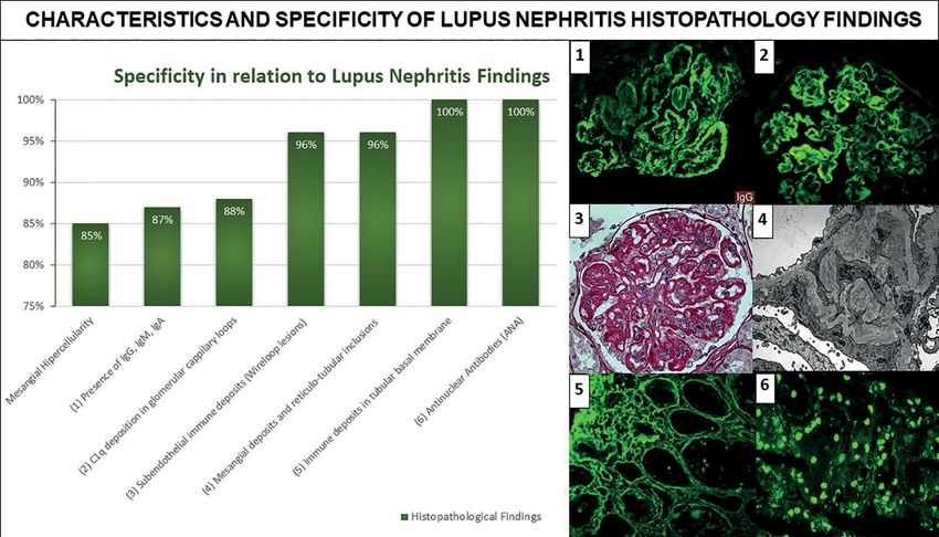
Figure 2. Characteristics and specificity of the histopathology of lupus nephritis. Adapted from Jennette et al. (1983).68
258 Braz. J. Nephrol. (J. Bras. Nefrol.) 2019;41(2):252-265Pediatric lupus nephritis
Figure 3. Histopathology classification of lupus nephritis according to the criteria established by the International Society of Nephrology and the
Renal Pathology Society (ISN/RPS) in 2003 revised in 2018.48,49,51
Table 2 Modifications proposed by the National Institutes of Health (NIH) to the system used to score
lupus nephritis activity and chronicity
Activity Index Definition Score
Endocapillary Endocapillary hypercellularity in < 25% (1+), 25-50% (2+), or > 50% (3+) of the
0-3
hypercellularity glomeruli
Neutrophils and/or karyorrhexis in < 25% (1+), 25-50% (2+), or > 50% (3+) of
Neutrophils/karyorrhexis 0-3
the glomeruli
Fibrinoid necrosis Fibrinoid necrosis in < 25% (1+), 25-50% (2+), or > 50% (3+) of the glomeruli (0-3)x2
Wire loop lesions and/or hyaline thrombi in < 25% (1+), 25-50% (2+), or > 50% (3+)
Hyaline deposits (0-3)x2
of the glomeruli
Cellular/fibrocellular Cellular and/or fibrocellular crescents in < 25% (1+), 25-50% (2+), or > 50% (3+)
0-3
crescents of the glomeruli
Interstitial inflammation Interstitial leukocytes in < 25% (1+), 25-50% (2+), ou > 50% (3+) of the cortex 0-3
Total 0-24
Chronicity Index 0-3
Global and/or segmental sclerosis in < 25% (1+), 25-50% (2+), or > 50% (3+) of
Glomerulosclerosis score 0-3
the glomeruli
Fibrous crescents Fibrous crescents in < 25% (1+), 25-50% (2+), or > 50% (3+) of the glomeruli 0-3
Tubular atrophy in < 25% (1+), 25-50% (2+), or > 50% (3+) of the cortical
Tubular atrophy 0-3
tubules
Interstitial fibrosis Interstitial fibrosis in < 25% (1+), 25-50% (2+), or > 50% (3+) of the cortex 0-3
Total 0-12
Source: Adapted from Bajema et al., 2018.51
Braz. J. Nephrol. (J. Bras. Nefrol.) 2019;41(2):252-265 259Pediatric lupus nephritis
with immunosuppressants is generally recommended production of antibodies and consequent in-
to manage extrarenal manifestations.48 However, they creases in inflammatory response and for-
may indicate the onset of progressive early stage in- mation of tertiary lymphoid tissue.24,48-51
jury, which warrants additional renal biopsies as pro- Deposition of IC along the tubular basement
teinuria increases or as the glomerular filtration rate membrane also occurs. These injuries may
(GFR) decreases.48,51 help identify patients responsive to thera-
pies targeting B cells, such as treatment with
Class III (focal proliferative LN) and Class IV (diffuse rituximab.
proliferative LN)
• Vascular injuries are common and may affect
Proliferative LN (classes III and IV) is caused by the patient prognosis.24,48-51,54,55 They originate
deposition of IC in the subendothelial space of the from the deposition of IC in vascular smooth
glomerular capillaries, either alone or in combina- muscle cells and endothelial cells or by local
tion with the deposition of IC in the mesangial re- complement activation. Five types of vascular
gion.23-25,48-51 Subendothelial deposition triggers the injuries are often observed: vascular IC de-
production of IFN-gamma by endothelial cells and, posits, arterionephrosclerosis, thrombotic mi-
consequently, local inflammation and endocapillary croangiopathy, noninflammatory necrotizing
hypercellularity.51 Reticular aggregates - ultrastruc- vasculopathy, and vasculitis. Other possible
tural findings characteristically seen in scenarios of events include endothelial edema, transmural
elevated IFN-gamma secretion - may also form.53 vasculitis with fibrinoid necrosis, mesangioly-
Severe modes of the disease have been associated wi- sis or fibrin thrombi and, enlargement of the
th crescentic formations stemmed from the rupture lamina rara interna of the glomerular base-
of glomerular capillary loops and leakage of mito- ment membrane seen with the aid of electron
genic proteins (mainly fibrinogen) into the urinary microscopy.56 Some of these injuries may be
space, with subsequent involvement of the parietal related to manifestations of LN, including sys-
epithelium. Proliferative LN presents lesions that temic hypertension, dyslipidemia, and throm-
characterize activity and chronicity.24,48-51 According boembolism.24,30,48-51 Vascular injuries may
to the ISN/RPS classification (2018), the criteria for help identify patients potentially responsive to
activity are: endocapillary hypercellularity; glomeru- eculizumab and thrombomodulin.57
lar neutrophils/karyorrhexis; fibrinoid necrosis; wire • Podocyte injuries are common and stem from
loop lesions and/or hyaline thrombi in the glomeruli; the loss of expression of the proteins present
cellular and/or fibrocellular crescents; and interstitial in the slit diaphragm (nephrin and podocin)
inflammation.51 The criteria for chronicity include: and the disorganization of the podocyte cy-
total score of segmental or global glomerulosclerosis; toskeleton, culminating with the flattening,
fibrous crescents; tubular atrophy and interstitial fi- effacement, and microvillus transformation
brosis (Table 2).51 of the foot processes.58 These changes can be
Involvement with active (A) and/or chronic (C) le- viewed only through an electron microscope.58
sions in less than 50% of the glomeruli is seen in LN Affected patients develop marked proteinuria.
class III.24,48-51 Involvement of more than 50% of the Podocyte injuries may be used to identify pa-
glomeruli indicates LN class IV, which is subdivided tients potentially responsive to calcineurin
into “S” - segmental glomerular injury, i.e., injuries inhibitors.
affecting less than half of the glomerular tufts - and • Crescentic injuries arise from immune depos-
“G” - global glomerular injury, i.e., injuries affecting its or direct attack by inflammatory cells.59
more than half of the glomerular tufts.24,30,48-51 Between 30-100% of the patients with diffuse
Although other injuries may occur with LN, they crescentic injury are positive for ANCA and/
are not used for classification purposes. Nevertheless, or anti-myeloperoxidase antibodies, showing
they may affect the choice of treatment. overlapping SLE and ANCA-positive vasculi-
tis.60,61 This group of injuries may help identify
• Tubulointerstitial injury: clonal expansion patients potentially responsive to plasmapher-
of B cells and plasma cells may trigger local esis and monoclonal anti-C5aR antibody.
260 Braz. J. Nephrol. (J. Bras. Nefrol.) 2019;41(2):252-265Pediatric lupus nephritis
Class V (membranous LN) Renal biopsy is required in the development of
LN class V originates from the subepithelial IC depo- LN therapy. However, extremely ill individuals can-
sition of either immune complexes transiting throu- not always undergo renal biopsies. Difficult-to-treat
gh the glomerular basement membrane or immune hypertension, massive proteinuria, and/or impaired
complexes formed locally to deal with podocyte an- function are indications of LN classes III and IV and,
tigens.23-25,51 The complement system is then activated as such, must be treated even in cases where the pa-
locally, usually with the formation of membrane at- tient cannot undergo a renal biopsy.4,27,18,40,63
tack complex (C5b-9), thickening of the glomerular
basement membrane, and destabilization of the po- Treatment of lupus nephritis classes I and II
docyte cytoskeleton.25 LN class V is often associated The treatment of pediatric LN classes I and II consists
with nephrotic-range proteinuria with or without he- of low-dose oral corticosteroids (prednisone/predni-
maturia. This class of the disease may occur in asso- solone < 0.5 mg/kg/day, no more than of 30 mg/day)
ciation with proliferative LN (Class III or IV). for 3-6 months, followed by gradual withdrawal of
medication.4,27,40,63 HCQ is also recommended for pa-
Class VI (advanced sclerosing LN) tients with LN class II, while other DMARDs (MTX
LN class VI results from the progression of lupus or AZA) should be considered in cases of severe ex-
nephritis.24 In this disease class, glomerular, vascu- trarenal manifestations.4,27,63,64 If proteinuria persists
lar, and tubulointerstitial injuries from glomerulos- after three months of treatment, a new renal biopsy
clerosis are seen in more than 90% of the analyzed should be considered.63 If LN progresses, some au-
glomeruli.24,48-51 thors have suggested the use of mycophenolate mofe-
til (MMF), tacrolimus (TAC), and cyclophosphamide
Treatment (CP).4,27,63
The therapeutic regimens tested for adults with SLE,
Treatment of lupus nephritis III and IV, associated or
although broadly recommended for juvenile SLE, may
not with class V
not be enough to manage the disease in pediatric pa-
tients. However, recent guidelines for the treatment of LN classes III and IV are the most common and se-
LN in children and adolescents are broadly based on vere forms of LN in children and adolescents. The
consensus documents developed for the adult popu- combination of proliferative LN and LN class V is
lation.4,18,27,38,40,62,63 The goals of LN therapy are: pro- highly prevalent in the pediatric population.4,27,63
duce complete remission from the disease; produce Since proliferative LN is usually linked to less-
maximal decreases in disease activity; minimize drug -favorable prognoses, treatment strategies do not
toxicity; prevent recurrences; prevent chronic kidney rely on the presence of an association with disea-
impairment; improve patient quality-of-life; and pro- se class V.4,24,27 The treatment of proliferative LN
vide advice to patients and family members on the is divided into two stages. The first stage inclu-
disease.40,63 Complete remission is characterized by des induction therapy, with the purpose of attai-
significant drops in proteinuria and improvement of ning remission from the acute manifestations of
the GFR after six to twelve months of treatment.40,64 LN.4,27,40,63 The second stage is called maintenance
Partial remission is characterized by a reduction of therapy, whose purpose is to prevent recurrence
50% or greater in proteinuria and by the partial re- and manage the disease in the long term.4,27,40,63
covery of the GFR after six to twelve months of treat- The main options for induction therapy are
ment.40,64 Table 3 summarizes the therapeutic schemes MMF and CP administered together with corticoste-
for the different classes of the disease.4,27,40,45,62,63 roids.4,27,40,63 MMF and CP are equivalent in terms of
Pediatric patients with SLE must be prescribed efficacy and adverse events, although intravenous CP
disease-modifying antirheumatic drugs (DMARDs) is more efficacious in the long term for children with
such as hydroxychloroquine (HCQ), methotrexate severe SLE.65 The long-term safety of intravenous CP
(MTX) or azathioprine (AZA).4,18,27,38,40,62,63 HCQ is in children is not entirely established. Gonadal toxic-
the most commonly prescribed drug for patients with ity by oral CP therapy is greater in sexually mature
juvenile SLE. The dosage for children is ≤ 5 mg/kg/ males and lesser in prepubertal children.4,27,63 MMF
day.65 Children on HCQ must be examined regularly is particularly useful when there is significant risk of
by an ophthalmologist.4,63,64 gonadal toxicity.64 Intravenous CP may be the first
Braz. J. Nephrol. (J. Bras. Nefrol.) 2019;41(2):252-265 261Pediatric lupus nephritis
Table 3 Summary list of treatment protocols for pediatric lupus nephritis according to histopathology
classification4,27,40,45,60-62
TREATMENT SUMMARY
A) Prednisone/prednisolone (< 0.5 mg/kg/day - no more than 30 mg/day).
LN Class I B) HCQ is generally not needed, but as other DMARDs, it is recommended based on the clinical
manifestation of SLE.
A) Prednisone/prednisolone (0.25-0.5 mg/kg/day - no more than 30 mg/day), with gradual decrease.*
LN Class II B) HCQ (or another DMARD) is generally needed in case of persistent proteinuria, if there is
no remission after three months of low-dose prednisone/prednisolone, or to manage extrarenal
manifestations.
Induction therapy: MMF or CP + corticosteroids
Chemotherapy regimen (MMF or CP) - 3 options
A) Euro-Lupus: intravenous CP (fixed 500 mg doses, every 15 days for three months - total dose of
3000 mg) followed by maintenance therapy with AZA.
B) NIH: intravenous CP (500 mg/m2, increased to 750 mg/m2 if tolerated, every 30 days for six
months - no more than 1 g) followed by trimestral administrations for another 18 months.
C) SHARE: oral MMF (1200 mg/m2/day, adjusting dose to a maximum of 1800 mg/m2/day, for six
LN Classes III and months - no more than 3000 mg/day).
IV, associated or
Corticosteroid therapy - 2 options
not with LN Class
V A) Intravenous methylprednisolone (30 mg/kg/dose for three consecutive days - maximum dose 1
g) followed by oral prednisolone/prednisone (0.5-1 mg/kg/day - no more than 40 mg/day, for four
weeks) with gradual withdrawal.*
B) High-dose oral prednisone/prednisolone (1-2 mg/kg/day - no more than 60 mg/day, for four
weeks), with gradual withdrawal.*
Maintenance therapy
A) Oral AZA: doses of 2-3 mg/kg/day, no more than 150 mg/day.
B) Oral MMF: doses of 500-3000 mg/day (teratogenic).
Induction therapy
A) SHARE: oral MMF + prednisone/prednisolone in doses of 0.5 mg/kg/day, wth gradual withdrawal.*
LN Class V B) CP, CNI (cyclosporine /tacrolimus) or rituximab must be considered as options for non-responders.
Maintenance therapy
A) SHARE: oral MMF or oral AZA.
Nephroprotection A) ACEi or ARBs to manage systemic hypertension and proteinuria.
LN Classes III or IV associated or not with LN Class V
Mild surge
A) Increase prednisone and consider changing DMARDs (HCQ, AZA, MTX).
Severe surge
A) Intravenous methylprednisolone.
Recurrence and
refractory cases B) High-dose oral prednisolone/prednisone (1-2 mg/kg/day - no more than 60 mg/day), with gradual
withdrawal after response.*
Refractory disease
A) Check compliance to treatment and keep current therapy in case of poor compliance.
B) Replace therapeutic agent (MMF, intravenous CP or rituximab).
C) Consider CNI (cyclosporine or tacrolimus) in selected cases.
262 Braz. J. Nephrol. (J. Bras. Nefrol.) 2019;41(2):252-265Pediatric lupus nephritis
Continued Table 3.
A) Use sun screen daily;
B) Routine lab workup for lupus activity;
C) Periodic eye examination for patients on antimalarial medication;
D) Daily exercises to help prevent cardiovascular disease;
E) Balanced diet, rich in calcium and low in salt content;
F) Supplementation with vitamin D, so that serum levels of 25-OH-vitamin D are above 30 ng/mL;
Adjuvant therapy G) Rigorous management of blood pressure and proteinuria with ACEi and/r ARBs when possible;
H) Control dyslipidemia;
I) Avoid nephrotoxic drugs (e.g.: non-steroid anti-inflammatory drugs - NSAIDs);
J) Discuss reproductive health with the patient, including birth control, contraceptive medication,
and sexually transmitted diseases;
K) Consider administration of influenza, pneumococcal, and meningococcal vaccines;
L) Assess changes in cognitive performance at school and at home.
Notes: * Gradual withdrawal of prednisone/prednisolone: gradual decreases of 10-20% from the initial dose every one or two weeks to attain doses
of 5-10 mg/day after six months. AZA: azathioprine; ARBs: angiotensin II receptor blockers; CP: cyclophosphamide; CNI: calcineurin inhibitors;
DMARDs: disease-modifying antirheumatic drugs; ACEi: angiotensin-converting-enzyme inhibitors; SLE: systemic lupus erythematosus; MMF:
mycophenolate mofetil; LN: lupus nephritis; SHARE: Single Hub and Access point for paediatric Rheumatology in Europe.
choice when there is risk of poor compliance to orally methylprednisolone and oral prednisone/predniso-
administered medication.63 lone, methylprednisolone should be preferred in more
There are two regimens for intravenous CP: the severe cases.4,40,63 Oral corticosteroids must be kept
low-dose (intravenous pulses of 500 mg every 15 for 3-4 weeks; good responders may be weaned from
days, in a total of six pulses through a period of the medication in steps of 10-20% of the initial dose
three months); and the high-dose protocol (intra- every one to two weeks, reaching doses of 5-10 mg/
venous pulses of 500-750 mg/m2/pulse; if 750 mg/ day after six months.40,63
m2/pulse is tolerated, a maximum dose of 1000- The most indicated medication for maintenance
1200 mg/pulse may be attained, with a total of six therapy for proliferative LN are AZA (2-3 mg/kg/day
monthly pulse injections).63 The long-term out- orally, no more than 150 mg/day) or MMF (initial
comes of these regimens are comparable in terms dose of 1200 mg/m2/day orally, no more than 2000
of safety and efficacy.66 The low-dose protocol mg/day, increased to 1800 mg/m2/day, no more than
may be preferred for Caucasian patients.38,63 The 3000 mg/day, if response is not good), with similar
SHARE group recommended that children and efficacy and adverse effects observed in children and
adolescents with proliferative LN be treated with adolescents.40,64 Some authors have indicated that
oral MMF for six months (initial dose of 1200 mg/ MMF is superior to AZA in adults.62,63,66,67 MMF has
m2/day, no more than 2000 mg/day, increased to teratogenic effects, while AZA may be used during
1800 mg/m2/day, no more than 3000 mg/day, if re- pregnancy. The ideal length of maintenance therapy
sponse is not good).63 is unknown. Consensus documents have indicated a
Regardless of the choice of CP or MMF, the in- minimum duration of three years.63
duction scheme must be administered jointly with
corticosteroids. The most commonly used cortico- Treatment of lupus nephritis class V
steroid protocols are: intravenous pulse of methyl- The prognosis of membranous LN (class V) is bet-
prednisolone (30 mg/kg/dose for three consecutive ter than that of proliferative LN.24,28 There is no con-
days, no more than de 1000 mg/dose), followed by sensus around the treatment of LN class V in adults.
oral prednisolone/prednisone (0.5-1.0 mg/kg/day); Immunosuppression therapy with CP or MMF has
or high dosage oral prednisone/prednisolone (1-2 been advocated, particularly for patients with ne-
mg/kg/day, no more than 60 mg/day).40,63 Although phrotic-range proteinuria.40,63 Some patients may
there is no difference in efficacy between intravenous respond to monotherapy with corticosteroids.4 The
Braz. J. Nephrol. (J. Bras. Nefrol.) 2019;41(2):252-265 263Pediatric lupus nephritis
SHARE recommends oral MMF combined with low- 3. Davidson A. What is damaging the kidney in lupus nephritis?
Nat Rev Rheumatol 2016;12:143-53.
-dose oral prednisone/prednisolone as induction the- 4. Mohan C, Putterman C. Genetics and pathogenesis of systemic
rapy for membranous LN in individuals with juvenile lupus erythematosus and lupus nephritis. Nat Rev Rheumatol
2015;11:329-41.
SLE, followed by maintenance therapy with MMF
5. Sinha R, Raut S. Pediatric lupus nephritis: Management up-
or AZA.63 The long-term prognosis for patients with date. World J Nephrol 2014;3:16-23.
subnephrotic-range proteinuria and normal GFR is 6. Malattia C, Martini A. Paediatric-onset systemic lupus erythe-
matosus. Best Pract Res Clin Rheumatol 2013;27:351-62.
generally favorable, and treatment may be initiated 7. Stojan G, Petri M. Epidemiology of systemic lupus erythemato-
with nephroprotective measures.63 sus: an update. Curr Opin Rheumatol 2018;30:144-50.
8. Vilar MJP, Rodrigues JM, Sato EI. Incidência de lúpus er-
itematoso sistêmico em Natal, RN - Brasil. Rev Bras Reumatol
Recurrent and refractory lupus nephritis 2003;43:343-6.
9. Wenderfer SE, Ruth NM, Brunner HI. Advances in the care of
Therapy failure occurs mostly due to poor complian- children with lupus nephritis. Pediatr Res 2017;81:406-14.
ce to treatment.40,46,63 Patient serum immunosuppres- 10. Kamphuis S, Silverman ED. Prevalence and burden of pediat-
ric-onset systemic lúpus erythematosus. Nat Rev Rheumatol
sant level monitoring is recommended. Therapy chan- 2010;6:538-46.
ges may be introduced if the patient fails to respond 11. Yu Y, Du L, Pan J, Zheng J, Chen A, Chen L. A 10-year retro-
after three months of treatment and poor compliance spective study of neonatal lupus erythematous in China. Asian
Pac J Allergy Immunol 2016;34:174-8.
has been ruled out.63 If the patient responds partially, 12. Klein-Gitelman MS. Neonatal Lupus: What we have learned
wait for an additional 3-6 months for complete re- and current approaches to care. Curr Rheumatol Rep
2016;18:60.
mission before changing the immunosuppressant 13. Gomes RC, Silva MF, Kozu K, Bonfá E, Pereira RM, Terreri MT,
regimen.63 The reintroduction or increased doses of et al. Features of 847 Childhood-Onset Systemic Lupus Erythema-
tosus Patients in Three Age Groups at Diagnosis: A Brazilian Mul-
corticosteroids combined with DMARDs should be ticenter Study. Arthritis Care Res (Hoboken) 2016;68:1736-41.
considered.40,63 In cases of persistent, active or refrac- 14. Silva CA, Avcin T, Brunner HI. Taxonomy for systemic lupus
erythematosus with onset before adulthood. Arthritis Care Res
tory proliferative LN - with or without membranous
(Hoboken) 2012;64:1787-93.
LN - MMF may be replaced with rituximab or intra- 15. Oliver JE, Silman AJ. Why are women predisposed to autoim-
venous CP; or intravenous CP may be replaced with mune rheumatic diseases? Arthritis Res Ther 2009;11:252.
16. Pereira MV, Revelo MP, Bambirra EA. Lupus nephropathy in
MMF.64 Although the efficacy of rituximab has not childhood: morphologic analysis of 18 cases. J Pediatr (Rio J)
been confirmed in clinical trials, cohort studies with 1996;72:32-4.
17. Aggarwal A, Srivastava P. Childhood onset systemic lupus ery-
adults and children suggested the drug should be used thematosus: how is it different from adult SLE? Int J Rheumatic
in cases of refractory LN.63 Dis 2015;18:182-91.
18. Almaani S, Meara A, Rovin BH. Update on Lupus Nephritis.
Clin J Am Soc Nephrol 2017;12:825-35.
Nephroprotection 19. Hiraki LT, Benseler SM, Tyrrell PN, Harvey E, Hebert D, Sil-
verman ED. Ethnic differences in pediatric systemic lúpus ery-
Despite the lack of consensus on the matter concerning thematosus. J Rheumatol 2009;36:2539-46.
pediatric patients, prescription of angiotensin-conver- 20. Lewandowski LB, Schanberg LE, Thielman N, Phuti A, Kalla
ting-enzyme inhibitors and/or angiotensin II receptor AA, Okpechi I, et al. Severe disease presentation and poor out-
comes among pediatric systemic lupus erythematosus patients
blockers helps control proteinuria in adults with LN.4,40,63 in South Africa. Lupus 2017;26:186-94.
21. Bennett M, Brunner HI. Biomarkers and updates on pediatrics
lupus nephritis. Rheum Dis Clin North Am 2013;39:833-53.
Conclusion 22. Muñoz LE, Lauber K, Schiller M, Manfredi AA, Herrmann M.
The role of defective clearance of apoptotic cells in systemic
Although SLE is a rare disease in pediatric popula- autoimmunity. Nat Rev Rheumatol 2010;6:280-9.
tions, its consequences may be severe and even fatal. 23. Nowling TK, Gilkeson GS. Mechanisms of tissue injury in lu-
pus nephritis. Arthritis Res Ther 2011;13:250.
Although the etiopathogenesis of LN in children and 24. Anders HJ, Fogo AB. Immunopathology of lupus nephritis. Se-
adults is similar, the disease is more severe in pedia- min Immunopathol 2014;36:443-59.
tric populations. Studies on LN affecting children and 25. Flores-Mendoza G, Sansón SP, Rodríguez-Castro S, Crispín
JC, Rosetti F. Mechanisms of Tissue Injury in Lupus Nephritis.
adolescents are required to detect new prognostic Trends Mol Med 2018;24:364-78.
markers and define specific therapeutic schemes for 26. Anders HJ. Pseudoviral immunity - a novel concept for lupus.
Trends Mol Med 2009;15:553-61.
individuals in this age range. 27. Vachvanichsanong P, McNeil E. Pediatric lupus nephritis: more
options, more chances? Lupus 2013;22:545-53.
28. Bernatsky S, Boivin JF, Joseph L, Manzi S, Ginzler E, Gladman
References DD, et al. Mortality in systemic lupus erythematosus. Arthritis
1. Levy DM, Kamphuis S. Systemic lupus erythematosus in chil- Rheum 2006;54:2550-7.
dren and adolescents. Pediatr Clin North Am 2012;59:345-64. 29. Petri M, Orbai AM, Alarcón GS, Gordon C, Merrill JT, Fortin
2. Bao L, Cunningham PN, Quigg RJ. Complement in Lupus Ne- PR, et al. Derivation and Validation of the Systemic Lupus Inter-
phritis: New Perspectives. Kidney Dis (Basel) 2015;1:91-9. national Collaborating Clinics Classification Criteria for Systemic
Lupus Erythematosus. Arthritis Rheum 2012;64:2677-86.
264 Braz. J. Nephrol. (J. Bras. Nefrol.) 2019;41(2):252-265Pediatric lupus nephritis
30. Hahn BH, McMahon MA, Wilkinson A, Wallace WD, Daikh 51. Bajema IM, Wilhelmus S, Alpers CE, Bruijn JA, Colvin RB,
DI, Fitzgerald JD, et al.; American College of Rheumatology. Cook HT, et al. Revision of the International Society of Ne-
American College of Rheumatology guidelines for screening, phrology/Renal Pathology Society classification for lupus ne-
treatment, and management of lupus nephritis. Arthritis Care phritis: clarification of definitions, and modified National In-
Res (Hoboken) 2012;64:797-808. stitutes of Health activity and chronicity indices. Kidney Int
31. Anders HJ, Weening JJ. Kidney disease in lupus is not always 2018;93:789-96. DOI: 10.1016/j.kint.2017.11.023
‘lupus nephritis’. Arthritis Res Ther 2013;15:108. 52. Wang Y, Yu F, Song D, Wang SX, Zhao MH. Podocyte involve-
32. Soliman S, Mohan C. Lupus nephritis biomarkers. Clin Immu- ment in lupus nephritis based onthe 2003 ISN/RPS system: a
nol 2017;185:10-20. large cohort study from a single centre. Rheumatology (Ox-
33. Thurman JM, Nester CM. All things complement. Clin J Am ford) 2014;53:1235-44.
Soc Nephrol 2016;11:1856-66. 53. Rich SA. Human lupus inclusions and interferon. Science
34. Yin Y, Wu X, Shan G, Zhang X. Diagnostic value of serum 1981;213:772-5.
anti-C1q antibodies in patients with lupus nephritis: a meta- 54. Wu LH, Yu F, Tan Y, Qu Z, Chen MH, Wang SX, et al. Inclu-
analysis. Lupus 2012;21:1088-97. sion of renal vascular lesions in the 2003 ISN/RPS system for
35. Eggleton P, Ukoumunne OC, Cottrell I, Khan A, Maqsood S, classifying lupus nephritis improves renal outcome predictions.
Thornes J, et al. Autoantibodies against C1q as a diagnostic Kidney Int 2013;83:715-23.
measure of Lupus Nephritis: Systematic review and meta-anal- 55. Barber C, Herzenberg A, Aghdassi E, Su J, Lou W, Qian G,
ysis. J Clin Cell Immunol 2014;5:210. et al. Evaluation of clinical outcomes and renal vascular pa-
36. Goilav B, Putterman C, Rubinstein TB. Biomarkers for kidney thology among patients with lupus. Clin J Am Soc Nephrol
involvement in pediatric lupus. Biomark Med 2015;9:529-43. 2012;7:757-64.
37. Qi S, Chen Q, Xu D, Xie N, Dai Y. Clinical application of 56. Goodship TH, Cook HT, Fakhouri F, Fervenza FC, Frémeaux-
protein biomarkers in lupus erythematosus and lupus nephritis. Bacchi V, Kavanagh D, et al.; Conference Participants. Atypical
Lupus 2018;27:1582-90. DOI: 10.1177/0961203318773643 hemolytic uremic syndrome and C3 glomerulopathy: conclusions
38. Houssiau FA, Vasconcelos C, D'Cruz D, Sebastiani GD, de Ramon from a "Kidney Disease: Improving Global Outcomes" (KDIGO)
Garrido E, Danieli MG, et al. Early response to immunosuppres- Controversies Conference. Kidney Int 2017;91:539-51.
sive therapy predicts good renal outcome in lupus nephritis: lessons 57. de Holanda MI, Pôrto LC, Wagner T, Christiani LF, Palma
from long-term follow-up of patients in the Euro-Lupus Nephritis LMP. Use of eculizumab in a systemic lupus erythemathosus
Trial. Arthritis Rheum 2004;50:3934-40. patient presenting thrombotic microangiopathy and heterozy-
39. Moreno JA, Yuste C, Gutiérrez E, Sevillano ÁM, Rubio-Navar- gous deletion in CFHR1-CFHR3. A case report and systematic
ro A, Amaro-Villalobos JM, et al. Haematuria as a risk factor review. Clin Rheumatol 2017;36:2859-67.
for chronic kidney disease progression in glomerular diseases: 58. Bomback AS, Markowitz GS. Lupus Podocytopathy: A Distinct
A review. Pediatr Nephrol 2016;31:523-33. Entity. Clin J Am Soc Nephrol 2016;11:547-8.
40. Klumb EM, Silva CA, Lanna CCD, Sato EI, Borba EF, Brenol 59. Yu F, Haas M, Glassock R, Zhao MH. Redefining lupus ne-
JCT, et al. Consensus of the Brazilian Society of Rheumatology phritis: clinical implications of pathophysiologic subtypes. Nat
for the diagnosis, management and treatment of lupus nephri- Rev Nephrol 2017;13:483-95.
tis. Rev Bras Reumatol 2015;55:1-21. 60. Nasr SH, D'Agati VD, Park HR, Sterman PL, Goyzueta JD,
41. Fathallah-Shaykh SA. Proteinuria and progression of pediatric Dressler RM, et al. Necrotizing and crescentic lupus nephritis
chronic kidney disease: lessons from recent clinical studies. Pe- with antineutrophil cytoplasmic antibody seropositivity. Clin J
diatr Nephrol 2017;32:743-51. Am Soc Nephrol 2008;3:682-90.
42. Brunner HI, Bennett MR, Mina R, Suzuki M, Petri M, Kiani 61. Kettritz R. Vasculitis: A CLEAR argument for targeting comple-
AN, et al. Association of noninvasively measured renal protein ment in ANCA vasculitis. Nature Rev Nephrol 2017;13:448-
biomarkers with histologic features of lupus nephritis. Arthritis 50.
Rheum 2012;64:2687-97. 62. Austin HA 3rd, Klippel JH, Balow JE, le Riche NG, Steinberg
43. Smith EM, Jorgensen AL, Midgley A, Oni L, Goilav B, Putter- AD, Plotz PH, et al. Therapy of lupus nephritis. Controlled
man C, et al. International validation of a urinary biomarker trial of prednisone and cytotoxic drugs. New Engl J Med
panel for identification of active lupus nephritis in children. Pe- 1986;314:614-9.
diatr Nephrol 2017;32:283-95. 63. Groot N, de Graeff N, Marks SD, Brogan P, Avcin T, Bader-
44. Brunner HI, Bennett MR, Abulaban K, Klein-Gitelman MS, Meunier B, et al. European evidence-based recommendations
O'Neil KM, Tucker L, et al. Development of a novel renal ac- for the diagnosis and treatment of childhood-onset lupus ne-
tivity index of lupus nephritis in children and young adults. phritis: the SHARE initiative. Ann Rheum Dis 2017;76:1965-
Arthritis Care Res (Hoboken) 2016;68:1003-11. 73.
45. Kashgarian M. Clinical significance of renal biopsy in subacute 64. Ruiz-Irastorza G, Ramos-Casals M, Brito-Zeron P, Khamashta
lupus erythematosus. Transfusion Sci 1992;13:135-44. MA. Clinical efficacy and side effects of antimalarials in sys-
46. Sprangers B, Monahan M, Appel GB. Diagnosis and treat- temic lupus erythematosus: a systematic review. Ann Rheum
ment of lupus nephritis flares--an update. Nat Rev Nephrol Dis 2010;69:20-8.
2012;8:709-17. 65. Appel GB, Contreras G, Dooley MA, Ginzler EM, Isenberg
47. Zickert A, Sundelin B, Svenungsson E, Gunnarsson I. Role of D, Jayne D, et al.; Aspreva Lupus Management Study Group.
early repeated renal biopsies in lupus nephritis. Lupus Sci Med Mycophenolate Mofetil versus Cyclophosphamide for in-
2014;1:e000018. duction treatment of Lupus Nephritis. J Am Soc Nephrol
48. Wilhelmus S, Alpers CE, Cook HT, Ferrario F, Fogo AB, Haas 2009;20:1103-12.
M, et al. The revisited classification of GN in SLE at 10 years: 66. Houssiau FA, D’Cruz D, Sangle S, Remy P, Vasconcelos C,
time to re-evaluate histopathologic lesions. J Am Soc Nephrol Petrovic R, et al.; MAINTAIN Nephritis Trial Group. Aza-
2015;26:2938-46. thioprine versus mycophenolate mofetil for long-term immu-
49. Weening JJ, D'Agati VD, Schwartz MM, Seshan SV, Alpers CE, nosuppression in lupus nephritis: results from the MAINTAIN
Appel GB, et al. The classification of glomerulonephritis in systemic Nephritis Trial. Ann Rheum Dis 2010;69:2083-9.
lupus erythematosus revisited. J Am Soc Nephrol 2004;15:241-50. 67. Dooley MA, Jayne D, Ginzler EM, Isenberg D, Olsen NJ, Wof-
50. Weening JJ, D'Agati VD, Schwartz MM, Seshan SV, Alpers CE, sy D, et al.; ALMS Group. Mycophenolate versus Azathioprine
Appel GB, et al.; International Society of Nephrology Work- as Maintenance Therapy for Lupus Nephritis. New Engl J Med
ing Group on the Classification of Lupus Nephritis; Renal Pa- 2011;365:1886-95.
thology Society Working Group on the Classification of Lupus 68. Jennette JC, Iskandar SS, Dalldorf FG. Pathologic differentia-
Nephritis. The classification of glomerulonephritis in systemic tion between lupus and nonlupus membranous glomerulopa-
lupus erythematosus revisited. Kidney Int 2004;65:521-30. thy. Kidney Int 1983;24:377-85.
Braz. J. Nephrol. (J. Bras. Nefrol.) 2019;41(2):252-265 265You can also read