Congenital Uterine Anomaly and Pelvic Organ Prolapse: A Rare Case of Pelvic Organ Prolapse in a Complete Bicornuate Uterus with Successful ...
←
→
Page content transcription
If your browser does not render page correctly, please read the page content below
https://doi.org/10.6118/jmm.21007
J Menopausal Med 2021;27:32-36
pISSN: 2288-6478, eISSN: 2288-6761
BRIEF COMMUNICATION
Congenital Uterine Anomaly and Pelvic Organ Prolapse:
A Rare Case of Pelvic Organ Prolapse in a Complete
Bicornuate Uterus with Successful Pregnancy Outcomes
Undiagnosed until the Time of Sacrocolpopexy
Gina Nam1, Sa Ra Lee2
1
Department of Obstetrics and Gynecology, Chung-Ang University Hospital, Chung-Ang University College of Medicine, Seoul, Korea,
2
Department of Obstetrics and Gynecology, Asan Medical Center, University of Ulsan College of Medicine, Seoul, Korea
Müllerian development anomalies (MDAs) are most commonly diagnosed in the reproductive period. A bicornuate uterus is the result
of a fusion defect of the Müllerian ducts, causing an abnormal fundal outline. Most of the cases are diagnosed early in life and present
with obstetrical complications, such as recurrent pregnancy loss, preterm birth, intrauterine growth restriction, placental abruption, and
cervical incompetence. Pelvic organ prolapse (POP) in women with MDAs has been reported; however, all reported cases were when
MDAs are diagnosed before or simultaneously with the development of POP in premenopausal young women aged < 35. A 52-year-
old menopausal woman, who successfully delivered vaginally at term, was presented with protruding mass through vaginal introitus.
On POP-Q examination, the cervix was elongated and descended to 1 cm out of the hymen during bearing down; however, the uterine
bodies were confined in the pelvic cavity, which is commonly encountered among POP patients with large uterus due to uterine fibroids
or adenomyosis. She also diagnosed for complete bicornuate uterus and underwent robotic sacrocolpopexy for advanced stage POP. It
is presumed to have been caused by the bicornuate uterus that prevented the total uterine prolapse with the effect of extending both
uterine horns bilaterally inside the pelvic cavity and trapping the uterus within the pelvis. Herein, we report a rare case of complete
bicornuate uterus with multiple successful vaginal deliveries at term without obstetric complications, which remained undiagnosed until
she was managed for the POP in her postmenopausal period.
Key Words: Pelvic organ prolapse, Pregnancy, Robotic surgical procedures, Uterine anomalies
INTRODUCTION incomplete development leads to uterine hypoplasia or
a unicornuate uterus. Incomplete fusion of the MDs re-
Congenital malformation of female genital tract is sults in uterus didelphys, bicornuate uterus, and arcuate
resulted from embryological maldevelopment of the uterus [1]. MDAs are rare with a prevalence of 4%–7%
Müllerian ducts (MDs) or paramesonephric ducts. [1]. Bicornuate uterus represents one-fourth of MDAs
Müllerian development anomalies (MDAs) arise [2]. MDAs have been found to be associated with preg-
whenever there is dysregulation in complex process of nancy loss, preterm birth (PTB), malpresentation, and
cellular differentiation, migration, fusion and canaliza- cesarean delivery [2]. In women with bicornuate uter-
tion [1]. MDs differentiate to uterus, fallopian tubes, us, conception does not appear to be affected; however,
cervix, and upper one-third of the vagina. Failure in the there is an increased risk of obstetrical complications
development of MDs results in uterine agenesis, and such as recurrent pregnancy loss, PTB, intrauterine
Received: February 22, 2021 Revised: March 4, 2021 Accepted: March 7, 2021
Address for Correspondence: Sa Ra Lee, Department of Obstetrics and Gynecology, Asan Medical Center, University of Ulsan College of
Medicine, 88 Olympic-ro 43-gil, Songpa-gu, Seoul 05505, Korea
Tel: 82-2-3010-3648, E-mail: leesr@amc.seoul.kr, ORCID: https://orcid.org/0000-0002-7890-8348
Copyright © by The Korean Society of Menopause
This is an Open Access article distributed under the terms of the Creative Commons Attribution Non-Commercial License (http://creativecommons.org/licenses/by-nc/4.0/).
32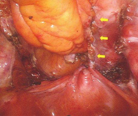
Müllerian Anomaly and Pelvic Organ Prolapse

growth restriction (IUGR), placental abruption, and                                                 CASE REPORT
cervical incompetence [2].
  In terms of pelvic organ prolapse (POP), the main                           This study was conducted according to the guidelines
cause of POP is weakening of the supportive ligaments.                      of the Declaration of Helsinki (2013) and was approved
The multiparity and increased age certainly are risk                        by the Asan Medical Center Institutional Review Board
factors in the development of genital prolapse [3]. Clas-                   (approval No. 2021-0292).
sification of POP consists of anterior compartment,                           A 52-year-old menopausal woman (gravida 5, para
apical compartment, posterior compartment prolapse.                         2) presented with intermittent dysuria, stress uri-
Anterior compartment prolapse is the most common                            nary incontinence and bothersome protruding mass
form and the old aged women usually manifested as                           through the vaginal introitus for several years which
multi-compartment POP, whereas young aged women                             was aggravated for 1 year. She got pregnant naturally
have mostly one compartment prolapse usually an api-                        and had a history of two uncomplicated spontaneous
cal compartment POP [3].                                                    vaginal deliveries without dystocia, malpresentation
  Diagnosis of MDAs associated with POP in the old                          of baby, and IUGR at term. Her obstetric history was
age is rare. Among the publications reported MDAs as-                       2-0-3 (1 spontaneous abortion at early pregnancy, 2
sociated with POP to date, 35-year-old patient was the                      artificial abortion)-2. When she looked back, she only
most oldest woman [2,4]. MDAs are usually diagnosed                         remember one obstetrician’s opinion that her uterus,
during evaluation due to symptoms such as abdominal                         which seems something different from normal uterus.
pain and amenorrhea, and without symptoms, prob-                            However, successful normal pregnancies and normal
lems with obstetric outcome may have occurred. Here                         deliveries in women with complete bicornuate uterus
we report a rare case of complete bicornuate uterus                         are extremely rare. At the first visit, pelvic examina-
which was not diagnosed even in pregnancy and child-                        tion revealed a one vagina, one cervix and POP-Q
birth. She carried her pregnancies to term and deliv-                       stage III (+3, +5, +1, 0, 0, –3) (6, 4, 8) based on the
ered by vaginal normal deliveries without any obstetric                     Pelvic Organ Prolapse Quantification System (POP-
problems or complications. She first became aware of                        Q). The anterior vaginal wall was fully prolapsed and
bicornuate uterus when diagnosed as POP with apical                         the most distal portion of the cervix descent 1 cm
prolapse. An uneventful robotic sacrocolpopexy was                          below the plane of the hymen. There were no signs of
done and POP has been completely resolved at her                            rectocele and enterocele on translabial sonography.
follow-up at postoperative 6 weeks. This is a first case                    On transvaginal sonography, we found two horns of
report of POP associated with bicornuate uterus in a                        uterus, which the patient recognized it for the first
multiparous, postmenopausal woman without any ob-                           time. Urodynamic study revealed the urodynamic
stetric complications.                                                      stress incontinence. The patient underwent robotic

                       RVL

                                                            Lt.U                     Rt.U

        Lt.U                     Rt.U

                                            A                                                B                                                  C

Fig. 1. Laparoscopic view. (A) The rectovesical ligament was crossing the junction between two horns of uterus. (B) After dissection of rectovesical
ligament from bladder and rectum, indentation at the level of uterine midline fully dividing uterine corpus was seen. (C) Robotic subtotal
hysterectomy, bilateral salpingo-oophorectomy, and sacrocolpopexy with retroperitoneal tunneling technique (arrows) were performed. Lt.U: left
hemiuterus, Rt.U: right hemiuterus, RVL: rectovesical ligament.

                                                                                                            https://doi.org/10.6118/jmm.21007       33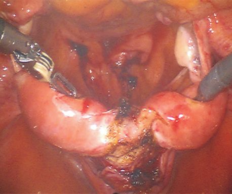
Gina Nam and Sa Ra Lee
body deformity [8]. The European Society of Human
Reproduction and Embryology (ESHRE) developed an
updated classification system [1]. Class U3b or com-
plete bicorporeal uterus is defined the uterus with an
Lt.U Rt.U
external fundal indentation completely dividing the
LS
LO uterine corpus up to the level of the cervix, as in this
RS case. The obstructive MDAs are mostly undiagnosed
RO until the patient presents with hematometra or hema-
tocolpos; however, a septate uterus or bicornuate uterus
are mostly asymptomatic and diagnosed in patients
with infertility or pregnancy. In addition, the diagnosis
can be missed at the time of antenatal care, if nongravid
uterus is positioned posterior to gravid uterus, as in
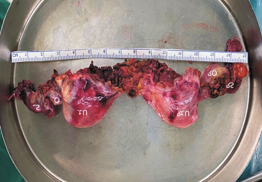
Fig. 2. Postoperative specimen showing bilateral uterine horns with this case [2].
normal ovaries and fallopian tubes. LS: left salpinx, LO: left ovary, It has been established that MDAs are related to ad-
Lt.U: left hemiuterus, Rt.U: right hemiuterus, RO: right ovary, RS: verse pregnancy outcomes. The presence of uterine
right salpinx. anomalies increased risk in recurrent pregnancy loss,
PTB, preterm premature rupture of membranes, breech
supracervical hysterectomy, bilateral salpingo-oopho- presentation, cesarean delivery, placenta previa, pla-
rectomy, sacrocolpopexy and transobturator vaginal cental abruption, and IUGR [9]. It is hypothesized that
tape insertion. Robotic sacrocolpopexy was performed abnormal uterine blood flow and decreased muscle
as described in our previous reports [5-7] using the mass would be reasons for PTB or IUGR [9]. Grim-
da Vinci Xi system (Intuitive Surgical, Sunnyvale, CA, bizis et al. [10] reported a spontaneous abortion rate
USA). Intraoperative findings included a rectovesical of bicornuate uterus as 36% and a PTB rate of 23%. In
ligament crossing the junction between two horns of addition, Heinonen et al. [11] identified that the deeper
uterus and normal fallopian tubes and ovaries (Fig. the bifurcation the poorer the obstetric performance.
1A). After dissection of rectovesical ligament from The incidence of PTB was 29% in partial and 66% in
bladder and rectum, indentation at the level of uterine complete bicornuate uterus. Breech presentation of
midline fully dividing uterine corpus was seen (Fig. baby was 0% in partial and 50% in complete bicornuate
1B). In the postoperative specimen, we could definite- uterus, and cesarean delivery rate was 20% and 36%.
ly identify two uterine horns with normal ovaries and However, there is a disputing result that patients with
fallopian tubes (Fig. 2). The postoperative course was the partial type of bicornuate uterus showed favorable
uneventful. At the 6 weeks of follow-up visits, the pa- outcomes than complete type for achieving pregnancy
tient had complete resolution of prolapse with POP- and childbirth following metroplasty procedure [12].
Q stage 0 and reported that dysuria and stress urinary Our patient had a first trimester spontaneous abortion,
incontinence had disappeared. but there were no adverse pregnancy outcomes such
as uterine rupture, PTB, and cesarean delivery without
DISCUSSION correction of the bicornuate uterus by metroplasty sur-
gery. Cesarean section is commonly recommended to
Congenital genital tract malformations are not com- women with bicornuate uterus for the risk of uterine
mon, and bicornuate uterus is one of the common type rupture before or during the labor. However, in this
of MDAs [1]. The reliable classifications have been case, successful vaginal deliveries were accomplished at
proposed because MDAs are associated with health and term.
reproductive problems depending on the type and de- It is well-known that multiparity and advanced age
gree of anomalies. The American Fertility Society (AFS) are major risk factors for POP [13]. Despite these
categorized female genital tract anomalies in 7 classes associations, the primary site of older age group
[8]. Bicornuate uterus is defined as double uterus with was distributed evenly between different sites. The
a single cervix, and divided into two subclasses, partial multicompartment prolapse and a higher grade of
and complete, according to the degree of the uterine prolapse in older women may represent that the first
34 www.e-jmm.org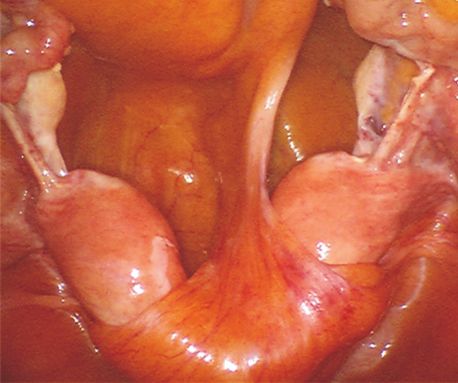
Müllerian Anomaly and Pelvic Organ Prolapse

site of prolapse is at the apical support, and the other                              REFERENCES
connective tissues fail to support over time [3]. Some
authors explain that the upper portion of the vagina           1. Grimbizis GF, Gordts S, Di Spiezio Sardo A, Brucker S, De Angelis
is derived from Müllerian structures, so that loss of             C, Gergolet M, et al. The ESHRE/ESGE consensus on the classifi-
apical support in young patients with MDAs was re-                cation of female genital tract congenital anomalies. Hum Reprod
lated to aberrant uterine anatomy [14]. However, in               2013; 28: 2032-44.
our patient, the primary site of prolapse was anterior         2. Stearns K, Al Khabbaz A. Bicornuate bicollis uterus with ob-
compartment with elongated cervix. The elongated                  struction of the lower uterine segment and cervical prolapse
cervix with uterine fundus remained in the pelvis was             complicating pregnancy. Case Rep Obstet Gynecol 2018; 2018:
noted in this case. It is probable that bicornuate uterus         8910976.
made an effect of support by uterine horns extending           3. Strohbehn K, Jakary JA, Delancey JO. Pelvic organ prolapse in
to both sides of pelvis and trapped in the pelvis.                young women. Obstet Gynecol 1997; 90: 33-6.
  In this case, the uterine anomaly was diagnosed late in      4. Rajamaheswari N, Seethalakshmi K, Gayathri KB. Case of longi-
her age during management of POP. When a search of                tudinal vaginal septum with pelvic organ prolapse. Int Urogynecol
PubMed was performed for all studies published from               J Pelvic Floor Dysfunct 2009; 20: 1509-10.
January 1986 to January 2021 using terms ‘Müllerian            5. Lee SR. Robotic Single-Site® sacrocolpopexy: first report and tech-
development anomalies’ and ‘pelvic organ prolapse’,               nique using the Single-Site® wristed needle driver. Yonsei Med J
there were 19 reports of MDAs diagnosed before age of             2016; 57: 1029-33.
35 associated with POP. Fourteen of the 19 reports were        6. Lee SR, Roh AM, Jeong K, Kim SH, Chae HD, Moon HS. First
cases in which neovagina was prolapsed after being di-            report comparing the two types of single-incision robotic sacro-
agnosed and treated as MDAs. The neovaginal prolapse              colpopexy: single site using the da Vinci Xi or Si system and single
is considered that the surgical creation of vagina allows         port using the da Vinci SP system. Taiwan J Obstet Gynecol 2021;
for normal menstruation and sexual function but does              60: 60-5.
not provide endopelvic facial support of neovagina [15].       7. Kim JH, Lee SR, Lee ES, Kim SH, Chae HD. Robot-assisted
POP and MDAs were diagnosed simultaneously in five                laparoscopic surgery for pelvic organ prolapse among peri-
of the 19 reports. Longitudinal vaginal septum associ-            and post-menopausal women. J Menopausal Med 2020; 26:
ated with POP in a 35-year-old multiparous woman                  154-8.
was reported [4]. The patient was not aware of the             8. The American Fertility Society. The American Fertility Society
vaginal anomaly. Her uneventful three normal vaginal              classifications of adnexal adhesions, distal tubal occlusion, tubal
deliveries were conducted with untrained birth at-                occlusion secondary to tubal ligation, tubal pregnancies, müllerian
tendant. There is the case of a 17-year-old primigravid           anomalies and intrauterine adhesions. Fertil Steril 1988; 49: 944-
patient with bicornuate uterus and cervical prolapse at           55.
38 weeks of gestation. She was delivered with cesarean         9. Hua M, Odibo AO, Longman RE, Macones GA, Roehl KA, Cahill
section due to obstruction of the lower uterine segment           AG. Congenital uterine anomalies and adverse pregnancy out-
of the gravid uterus [2].                                         comes. Am J Obstet Gynecol 2011; 205: 558.e1-5.
  To our knowledge, this is the first report of POP oc-       10. Grimbizis GF, Camus M, Tarlatzis BC, Bontis JN, Devroey P.
curred in a bicornuate uterus with successful pregnancy           Clinical implications of uterine malformations and hysteroscopic
outcomes without surgical correction of MDAs. In this             treatment results. Hum Reprod Update 2001; 7: 161-74.
case, women with a complete bicornuate uterus can             11. Heinonen PK, Saarikoski S, Pystynen P. Reproductive perfor-
conceive spontaneously and successfully give a birth              mance of women with uterine anomalies. An evaluation of 182
vaginally without midterm adverse pregnancy out-                  cases. Acta Obstet Gynecol Scand 1982; 61: 157-62.
comes. Anatomical variants of MDAs such as complete           12. Nishida M, Otsubo Y, Arai Y, Ichikawa R, Sakanaka M. Dif-
type of bicornuate uterus may contribute to the clinical          ference in reproductive performance between two subtypes
manifestations of the POP.                                        of bicornuate uterus. Arch Gynecol Obstet 2016; 293: 1335-
                                                                  8.
            CONFLICT OF INTEREST                              13. Gleason JL, Richter HE, Varner RE. Pelvic organ prolapse. In: Ber-
                                                                  ek JS, editor. Berek and Novak’s gynecology. 15th ed. Philadelphia:
 No potential conflict of interest relevant to this article       Wolters Kluwer; 2015. p. 912-5.
was reported.                                                 14. Hullfish KL, Roth JA. Cloacal malformation, müllerian dys-

                                                                                              https://doi.org/10.6118/jmm.21007    35
Gina Nam and Sa Ra Lee
genesis, pelvic organ prolapse, and urethrovaginal fistula in a 15. Hao Z, Yang S. Neovaginal prolapse treated with sacrospinous
17-year-old girl. Female Pelvic Med Reconstr Surg 2004; 10: ligament suspension: a case report and review of the literature. J
223-5. Pediatr Adolesc Gynecol 2017; 30: 505-7.
36 www.e-jmm.orgYou can also read