Paciente masculino de 57 años portador de probable miocardiopatía hipertrófica apical
←
→
Page content transcription
If your browser does not render page correctly, please read the page content below
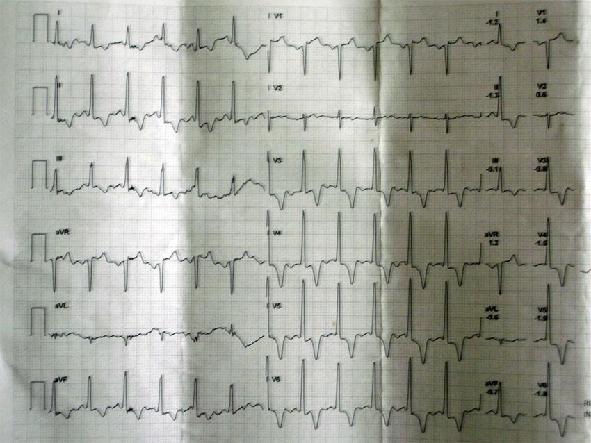
Paciente masculino de 57 años
         portador de probable
   miocardiopatía hipertrófica apical
                – 2008
                         Dr. Feliciano Pérez Casar

Estimado Edgardo, te envío este ECG por si crees de interés publicarlo en el Foro, en la
forma que creas oportuna. Se trata de un varón de 57 años que acudió a chequeo de
empresa. Ningún antecedente familiar o personal de interés. Exploración cardíaca normal.
No hipertensión. No factores de riesgo. Ante el ECG le realizan ECO que es normal.
Coronariografía que es normal. Gammagrafía isotópica que es normal. Me envían el ECG
que está realizado en posición supina porque le van a hacer un test de esfuerzo para
saber mi opinión antes de realizalo, ocultándome los datos que antes te he expuesto.
Opino que debe de realizarse y quedo pendiente de su resultado informándome a
posteriori que la PEG es normal pero no veo el trazado.
El examen del mismo a mi juicio corresponde a un PR corto, con las alteraciones en la
repolarización secundarias al mismo, despolarización y repolarización acelerada. Dado
que no son frecuentes dichas alteraciones en el PR corto, espero de la audiencia otras
opiniones.

Cordialmente
Feliciano Pérez Casar

PD: el Dr. Federico Curra ha colaborado en el tratamiento de la imagen
OPINIONES DE COLEGAS Parece un ECG de Miocardiopatía Hipertrófica, es raro que el ECO haya sido normal. No veo lo del PR corto . Dr. Amilcar Lezcano
Opino lo mismo que el Dr. Lezcano. Esos altos voltajes del QRS sobre todo en
precordiales junto a esas T negativas, simétricas y profundas con ascenso del ST en V1 y
aVR van a favor de miocardiopatía hipertrófica.

Saludos,

Javier García Niebla.

Queridos amigos tenho forte suspeita que o “ecocardiografista” que fez o ECHO no
detecto o problema. Isto - na nossa experiência - não é raro de acontecer nas
cardiomiopatias hipertróficas apicais não obstrutivas. Existem forças anteriores
proeminentes predominantes nas derivações intermediárias. Nestes casos, a parede livre
do VE não possui o normal decréscimo da espessura da base ao ápice fato que em
ecocardiografistas não muito experientes passa desapercebido. Por outra parte o septo
apresenta-se com maior espessura na parte inferior (apical) o que oculta os fatos em
mãos não mutio experientes. O ECG reflete SVE com “strain pattern” com ângulo QRS/T
próximo dos 180º e ondas R proeminentes nas precordiais intermediárias. Concordo com
Lescao e Javier.
Edgardinho gostaria comentar outras coisinhas mas meu sobrinho esteve usando o
computador e deve ter deletado o traçado.
Poderias me reenviar?
Envio-lhes estes comentários interessantes sem as figuras que são muito pesadas. Mas
tarde se desejam mando as figuras. Peço disculpa porque este ressum o escrevi em
english.
                      Apical Hypertrophic CardioMyopathy ApHCM,
This entity may cause prominent anterior forces (PAF) translated by R waves with
increased voltage in right precordial (V1-V2) and/or intermediary leads (V3-V4)(1).
Prominent R waves in right precordial leads (V3R, V1 and V2) may be observed and
mistakenly attributed to right ventricular hypertrophy (RVH), as PAF may be due to
hypertrophy in the left septal mass, which causes increase of magnitude of the 1AM
vector. Concomitantly, deep q or Q waves may be found; however with duration
(20 to 50% of cases) in inferior leads and/or from V4 through V6, because the septal vector frequently is heading upward, to the front and the right. A conclusive proof that wide R wave of right precordial leads may be due to left septal mass hypertrophy, is its disappearance after surgical myectomy on the left ventricular outflow tract (LVOT) area in patients with severe obstructive HCM, non responsive to drugs. Exceptionally, patterns of true RVH have been described in HCM, originating PAF, right anterior potentials, predominant and not dependent on left septal mass hypertrophy, since the echocardiographic study reveals RVH (2) In nonobstructive forms hypertrophic nonobstructive cardiomyopathy forms (NO-HCM)), Japanese researchers (3) have highlighted the relative frequency with which the typical electrovectocardiographic features of LSFB are observed: 1) R waves with great voltage in intermediary right precordial leads (mid-precordial changes); 2) R wave "in crescendo" from V2 through V4 and decreasing from V5 through V6; 3) Absence of initial q wave in left leads DI (87%) and V5 (91%); 4) Marked anterior and left shifts of QRS loop in the HP (74%) (> 2/3 of the QRS loop area located in the left anterior quadrant); 5) T loop located in the right posterior quadrant (91%). The intraventricular septum (IVS) is thicker in its inferior part (absence of normal decrease in septal thickness at the base of the apex). Additionally, the free wall of the left ventricle is hypertrophic. Anterior and left shift of QRS loop is marked in the HP, and below and to the left in the FP, translated by R waves of greater voltage in V4 and DII, absence of q waves in the left leads and greater inversion of T wave in V4 and DII. The authors attribute the ECG-VCG modifications to selective hypertrophy of the inferior third or apex of IVS (involvement of the distal IVS and the apex). The following is observed: 1) Anterior and left shift of QRS loop in the HP and below and to the left in the FP, translated by R waves with greater voltage in V4 and DII; 2) The absence of q waves in left leads is attributed by the authors to ILBBB or LSFB; 3) Greater T wave inversion in V4 and DII, and 4) T loop located back and to the right (this would be the result of delayed repolarization in the apical region). Nakaya et al (4) observed PAF in HCM, particularly in NO-HCM as broad R from V2 through V4 and QRS loop with more than 2/3 of the area located in the left anterior quadrant, associated to absence of convexity at the right of the vector of the first 20 ms. The authors suggest that the phenomenon may be due to true LSFB and attribute it to LSF involvement by fibrosis of the septum.
Acase reported from Nakaya et al, of a patient carrier of NO-HCM, where interventricular
septum IVS has a very increased thickness in echocardiogram, and QRS loop of VCG in
the HP with typical characteristics of LSFB: 1) Absence of initial convexity to the right of
the first 20 ms of QRS loop; 2) 2/3 of the area located in the left anterior quadrant (at the
front of the X orthogonal lead (0º to ±180º)

In this case the diagnosis of LSFB is ruled out by the presence, although minimal, of initial
q wave in left leads.
Conclusion: SEVERE ApHCM, GREAT NON OBSTRUCTIVE SEPTAL HYPERTROPHY
IN THE APICAL REGION.

NO-HCM localized to the cardiac apex (wall thickening is confined to the most distal region
at the apex,) or apical hypertrophic cardiomyopathy (ApHCM) is a specific variant of HCM.
This disease has been first described in where the prevalence is much higher than in the
western world.
ApHCM, occurs in only 1 to 2% of the non-Japanese population. Only a limited number of
sarcomere gene defects (eg, cardiac actin Glu101Lys) consistently produce ApHCM (5). A
single amino acid substitution in actin causes either CHF or maladaptive cardiac
hypertrophy, depending on its effect on actin structure and function. De novo mutations in
cardiac actin gene were identified in two patients with sporadic HCM who presented with
syncope in early childhood. Patients were heterozygous for missense mutations resulting
in Pro164Ala and Ala331Pro amino acid substitutions, adjacent to regions of actin-actin
and actin-myosin interaction, respectively. A mutation that cosegregated with familial HCM
was also found, causing a Glu99Lys substitution in a weak actomyosin binding domain.
The cardiac phenotype in many affected patients was characterized by an ApHCM (6).
The typical features of AHC include:
       1) Giant negative T waves in the precordial ECG leads Giant negative T waves
       negativity mayor or equal 1.0 mV (). Giant negative T waves are more
       common in Japanese patients than American patients: 15% invs 3% in US (7);
       2) Sometimes R-wave voltage and T-wave negativity progressively decreased
       in magnitude at serial electrocardiograms;
       3) Non-sustained or sustained VT in patients that developed apical aneurysm
       with normal coronary arteries;
       4) A spade-like configuration of the left ventricle at end-systole in the right
       anterior oblique projection. Non-spade ApHCM was newly identified on
       cardiac magnetic resonance (CMR) short-axis images, and this could be an
       additional, important underlying cause of moderately to severely inverted T
       waves. The area of hypertrophied myocardium is confined to a narrow region
       of the septum or the anterior or lateral wall at the apical level (non-spade
       apical hypertrophic cardiomyopathy (8);
       5) The absence of an outflow tract pressure gradient;
       6) Mild symptoms;
7) The prognosis of ApHCM with regard to SCD is believed to be better than
that of common HCM. Patients with the ApHCM had a benign clinical course.
Howeber, the mutation Arg719Trp in the cardiac beta-myosin heavy chain
(beta MHC) gene is a high risk factor for sudden death and can be associated
with an unusual ApHCM (9);
8) Progressing to myocardial necrosis and aneurysm formation because of
the chronic myocardial ischemia at the apex eventually is observed (10);
9) 123I-MIBG imaging revealed regional sympathetic denervation in the
inferior and lateral regions.
Recent observations suggest that the risk of SCD might be increased not only in common
HCM, but also in Japanese-type ApHCM (11).
PES demonstrated reproducible induction of VF in aborted SD and presyncopal patients,
resulting in the need for an ICD and amiodarone.
Patients with refractory atrial fibrillation with a rapid ventricular response suffered from
serious congestive heart failure (HF). A prudent assessment and strategy in patients with
this disease would be indispensable in avoiding a disastrous outcome.
To clarify the mechanisms of ECG abnormalities in hypertrophic cardiomyopathy, 102
patients were examined with CMR. Distribution and magnitude of hypertrophy and late-
enhancement were correlated with ECG abnormalities:
1) Abnormal Q waves reflect the interrelation between upper anterior septal
thickness and other regions of the left and right ventricles, and wider Q waves are
associated with late-enhancement;
2) Conduction disturbances and absent septal Q waves are associated with late-
enhancement;
3) The depth of negative T waves is related to craniocaudal asymmetry and apical
late-enhancement (12).
As many as 25% of Japanese patients with HCM have predominately apical involvement.
Despite its low incidence, physicians caring for patients with chest pain need to consider
ApHCM, in their differential diagnosis (13).
In ApHCM, sustained cavity obliteration is an important pathophysiologic condition as well
as hypertrophy, ischemia, and prolonged QTc, which are considered jointly related to the
development of aneurysm through interactions (14).
References
1) Maron BJ, Wolfson JK, Ciro E, et al.Relation of electrocardiographic
abnormalities and patterns of left ventricular hypertrophy identified by 2-dimensional
echocardiography in patients with hypertrophic cardiomyopathy.Am J Cardiol. 1983;
51:189-194.
2) Comella A, Magnacca M, Gistri R, et al. Right ventricular involvement in
hypertrophic cardiomyopathy. A case report and brief review of the literature
Ital2004; 5:154-159.3) Cheng CH, Nobuyoshi M, Kawai C, et al. ECG pattern of left ventricular
hypertrophy in non obstrutive hypertrophic cardiomiopathy: The significance of the
mid-precordial changes. Am; 97:687-695.
4) Nakaya Y, Hiasa Y, Murayama Y, et al. Prominent anterior QRS force as a
manifestation of left septal fascicular block J Electrocardiol 1978; 11:39-46.
5) Arad M, Penas-Lado M, Monserrat L, et al. Gene mutations in apical hypertrophic
cardiomyopathy. Circulation. 2005; 112: 2805-2811.
6) Olson TM, Doan TO, Kishimoto NY, et al. Inherited and de novo mutations in the
cardiac actin gene cause hypertrophic cardiomyopathy. J Mol Cell Cardiol. 2000;
32: 1687-1694.
7) Kitaoka H, Doi Y, Casey SA, Comparison of prevalence of apical hypertrophic
cardiomyopathy inand the. Am J Cardiol. 2003; 92:1183-1186.
8) Suzuki J, Watanabe F, Takenaka K, et al. New subtype of apical hypertrophic
cardiomyopathy identified with nuclear magnetic resonance imaging as an
underlying cause of markedly inverted T waves J Am Coll Cardiol. 1993; 22: 1175-
1181.
9) Dohlemann C, Hebe J, Meitinger T, Apical hypertrophic cardiomyopathy due to a
de novo mutation Arg719Trp of the beta-myosin heavy chain gene and cardiac
arrest in childhood. A case report and family study. J Am Coll Cardiol, 2002; 39:638-
645.
10) Marcus CB, Kapoor A, Donohue TJ Apical aneurysm in a patient with apical
hypertrophic cardiomyopathy. Conn Med. 2006; 70:297-300.
11) Ridjab D, Koch M, Zabel M, Schultheiss HP, Morguet AJ. Cardiac Arrest and
Ventricular Tachycardia in Japanese-Type Apical Hypertrophic
Cardiomyopathy.Cardiology. 2006; 107:81-86.
12) Dumont CA, Monserrat L, Soler R, et al.Interpretation of electrocardiographic
abnormalities in hypertrophic cardiomyopathy with cardiac magnetic resonance. Eur
Heart J. 2006; 27:1725-1731.
13) Iskandar SB, Dittus K, Merrick D. Uncommon cause of a common disease.
South Med J.2003; 96:828-830.
14) Matsubara K, Nakamura T, Kuribayashi T, et al. Sustained cavity obliteration
and apical aneurysm formation in apical hypertrophic cardiomyopathy. J Am Coll
Cardiol. 2003; 42:288-295.
Abraço a todos
Andrés R. Pérez Riera.Sin ningún ánimo de defender al ecografista, y sólo de ser objetivo, es frecuente que en
mi país -y supongo que en muchos de la región también- ("en todas partes se cuecen
habas") se solicite un estudio ecocardiográfico sin ofrecer al ecocardiografista ningún
dato. Quien ve imágenes no es ningún adivino y debe tener al menos un breve resumen
del cuadro clínico y los exámenes previos efectuados (ECG y Rx de tórax, habitualmente).
En tal caso, buscará apuntar mejor para encontrar algún daño estructural cardiaco. Si yo
observara previamente ese trazado, por supuesto que buscaría algún segmento
hipertrófico del SIV. Si fuera en el caso un miocardiopatía hipertrófica apical no obstructiva
(como pareciera sugerir Andrés), ¿qué recomendaciones darían al paciente? Tengo la
impresión -no la certeza- que la estrategia terapéutica sería diferente que si fuera una
hipertrofia septal basal obstructiva del tracto de salida del ventrículo izquierdo
(miomectomía, "alcoholización" del segmento afectado).
Cordial saludo.
Luciano Pereira
Estimados colegas.
Pienso que se ha hecho un excelente análisis del ECG de este paciente y me sumo al
criterio de que las ondas T negativas y profundas en precordiales, a veces más difusas y
los grandes vectores se corresponden con una cardiomiopatía hipertrófica de la variedad
apical, que por ser del tipo no obstructivas, (No SAM, ni gradiente sistólico en TSVI),
tienen un curso más benigno, por lo que algunas de las investigaciones pueden resultar
negativas, como se expone. No obstante quería referirme al Ecocardiograma que
constituye mi perfíl básico y sin ánimo de criticar o ser poco ético, estaría más conforme
con una foto de una vista 4C apical ó longitudinal, para poder afirmar que este estudio
resulta totalmente negativo.
Saludos.
Dr. Francisco Rodríguez Martorell.You can also read