Skin Oncoplasties: Is Z-Plasty A Technique of Choice In Situation of Limited Resources? Case of Burkina Faso.
←
→
Page content transcription
If your browser does not render page correctly, please read the page content below
Skin Oncoplasties: Is Z-Plasty A Technique of
Choice In Situation of Limited Resources? Case of
Burkina Faso.
Nayi ZONGO ( nayizongo@yahoo.fr )
University of Ouagadougou, Division of General surgery https://orcid.org/0000-0002-0002-215X
NL Marie Ouédraogo
Saint Camille Hospital: Hopital Saint Camille
Windsouri Mamadou
CHU Tengandogo, Burrkina Faso
Laure SC Yameogo
CHU Tengandogo, Burkina Faso
Thierry R. KOUCHIKA CHABI
University Hospital Yalgado Ouedraogo: Centre Hospitalier Universitaire Yalgado Ouedraogo
Pascal Niamba
Université Joseph Ki-Zerbo: Universite Joseph Ki-Zerbo
Adama Traoré
Joseph Ki-Zerbo University: Universite Joseph Ki-Zerbo
Research Article
Keywords: cancer-skin, Z-plasty, healing
Posted Date: September 20th, 2021
DOI: https://doi.org/10.21203/rs.3.rs-876339/v1
License: This work is licensed under a Creative Commons Attribution 4.0 International License.
Read Full License
Page 1/11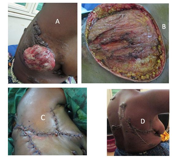
Abstract
Background: In developing countries, the long delays in consultation lead to a delay in diagnosis and
management of the skin tumours. The lesions are often large and brings the problem of skin coverage
after their resections. Several reconstruction techniques allow skin coverage. The objective of this study
is to describe the place of Z-plasty in the surgical treatment of skin cancers in Ouagadougou.

Patients and methods: It was a two-centre, retrospective, descriptive study on Z-plasty in skin cancers. It
included patients who underwent surgery between January 1st, 2013 and March 30th, 2021 in
Ouagadougou. Scar quality and healing time in Z-plasty were compared with those of secondary healing.

Results: In 8 years and 3 months, 171 skin cancers were identified. The mean time to consultation was
13.6 months. The average size of the tumours was 9 cm. A Z-plasty was performed in 42 cases, being
58.3% of the patients operated on. The average healing time was 15 days. It was four and a half times
shorter in Z-plasty than in secondary healing. Ischaemic necrosis of the Z-corner was noted in 7 cases.
The recurrence rate in Z-plasty and secondary healing was 7.1% and 9.1% respectively. Hypertrophic or
keloidal scars were noticed in 7 cases and hypochromia in 2 cases.

Conclusion: Z-plasty is a technique of choice for skin coverage after large resections in surgical oncology.
It reduces the healing time and the cost of postoperative care without increasing the risk of tumour
recurrence.

Introduction
Skin cancers are a public health problem. Their incidence has risen sharply in recent years worldwide,
making them the third most common type of cancer [1]. One in three new cancers in Switzerland is a skin
cancer [2]. They include basal cell carcinomas, squamous cell carcinomas, melanomas and cutaneous
sarcomas [3–5]. Surgery plays an important role in the management of skin cancers [3, 6, 7]. The
modalities of surgical treatment depend on the location, histological type, size of the lesion and the
patient's condition [6, 7]. In the West, the diagnosis of skin tumours is made at an early stage, with small
lesions allowing resection and suturing in majority of cases [8–10]. The situation is quite different in
developing countries, particularly in Burkina Faso [3, 7]. Indeed, the long delays in consultation lead to a
delay in diagnosis and management [7]. The lesions are often large and brings the problem of skin
coverage after their resections. Several reconstruction techniques allow skin coverage [10, 11]. Skin grafts
require a good basement and good hygiene conditions. Secondary healing involves numerous wounds
dressings over several months. The mobilisation of fascia-muscle flaps requires technical skills and a
trained team [7, 10–13]. Z-plasty, a technique which has being used for long in plastic surgery, is less
complex to perform and is therefore an alternative for skin coverage after large tumour resections [11, 13,
14]. In Burkina Faso, Z-plasty has been used for several years now, following skin cancers large
resections [6]. The objective of this study is to describe its indications, technique and results in
Ouagadougou.

                                                  Page 2/11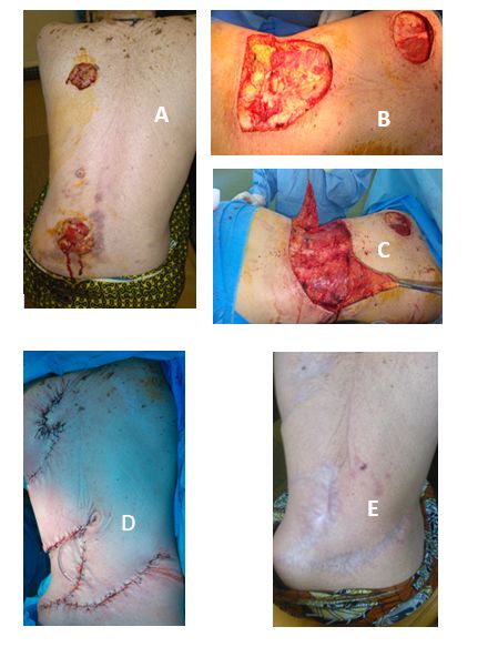
Patients And Methods
This is a two-centre, retrospective, descriptive study on Z-plasty in skin cancer, carried out between
January 1st, 2013 and March 30th, 2021 in Ouagadougou. The surgical departments of the Yalgado
Ouédraogo Teaching Hospital and the Schiphra Protestant Hospital were used as study sites. The study
population was represented by all patients with a skin tumour who had undergone excision. Patients who
had a Z-plasty after skin cancer surgery and were followed until healing were included in this study. Our
data sources were the referral forms, the operating theatre register, and the medical records of the
surgical department of the Yalgado Ouédraogo Teaching Hospital and the Schiphra Protestant Hospital.
The data was collected using a data collection form. We took into account variables such as age, sex of
patients, topography and size of tumours, histological type, closure technique after tumour resection and
complications. Data analysis was done by EPI info software.

Results
In 8 years 3 months, 171 skin cancers were identified. The average age of the patients was 48.5 years
with extremes of 15 and 75 years (Table I). Sixty percent of the patients were men. The average time to
consultation was 13.6 months. The cancers were located on the thorax in 24% of cases (Table I). The
average size of the tumours was 9 cm, with a size greater than 5 cm in 63.3% of cases (Table I).
Cutaneous carcinomas accounted for 60%, melanomas for 25% and sarcomas for 15% of cases (Table I).
Surgery was performed on 72 patients (42%). After lumpectomy or compartmental surgery, direct suture,
skin detachment, and offloading incision allowed skin closure in 14 cases (19.4%), three cases (4.2%),
and two cases (2.8%) respectively. Secondary wound healing was the option in 11 cases (15.3%) with an
average healing time of 67 days with extremes of 52 and 126 days. On average, two dressings were
made per week.

Reconstructive surgery was performed for carcinomas and sarcomas. A Z-plasty was performed in 42
cases, being 58.3% of the patients (Figs. 1, 2, 3, 4). The indication was a wide resection with an
impossibility of direct suture. We only performed this plasty when the resection was at least
macroscopically complete during surgery. The lateral margins of resection varied between 1.5 and 3 cm
with an average of 2.4 cm in carcinomas (34 cases), 3 to 5 cm in sarcomas (7 cases). The length of the
mobilised skin flaps was less than twice their width in all cases (Fig. 1, 2). The average healing time was
15 days with extremes of 12 and 33 days. Partial flap necrosis was noticed in 2 cases. Ischaemic
necrosis of the "Z" angles was noticed in 5 cases (Fig. 3). Healing was achieved by excision of the
necrotic tissue and further wound dressing.

Histology of the surgical specimen referred to pathology noted an R0 resection in 40/42 cases.
Recurrence was observed in 3 cases.

The time required for secondary healing compared to Z-plasty was 4.5 times longer. The recurrence rate
in Z-plasty and secondary healing was 7.1% and 9.1% respectively. Hypertrophic or keloidal scars were

                                                  Page 3/11
noticed in 7 cases (Fig. 4) and hypochromia in 2 cases (Fig. 1).
Discussion
Skin cancers are increasing in incidence and are ranked third among all cancers [1, 15]. They are among
the most common in the West [1, 15, 16]. In Australia, skin cancers have the highest incidence in the world
with 33.6 cases / 100,000 population [16]. This high frequency in the West contrasts with their relative
rarity in Africa. Indeed, skin cancers represent 7.5–11.8% of all cancers in Africa [4, 17]. Skin cancers,
although regularly diagnosed in Burkina Faso, remain relatively rare compared to the European and
American literature [7]. Despite their low frequency, these cancers present difficulties in their
management. Indeed, their diagnosis is late due to long delays in consultation (13.6 months) and
consequently tumour sizes are large with an average of 9 cm. This contrasts with the small tumour sizes
noted by some authors in developed countries, which range from 0.4 cm to 2 cm [8, 10]. When the tumour
is small, direct excision-suture is possible [18]. However, when the tumour is large, the resection leaves
large defects that can be filled by several procedures [16, 19]. There are many indications for skin
coverage, including post-traumatic skin defects, surgical excision for benign or malignant tumours, burns,
and deformities [18]. Skin coverage after large tumour resections remain a real challenge for healing.
Direct suturing helped by the intrinsic elasticity and plasticity of the skin is no more possible [18, 19]. The
prerequisite for direct suturing is an early diagnosis with small resections. This is far from being the case
in our series where the average size of the tumours was 9 cm. Several methods of skin coverage must
therefore be used [18, 19]. Pedicle flaps are used to fill in surgical defects [19]. Local skin or
musculocutaneous T- and H-shaped flaps are used to treat skin defects in the forehead [20]. Rhombic
flaps, which are local transposition flaps, are used to fill defects after skin cancer surgery in the head and
neck region [19]. Z-plasty is the most commonly used technique in precarious situations and has solved
58.3% of the skin coverage problems of the trunk and limbs in our series. In addition to the size of the
tumour, the indications for Z-plasty in our series were the absence of superinfection of the tumour, the
absence of bony relief making it difficult to mobilise the flaps, and the localisation of the tumour in an
area where flaps can be mobilised. The size of the mobilised flaps remains function of the width of the
surgical wound [18, 20]. However, for vitality of the flaps in the Z-plasty that are free, non-pediculised
flaps, we followed the 2:1 rule, meaning the length should not be more than twice the width.
In a situation of limited resources, diagnostic delays, poor results and inaccessibility of chemotherapy,
and the absence of radiotherapy give surgery a central place in the management of skin cancers. In case
of large tumour sizes, the surgeon has the choice between directed healing and mobilisation of skin flaps
or skin grafts [19]. Z-plasty was performed in 58.3% of our patients with tumours size between 5 and 20
cm. Z-plasty allowed skin closure after large skin resections. Unlike vascularised flaps, it does not require
a great technical skill, is fast to perform and accessible to most surgeons. The average healing time after
Z-plasty was 15 days. Min and col. in their series found 29 days [13]. In our series, this healing time is 4
times longer in secondary healing. In addition, with an average of two wound dressings per week, it
makes the total number of wound dressings to be 5 times higher in secondary wound healing than in Z-
plasty.
Page 4/11The flap does not increase the recurrence rate, nor does it interfere with other adjuvant oncological
treatments [19, 20]. The advantages of Z-plasty over secondary wound healing and skin grafting are short
healing times and low postoperative care costs. Z-plasty thus seems to us to be a technique of choice in
precarious situations for low-income countries such as Burkina Faso. In our series, the Z-plasty proved to
be practicable, simple to perform and with very few complications. The healing time was short compared
to secondary healing. It therefore reduces the number of dressings, trips to health centres, and the cost of
care. These skin oncoplasties also reduce the rate of recurrence because of the large resections they
allow the surgeon to perform without having to worry about compromising skin closure.
Conclusion
Cutaneous oncoplastic surgery is in its onset in Burkina Faso. Z-plasty allows skin coverage while
optimising healing. It reduces the healing time compared to secondary healing and consequently the
number of wound dressings and trips to health centres, in short the cost of care. It also reduces
recurrence rates because of the large resections it allows without the surgeon having to worry about
compromising skin closure. In addition to sarcomas and cutaneous carcinomas, it should also be used
for rare skin cancers such as melanomas. The promotion of oncoplasty, a larger cohort and sufficient
hindsight would allow a better appreciation of its advantages in the precarious situation of Burkina Faso.
Declarations
Ethics approval and consent to participate
Access to the information sources was authorised by the heads of departments and directors of the
hospitals concerned. The informations collected were kept confidential and anonymous.
Consent for publication : We have obtained the consent of each patient for the use of the data, especially
the illustrative photos
Availability of data and materials : The datasets used and/or analysed during the current study are
available from the corresponding author on reasonable request.
Competing interests : The authors declare that they have no competing interests" in this section.
Funding : None
Authors' contributions : All authors read and approved the final manuscript."
Concept and design: NZ, NLMO
Drafting of the manuscript: NZ, MW, LSCY, TRKC
Critical revision of the manuscript for important intellectual content: LSCY, AT, PN
Page 5/11All authors approved the final version ofthis publication.
Acknowledgements
We would like to thank Dr NDZANA DIANE for the French-English translation. We would also like to thank
the doctors and nurses working in the surgical departments of the Yalgado Ouédraogo Teaching Hospital
and the Schiphra Hospital for their collaboration.
Authors' information (optional)
Conflicts of interest: None
References
1. Bray F, Ferlay J, Soerjomataram I, Siegel RL, Torre LA, Jemal A. Global cancer statistics. 2018.:
GLOBOCAN estimates of incidence and mortality worldwide for 36 cancers in 185. countries. CA: A
Cancer Journal for Clinicians. nov 2018;68(6):394–424. doi: 10.1002/ijc.29210.
2. Bulliard J, Panizzon RG, Levi F. Epidémiologie des cancers épithéliaux de la peau. Revue. Médicale S.
2009;(5):882–8. PMID: 19438088.
3. Korsaga-Somé N, Zongo N, Ouangré E, et al. Aspects épidémiologique, clinique et.
anatomopathologique du mélanome CHU Yalgado Ouédraogo de Ouagadougou (Burkina Faso). Pan
Afr Med J. 2015;20:220. doi:10.1016/j.bulcan.2019.07.008.
4. Goumbri-Lompo O, Domagni OE, Sanou A, Konségré V. Soudré R. Aspects. épidémiologiques et
histopathologiques des cancers au Burkina Faso. J Afr Cancer. 2009;1:207–11.
https://doi.org/10.1007/s12558-009-0052-x.
5. Effi AB, N’dah KJ, N’Guiessan AA, et al. Épidémiologie descriptive des cancers en Côte d’Ivoire.
Journal Africain du Cancer. 2012;4:41–7. doi:10.1684/bdc.2013.1695.
6. Zongo N, Doamba RN, Somé OR, et al. Factors of Local Recurrence in Soft Tissue Sarcomas at
Yalgado Ouédraogo University Hospital (Burkina Faso). Journal of Cancer Treatment Research.
2017;5(4):66–70. doi:10.11648/j.jctr.20170504.1.
7. Windsouri M, Coulibaly S, Zongo N, et al. Les cancers de la peau et des tissus mous des membres:
épidémiologie, diagnostic et traitement au centre hospitalier universitaire Yalgado Ouédraogo (CHU
YO) au Burkina Faso. Burkina Medical. 2019;23(02):155–63.
8. Wernli KJ, Henrikson NB, Morrison CC, Nguyen M, Pocobelli G, Blasi PR. Screening for Skin Cancer in
Adults: Updated Evidence Report and Systematic Review for the US Preventive Services Task Force.
JAMA. 2016;316(4):436–47. doi:10.1001/jama.2016.5415.
9. Brunssen A, Waldmann A, Eisemann N, Katalinic A. Impact of skin cancer screening and secondary
prevention campaigns on skin cancer incidence and mortality: A systematic review. J Am Acad
Dermatol. 2017;76(1):129–39.e10. doi:10.1016/j.jaad.2016.07.045.
Page 6/1110. Behan JW, Sutton A, Wysong A. Management of Skin Cancer in the High-Risk Patient. Curr Treat
Options Oncol déc. 2016;17(12):60. doi:10.1007/s11864-016-0435-z.
11. Mikami T, Kagimoto S, Yabuki Y, et al. Deltopectoral flap revisited for reconstruction surgery in
patients with advanced thyroid cancer: a case report. BMC Surg 15 sept. 2017;17(1):101.
doi:10.1038/s41598-019-44735-w.
12. Young Kyoo C, Sung Gun B, Byung Chae C. Comparison between Z-plasty and V-Y Advancement for
the Surgical Correction of Cryptotia. Arch Craniofac Surg avr. 2014;15(1):7–13.
doi:10.7181/acfs.2014.15.1.7.
13. Kyunghyun M, Eun JC, Yeon HL, et al. Single vertical incision thoracoabdominal flap for chest wall
reconstruction following mastectomy of locally advanced breast cancer. Annals of Surgical
Treatment Research. 2019;4(97):168–74. doi:10.4174/astr.2019.97.4.168.
14. Saxena K, Manohar V, Bhakhar V, Bahl S. Adenoid basal cell carcinoma: a rare facet of basal cell
carcinoma. BMJ Case Rep. 2016. 10.1136/bcr-2015–214166. doi: 10.1136/bcr-2015-214166.
15. Defossez G, Le Guyader–Peyrou S, Uhry Z, et al. Estimations nationales de l’incidence et de la
mortalité par cancer en France métropolitaine entre 1990 et 2018. Tumeurs solides. Santé publique
France. 2019;1(1):372. https://www.santepubliquefrance.fr. Accessed 15 June 2021.
16. Australian Institute of Health and Welfare. Health system expenditures on cancer and other
neoplasms in Australia 2000–01. Australian Institute of Health and Welfare. AIHW. 2005;60.
https://www.aihw.gov.au. Accessed 15 June 2021.
17. Napo-Koura G, Pitche P, Tchangai-Walla K, et al. Les cancers cutanés au Togo: 223 observations.
Médecine tropicale. 2010;2(70):169–71. PMID: 20486355.
18. Ettalbi S, Droussi H, Ouahbi S, Ibnouzahir M, Boukind EH. LLL plasty: simple procedure for coverage
of cutaneous defects. Ann Chir Plast Esthet. 2013;58(4):367–72. doi:10.1016/j.anplas.2010.10.003.
19. Lebas D, Amici J-M. Introduction to tissue movements - Principles of flaps. Ann Dermatol Venereol.
2019;146(12):832–46. doi:10.1016/j.annder.2019.09.011.
20. Marco O, Boccara D, Mimoun M, Luini J, Chaouat M. La plastie en T pour la reconstruction des pertes
de substances cutanées du front. Annales de chirurgie plastique esthétique. 2005;60(3):208–13.
doi:10.1093/jbcr/irx005.
Tables
Table I : Clinico-pathological characteristics of patients who had a Z-plasty n=42
Page 7/11Number Percentage %
Age of patients (years)
[15–25[ 9 21
[25–50[ 23 56
[50–75] 10 23
Total 42 100
Topography of cancers
Thorax 10 24
Abdomen 8 19
Buttock 9 1
Thigh 7 17
Leg 6 14
Arm 2 5
Total 42 100
Tumour size (cm)
[0–5[ 4 10
[5–10[ 17 40
[10–15[ 11 25
[15–20[ 6 15
More than 20 4 10
Histological type
Darier and Ferrand Dermato-fibrosarcoma 2 4
Fibrosarcoma 4 10
Squamous cell carcinoma 29 72
Basal cell carcinoma 5 12
Undifferentiated sarcoma 2 4
Figures
Page 8/11Figure 1
Z-plasty and multifocal carcinoma of the back in an albino A: Bifocal squamous cell carcinoma of the
back B: Surgical wounds after tumour resection C: Mobilization of skin flaps D: Appearance after Z-plasty
E: Scar appearance one year after Z-plasty surgery
Figure 2
Z-plasty and skin sarcoma of the buttock A: Ulcerative burgeoning skin tumour of the right buttock B:
Operative wound after tumour resection C: Mobilisation of skin flaps D: Appearance after Z-plasty
allowing skin coverage of the wound
Page 9/11Figure 3
Corner necrosis after Z-plasty treated with rapture and directed healing A : Buttock wound after
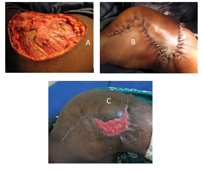
lumpectomy B : Z-plasty C : Necrotic Z-angle appearance after necrosectomy
Page 10/11Figure 4
Z-plasty complicated by keloids A: Cutaneous sarcoma B: Wound after resection C: Z-plasty D: Keloid
healing
Page 11/11You can also read