Ovarian cyst elevation using a metreurynter for laparoscopic cystectomy of a benign ovarian cyst during pregnancy
←
→
Page content transcription
If your browser does not render page correctly, please read the page content below
Kotani et al. BMC Pregnancy and Childbirth           (2021) 21:321
https://doi.org/10.1186/s12884-021-03774-w

 RESEARCH ARTICLE                                                                                                                                   Open Access

Ovarian cyst elevation using a metreurynter
for laparoscopic cystectomy of a benign
ovarian cyst during pregnancy
Yasushi Kotani* , Kosuke Murakami, Kiko Yamamoto, Risa Fujishima, Tamaki Yahata, Yoshie Yo,
Masao Shimaoka and Noriomi Matsumura

  Abstract
  Background: A uterine manipulator cannot be used to elevate the ovary in benign ovarian surgery during
  pregnancy. This report describes our method of elevation of the ovary using a metreurynter with the success rate
  of the procedure and a comparison of surgical results and pregnancy outcomes between the successful and
  unsuccessful cases.
  Methods: Between August 2003 and February 2020, 11 pregnant patients with a tumor found sunk in the Cul-de-
  sac underwent laparoscopic cystectomy for a benign ovarian cyst with a metreurynter. The surgical results, success
  and failure of the elevation by a metreurynter, pregnancy outcomes, and fetal status at delivery were evaluated.
  Results: Elevation of ovarian tumors with a metreurynter was successful in nine cases. However, it was unsuccessful
  in the remaining two cases wherein the ovary was lifted with forceps while the uterus was in a compressed state.
  The operative time was also longer in these cases. The pregnancy prognosis, however, was good for both,
  successful and unsuccessful cases.
  Conclusions: The metreurynter is an inexpensive and practical obstetric device, and its optimal use allows the
  performance of a procedure with minimal burden on a pregnant uterus. Therefore, we recommend the appropriate
  use of this method to enable effective laparoscopic cystectomy of ovarian tumors during pregnancy.
  Keywords: Laparoscopic surgery, Metreurynter, Pregnancy, Ovarian cyst elevation, Ovarian tumor

Background                                                                             unknown, including those associated with anesthesia, sur-
Ovarian tumor is one of the most common gynecological                                  gical infections, and pneumoperitoneum. Opinions thus
tumors. Most ovarian tumors are asymptomatic and de-                                   far on the safety of laparoscopic intervention during preg-
tected for the first time on ultrasonography. Benign ovar-                             nancy for ovarian tumor have varied [3].
ian tumors reportedly complicate 5–6% of all pregnancies                                 Some recent retrospective studies compared pregnancy
[1]. Because it is less invasive than open surgery, laparo-                            outcomes after open and laparoscopic surgeries and re-
scopic surgery is currently the gold standard for the treat-                           ported no significant differences in neonatal outcome
ment of benign ovarian tumors in non-pregnant women                                    [4–9]. The SAGES guideline also states that laparoscopic
[2]. However, the effects of surgery on ovarian tumors                                 surgeries are feasible at any time during pregnancy [8].
during pregnancy, particularly on the fetus, remain                                      Therefore, in our department, laparoscopic surgery is
                                                                                       performed under pneumoperitoneum even in pregnant
* Correspondence: y-kotani@med.kindai.ac.jp                                            cases if diagnosed as benign [7]. However, the uterine
Department of Obstetrics and Gynecology, Kindai University Faculty of                  manipulator cannot be used to elevate the uterus during
Medicine, 377-2 Ohno-higashi, Osaka-sayama, Osaka 589-8511, Japan

                                        © The Author(s). 2021 Open Access This article is licensed under a Creative Commons Attribution 4.0 International License,
                                        which permits use, sharing, adaptation, distribution and reproduction in any medium or format, as long as you give
                                        appropriate credit to the original author(s) and the source, provide a link to the Creative Commons licence, and indicate if
                                        changes were made. The images or other third party material in this article are included in the article's Creative Commons
                                        licence, unless indicated otherwise in a credit line to the material. If material is not included in the article's Creative Commons
                                        licence and your intended use is not permitted by statutory regulation or exceeds the permitted use, you will need to obtain
                                        permission directly from the copyright holder. To view a copy of this licence, visit http://creativecommons.org/licenses/by/4.0/.
                                        The Creative Commons Public Domain Dedication waiver (http://creativecommons.org/publicdomain/zero/1.0/) applies to the
                                        data made available in this article, unless otherwise stated in a credit line to the data.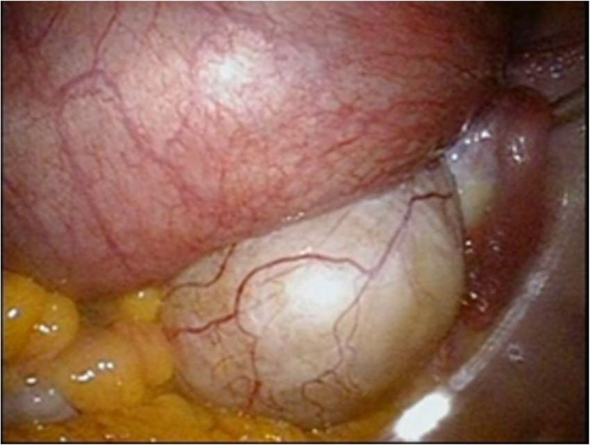
Kotani et al. BMC Pregnancy and Childbirth (2021) 21:321 Page 2 of 5
Fig. 1 Metreurynter (Fujimetro®) used in our department. This figure was taken from the website of Fuji Latex Co
pregnancy. In addition, the enlarged pregnant uterus in- week of pregnancy, operative time, blood loss, tumor
terferes with surgery for a tumor in the Cul-de-sac. In size, pathology, postoperative hospital stay, and success/
this case, the ovary lesion should be elevated with mini- failure of the elevation. Pregnancy outcomes and fetal
mum stimulation to the pregnant uterus. status at delivery, including the week of gestation at de-
Murakami et al. reported using a metreurynter to ele- livery, delivery style, infant birth weight, Apgar score
vate the ovary in a gasless surgery [10]. In our depart- and obstetric complications were also evaluated. This
ment, all laparoscopic surgeries are performed under study was approved by the Institutional Review Board of
pneumoperitoneum, both in pregnant and non-pregnant Kindai University Faculty of Medicine (No.RR01–29).
cases [6]. In pregnant cases, however, the enlarged All research was performed in accordance with Ethical
uterus makes it challenging even to elevate the ovary
tumor sunk in the Cul-de-sac. Lifting the ovaries with
forceps while compressing the uterus can damage the
pregnant uterus and cause heavy bleeding.
This report describes our method using a metreuryn-
ter in benign ovarian surgery during pregnancy. To the
best of our knowledge, there have been no published re-
ports of a large number of ovarian elevations using a
metreurynter during pregnancy.
Methods
We have performed laparoscopic surgery for 44 ovarian
tumors during pregnancy to date. Among them, 11 cases
involved a tumor found sunk in the Cul-de-sac and re-
ceived ovarian cyst elevation using a metreurynter. The
remaining 33 cases had tumors located in front of or
over the uterus and had not received an ovarian cyst ele-
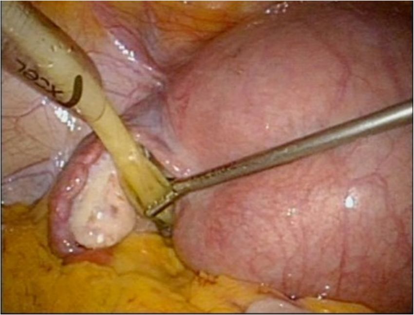
Fig. 2 Ovarian cyst elevation using a metreurynter. A metreurynter is
vation. For the 11 patients who received an ovarian cyst
inserted via the 12-mm trocar
elevation with a metreurynter, we evaluated the age,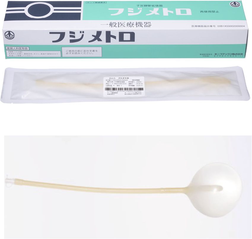
Kotani et al. BMC Pregnancy and Childbirth          (2021) 21:321                                                               Page 3 of 5

                                                                           using a closed method and then inserting three 5-mm
                                                                           trocars, one in the median of the lower abdomen and
                                                                           one each to its right and left, and one 12-mm trocar
                                                                           through the umbilicus [7]. During pregnancy, all trocars
                                                                           are inserted more cranially than usual considering the
                                                                           enlarged uterus [13, 14]. At the start of the surgery, the
                                                                           patient is positioned head down at an angle of approxi-
                                                                           mately 15°. A metreurynter (Fujimetro®, Fuji Latex Co.,
                                                                           Tokyo, Fig. 1) is placed in the Cul-de-sac via the12-mm
                                                                           trocar through the left side of the uterus. An important
                                                                           tip at this point is to insert the metreurynter from
                                                                           around the lateral side of the tumor in the Cul-de-sac,
                                                                           targeting the margin below the tumor. The drawback of
                                                                           this method is the short 30-cm length of the metreuryn-
                                                                           ter. This situation can be avoided by connecting an ex-
    Fig. 3 Saline (300 mL) injected in to the metreurynter in
    the Cul-de-sac                                                         tension intravenous infusion line to the metreurynter to
                                                                           extend the length. After inserting the metreurynter is in-
                                                                           flated with 300–500 mL of saline to elevate the tumor to
Guidelines for Medical and Health Research Involving                       the abdominal cavity (Figs. 2 and 3). At this point, trans-
Human Subjects.                                                            vaginal sonography is useful to ensure that the metreur-
                                                                           ynter is inserted correctly under the tumor. Once the
Surgical technique                                                         ovaries are elevated, the deflated balloon is removed,
When an ovarian tumor is diagnosed by ultrasonography                      and the metreurynter is then collected from the abdom-
during pregnancy, magnetic resonance imaging (MRI) is                      inal cavity. An ovarian tumorectomy is performed by
performed for the differentiation of diagnosing benign                     making an incision halfway around the tumor along the
and malignant lesions. This is the standard procedure in                   equatorial line starting from the opposite side of the
our hospital because MRI is considered to be more use-                     ovarian hilum, and the ovarian tumor alone is carefully
ful than ultrasonography in differentiation [11]. In our                   removed to prevent cyst rupture [7]. The tumor is then
department, MRI is performed later than the 12th week                      transferred outside the body after its contents are aspi-
of gestation. The optimal timing for laparoscopic surgery                  rated inside an Endo Catch™ pouch (Medtronic Japan,
is between the 12th and 16th weeks of gestation when                       Tokyo) inserted through the 12-mm trocar [7]. The pa-
the placental and organ development stages have ended                      tient is permitted to walk and eat on the day after sur-
but the uterus has not grown large enough to impede                        gery and is typically discharged 3–4 days after surgery.
the operative visual field [12].                                           In our hospital, if the patient has no subjective symptom,
  In our department, laparoscopy is performed by first                     tocolysis is not used and postoperative follow-up is man-
attaining pneumoperitoneum through the umbilicus                           aged in the same way as routine laparoscopic surgery.

Table 1 Surgical results of cases using a metreurynter
Case Age range Body mass Week of          Operative Blood                        Tumor     pathology            Postoperative       result
     (years)   index     pregnancy (week) time (min) loss (ml)                   size (cm)                      hospital stay (day)
1        24–37        25.4          14                   95         Small amount 5        Mature cystic teratoma 9                  Success
2                     18.4          16                   105        Small amount 4        Mature cystic teratoma 7                  Failure
3                     19.3          15                   105        37           6        Mature cystic teratoma 5                  Success
4                     17.0          16                   72         Small amount 5        Mature cystic teratoma 4                  Success
5                     19.1          16                   41         Small amount 7        Mature cystic teratoma 3                  Success
6                     20.7          16                   110        50           9        Mature cystic teratoma 4                  Failure
7                     20.4          13                   93         Small amount 10       Para ovarian cyst     4                   Success
8                     18.5          15                   108        Small amount 7        Mature cystic teratoma 4                  Success
9                     23.0          15                   107        Small amount 7        Mature cystic teratoma 4                  Success
10                    20.4          15                   96         Small amount 6        Mature cystic teratoma 5                  Success
11                    24.8          13                   100        Small amount 5        Mature cystic teratoma 5                  Success
Kotani et al. BMC Pregnancy and Childbirth (2021) 21:321 Page 4 of 5
Table 2 Pregnancy prognosis of cases using a metreurynter
Case Week of gestation Delivery style Infant weight Apgar score Obstetric complication
at delivery (week) at delivery (g) (1 min/5 min)
1 38 Natural delivery 3090 8/9
2 40 Natural delivery 2682 7/9
3 40 Natural delivery 3484 8/9
4 39 Natural delivery 2306 8/9
5 39 Natural delivery 3210 8/9
6 41 Natural delivery 3492 9/10
7 41 Natural delivery 3838 9/10
8 40 C-section 3195 9/9 cephalopelvic disproportion
9 40 C-section 3123 8/8 Fetal dysfunction
10 38 Natural delivery 3095 9/9
11 During pregnancy
Results beneath. There are also reports on the use of a rectal
Ovarian tumor elevation using a metreurynter was suc- probe (Rectal Sonde™ [Hakko Medical, Tokyo]) or
cessful in nine cases, while it was unsuccessful in two SAND balloon catheter™ (Hakko Medical, Tokyo) to ele-
cases in which we lifted the ovaries using forceps while vate the ovary [17]. However, SAND balloon catheter™
compressing the uterus. Table 1 shows the surgical can only be used after the ovaries have been raised ini-
results. The success rate was 81.8% (9/11). The two un- tially. In addition, the use of forceps accompanies the
successful cases had longer operative times. However, risk of hemorrhage, and the use of fingers and rectum
these cases included no bleeding or intraoperative probes may create considerable uterine pressure on the
complications. patient. In contrast, our method of using the metreuryn-
Table 2 shows the pregnancy prognoses. The preg- ter creates minimal pressure on the pregnant uterus.
nancy prognoses were favorable for both successful and The metreurynter was originally designed to be suffi-
unsuccessful cases. Case 8 and 9 involved a cesarean sec- ciently strong to be inserted into the pregnant uterus;
tion for obstetric reasons, but the birth prognosis was fa- hence, it had to be sufficiently strong. We experienced
vorable in all cases. no metreurynter deflation in any of the cases evaluated
in the present study.
Discussion The success rate of this technique for ovarian cyst ele-
The metreurynter is an obstetric device that is often vation was 81.8% in this study. Transvaginal ultrasound
used in Japan during delivery. Murakami et al. reported was not combined in the introduction stage of this tech-
the usefulness of the instrument for ovarian cyst eleva- nique. In the two unsuccessful cases (Cases 2 and 6), the
tion in sling surgeries [10]. There have been several re- metreurynter inflated without confirming the position by
ports confirming the efficacy of a sling procedure to transvaginal ultrasound and did not enter the Cul-de-sac
avoid the effects of general anesthesia and pneumoperi- despite repeated trials to elevate the ovary. Furthermore,
toneum [15]. However, other investigators have reported the failure was not due to adhesion or other causes since
the need to convert to open surgery because of the lim- these two cases had no prior surgical history.
ited field of view allowed by sling procedures [16]. Our All recent cases after Case 7 involved the use of trans-
institution uses pneumoperitoneum in all laparoscopic vaginal ultrasound to ensure that the metreurynter was
surgeries, and several recent studies reported no negative swollen in the Cul-de-sac. No failure has occurred since
influence of pneumoperitoneum on neonatal outcomes the use of transvaginal ultrasound, thus indicating the
[4–9]. In laparoscopic surgery for non-pregnant patients, importance and usefulness of this technique which en-
the uterine manipulator can be used to elevate the lesion sures that the tip of the metreurynter is well placed in
in the Cul-de-sac together with the uterus, and the the Cul-de-sac.
method presented in this report may be useful particu- Cases 1 and 2 were previous cases wherein tocolysis
larly during pregnancy, when the uterine manipulator agents were administered postoperatively. Recent reports
cannot be used. Other methods of elevating the ovary state, however, that it is not necessary to administer
during pregnancy include the use of forceps to create tocolysis agents after surgery [8, 18]. We allow patients
space and of fingers to push up the Cul-de-sac from to be discharged as in non-pregnancy cases.Kotani et al. BMC Pregnancy and Childbirth (2021) 21:321 Page 5 of 5
Conclusions 8. Pearl JP, Price RR, Tonkin AE, Richardson WS, Stefanidis D. SAGES guidelines
The metreurynter is an inexpensive and practical obstet- for the use of laparoscopy during pregnancy. Surg Endosc. 2017;31(10):
3767–82. https://doi.org/10.1007/s00464-017-5637-3.
ric device, and its optimal use allows the performance of 9. Koo FH, Wang KC, Chen CY, Chang WH, Yeh CC, Yang MJ, et al. An 11-year
a procedure with minimal burden to the pregnant experience with ovarian surgery during pregnancy. J Chin Med Assoc. 2013;
uterus. The procedure presented herein is likely to be a 76(8):452–7. https://doi.org/10.1016/j.jcma.2013.04.008.
10. Murakami T, Yoshinaga K, Konno R, Terada Y, Nabeshima H, Sawada R, et al.
useful method for removal of a tumor found sunk in the The cul-de-sac packing method with a metreurynter in gynecologic gasless
Cul-de-sac. We recommend the use of this method to laproscopy. Tohoku J Exp Med. 2002;197(3):133–8. https://doi.org/10.1620/
enable effective laparoscopic cystectomy of ovarian tu- tjem.197.133.
11. Yacobozzi M, Nguyen D, Rakita D. Adnexal masses in pregnancy.
mors during pregnancy. Semin Ultrasound CT MR. 2012;33(1):55–64. https://doi.org/10.1053/j.
sult.2011.10.004.
Abbreviation 12. Morice P, Louis-Sylvestre C, Chapron C, Dubuisson JB. Laparoscopy for
MRI: Magnetic resonance imaging adnexal torsion in pregnant women. J Reprod Med. 1997;42(7):435–9.
13. Chohan L, Kilpatrick CC. Laparoscopy in pregnancy: a literature review.
Acknowledgements Clin Obstet Gynecol. 2009;52(4):557–69. https://doi.org/10.1097/GRF.
Not applicable 0b013e3181bea92e.
14. Weiner E, Mizrachi Y, Keidar R, Kerner R, Golan A, Sagiv R. Laparoscopic
Authors’ contributions surgery performed in advanced pregnancy compared to early
YK, KM and NM were major contributors in writing the article. YK, KY, RF, TY, pregnancy. Arch Gynecol Obstet. 2015;292(5):1063–8. https://doi.org/10.1
YY and MS made clinical examinations and performed surgery. NM 007/s00404-015-3744-8.
supervised the project. All authors have read and approved the manuscript. 15. Sesti F, Pietropolli A, Sesti FF, Piccione E. Gasless laparoscopic surgery
during pregnancy: evaluation of its role and usefulness. Eur J Obstet
Funding Gynecol Reprod Biol. 2013;170(1):8–12. https://doi.org/10.1016/j.ejogrb.2
No funding 013.04.012.
16. Cravello L, D'Ercole C, Roger V, Samson D, Blanc B. Laparoscopic surgery in
Availability of data and materials gynecology: randomized prospective study comparing pneumoperitoneum
The datasets used and/or analysed during the current study available from and abdominal wall suspension. Eur J Obstet Gynecol Reprod Biol. 1999;
the corresponding author on reasonable request. 83(1):9–14. https://doi.org/10.1016/S0301-2115(98)00239-5.
17. Kurihara K, Minagawa M, Masuda M, Fukuyama M, Tanigaki K, Yamamoto A,
Declarations et al. The evaluation of laparoscopic surgery on pregnant patients with ovarian
cysts and its effects on pregnancy over the past 5 years. Gynecol Minim
Ethics approval and consent to participate Invasive Ther. 2018;7(1):1–5. https://doi.org/10.4103/GMIT.GMIT_12_17.
The study was performed in accordance with the Declaration of Helsinki 18. Stewart MK, Terhune KP. Management of pregnant patients undergoing
ethical standards and was approved by the institutional review boards of the general surgical procedures. Surg Clin North Am. 2015;95(2):429–42. https://
Kindai University Hospital after obtaining written informed consents from the doi.org/10.1016/j.suc.2014.10.007.
participants (No. RR01–29).
Consent for publication Publisher’s Note
Not applicable. Springer Nature remains neutral with regard to jurisdictional claims in
published maps and institutional affiliations.
Competing interests
The authors declare that they have no competing interests.
Received: 8 September 2020 Accepted: 5 April 2021
References
1. Naqvi M, Kaimal A. Adnexal masses in pregnancy. Clin Obstet Gynecol.
2015;58(1):93–101. https://doi.org/10.1097/GRF.0000000000000088.
2. Nezhat F, Nezhat C, Welander CE, Benigno B. Four ovarian cancers
diagnosed during laparoscopic management of 1011 women with adnexal
masses. Am J Obstet Gynecol. 1992;167(3):790–66. https://doi.org/10.1016/
S0002-9378(11)91591-9.
3. Al-Fozan H, Tulandi T. Safety and risks of laparoscopy in pregnancy. Curr
Opin Obstet Gynecol. 2002;14(4):375–9. https://doi.org/10.1097/00001703-2
00208000-00003.
4. Reedy MB, Källén B, Kuehl TJ. Laparoscopy during pregnancy: a study of five
fetal outcome parameters with use of the Swedish Health Registry. Am J
Obstet Gynecol. 1997;177(3):673–9. https://doi.org/10.1016/S0002-93
78(97)70163-7.
5. Oelsner G, Stockheim D, Soriano D, Goldenberg M, Seidman DS, Cohen SB,
et al. Pregnancy outcome after laparoscopy or laparotomy in pregnancy. J
Am Assoc Gynecol Laparosc. 2003;10(2):200–4. https://doi.org/10.1016/S1
074-3804(05)60299-X.
6. Chen L, Ding J, Hua K. Comparative analysis of laparoscopy versus
laparotomy in the management of ovarian cyst during pregnancy. J Obstet
Gynaecol Res. 2014;40(3):763–9. https://doi.org/10.1111/jog.12228.
7. Kotani Y, Shiota M, Umemoto M, Tobiume T, Tsuritani M, Shimaoka M, et al.
Laparoscopic versus open surgery for pregnancy complicated by benign
ovarian tumor. Jpn J Gynecol Obstet Endosc. 2011;27:489–94.You can also read