A Single Intramedullary K-Wire Is Sufficient for the Management of Nonthumb Metacarpal Shaft Fractures
←
→
Page content transcription
If your browser does not render page correctly, please read the page content below
Hindawi
Advances in Orthopedics
Volume 2021, Article ID 9963186, 7 pages
https://doi.org/10.1155/2021/9963186

Research Article
A Single Intramedullary K-Wire Is Sufficient for the
Management of Nonthumb Metacarpal Shaft Fractures

          Mohamed I. Abulsoud ,1 Mohammed Elmarghany ,1 Tharwat Abdelghany ,1
          Mohamed Abdelaal ,1 Mohamed F. Elhalawany ,1 and Ahmed R. Zakaria 2
          1
              Department of Orthopedic Surgery, Faculty of Medicine, Al-Azhar University, Cairo, Egypt
          2
              Department of Orthopedic Surgery, Helwan University, Helwan, Egypt

          Correspondence should be addressed to Mohamed I. Abulsoud; mohamedabulsoud@azhar.edu.eg

          Received 6 March 2021; Accepted 28 April 2021; Published 4 May 2021

          Academic Editor: Francesco Liuzza

          Copyright © 2021 Mohamed I. Abulsoud et al. This is an open access article distributed under the Creative Commons Attribution
          License, which permits unrestricted use, distribution, and reproduction in any medium, provided the original work is
          properly cited.

          Objective. This study aims to evaluate the outcome after the internal fixation of diaphyseal metacarpal fractures by a single
          intramedullary K-wire. Methods. In this prospective case series study, conducted from July 2017 to June 2019 in 23 adult patients
          with a single, unstable, diaphyseal metacarpal fracture, outcomes after internal surgical fixation using a single antegrade
          intramedullary K-wire were evaluated. The outcomes were evaluated by union rate, time to union, handgrip measurements at 6
          and 12 months, and the modified Disabilities of the Arm, Shoulder, and Hand (DASH) score at 12 months. Results. The study
          population consisted of 17 males and 6 females, with a mean patient age of 28.4 ± 8.5 years (range, 16–45 years). The median time
          to final follow-up was 14 ± 1.8 months (range: 12–24 months). The mean duration of the union was 7.3 ± 1.6 weeks (range: 5–11
          weeks), with a union rate of 95.7% (22 cases). The mean handgrip strength was 68% ± 12.8% of the strength of the uninjured hand
          after 6 months and 92.7% ± 6.9% after 12 months. The mean modified DASH score was 2.6 ± 0.26 after 12 months (range: 0–5.8).
          There were no cases of malrotation or infection. In conclusion, using a single 1.8–2.0 mm K-wire gives excellent functional
          outcomes and union rate without significant complications when used to treat an unstable metacarpal shaft fracture.

1. Introduction                                                         ulnarly and distally [8]. Surgical options for treatment show
                                                                        wide variabilities without a preference for the fixation
Metacarpal fractures are the third most common upper limb               method [9, 10].
injury in young adults. when combined with phalangeal                       As early as 1953, Vom [11] described intramedullary
fractures, they are the most common upper limb injury                   fixation of metacarpal fracture and introduced a K-wire
[1, 2]. Men and young adults are more vulnerable to these               through the head of the metacarpal. Foucher’s [12] bouquet
injuries, as are people of low socioeconomic status [1]. The            technique is the most popular approach for antegrade
leading mechanisms of injury are direct trauma and sports               K-wire fixation; it was initially restricted to the neck of fifth
trauma [1, 2]. Diaphyseal metacarpal fractures cause marked             metacarpal fractures, but has been applied to diaphyseal
angulation and shortening, impeding the function of ex-                 fractures with different modifications [13–17]. In surgical
tensor and flexor tendons [3–5]. Even small degrees of                   practice, ad hoc technological instruments (e.g., plates) often
malrotation are poorly tolerated, leading to digital overlap            are preferred as opposed to K-wires because they are sup-
and impairments of hand functions [6], as the deep trans-               posed to fix the fracture better. However, in adult upper limb
verse metacarpal ligament helps in maintaining shortening               fractures, a safe and effective fixation can be obtained with
and rotation [7]. Metacarpal fractures are more easily tol-             smooth wires and rods with very good functional outcome
erated and can be treated nonoperatively if they occur more             [18–20].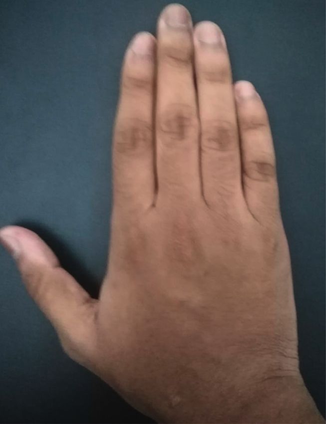
2                                                                                                     Advances in Orthopedics

1.1. Specific Aim and Hypothesis. This study aims to evaluate          Next, the rotation was assessed clinically and radio-
outcomes after internal fixation of diaphyseal metacarpal          graphically, with adjustments made until any malrotation
fractures using a single intramedullary K-wire.                   was addressed and appropriate reduction had been achieved.
     We hypothesized that a single intramedullary K-wire is       During this process, the K-wire was advanced into the distal
enough to fixate a displaced metacarpal fracture, leading to       segment until it reached the metacarpal head where it was
full union and a satisfactory outcome without major               adjusted to achieve the principle of three-point fixation.
complications.                                                    Violation of the articular surface of the metacarpophalangeal
                                                                  joint was carefully avoided. To allow skin closure, the K-wire
2. Methods                                                        was cut short proximally. To avoid any friction, which could
                                                                  lead to tendon rupture or could limit the range of motion in
A case series study was conducted to evaluate the outcome of      the finger, the bend in the K-wire was positioned away from
single antegrade intramedullary K-wire fixation on displaced       the track of the extensor tendon (Figure 3).
metacarpal fractures within 2 weeks of the initial injury. This       The skin incision was closed with simple stitches, and a
study included 23 consecutive patients treated from July          splint was applied below the elbow for 2 weeks.
2017 to June 2019.
    To be included, a patient had to be an adult older than 16
                                                                  2.2. Postoperative Program. Patients visited the outpatient
years with a single unstable diaphyseal fracture of the
                                                                  clinic 2 weeks postoperatively to have the stitches and splint
metacarpal. Unstable fractures were defined as having an-
                                                                  removed. X-rays were taken to ensure adequate reduction
gulation >40°, shortening >2 mm, or malrotation.
                                                                  and fixation. The patient was encouraged to move all joints
    Cases were excluded from the study if the patient had an
                                                                  of the hand actively and passively. Regular follow-up visits
open fracture, associated compartment syndrome of the
                                                                  were scheduled until full union had been achieved.
hand, intraarticular extension, multiple metacarpal frac-
tures, or severe comminution (AO/OTA types 77. 3.2C2 and
77. 3.2C3), or if the patient was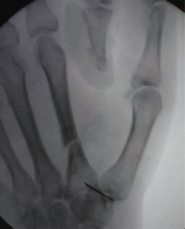
Advances in Orthopedics                                                                                                                        3

                                         (a)                                                     (b)

Figure 1: A 30-year-old male patient with spiral fracture at the fourth metacarpal: the medulla of the second and third metacarpal bones is
too narrow, while the medulla of the fourth metacarpal is patent which allows intramedullary fixation.

                                 (a)                                   (b)                                   (c)

Figure 2: (a) Fluoroscopic photo showing the identification of the incision site by a syringe needle. (b) Fluoroscopic photo for antegrade
fixation of a fracture of the second metacarpal shaft. A 2.5 drill bit is used for drilling of the dorsal cortex. (c) Fluoroscopic photo shows the
advancement of the blunt-tipped prebent K-wire through the entry hole in the dorsal cortex.

Dynamometer Grip Strength Measurement, measuring ca-                         (DASH) score, with scores ranging from 0 (best possible
pacity of 198 lbs/90 kgs by comparing the injured and un-                    score) to 100 (worst possible score). The DASH score
injured hands. The mean handgrip strength of the injured                     measures the severity of symptoms, including pain, stiffness,
hand was 68% ± 12.8% of the strength of the uninjured hand                   weakness, and tingling, as well as the ability to perform
after 6 months and 92.7% ± 6.9% of the strength of the                       activities of daily living, including opening a jar, turning a
uninjured hand after 12 months.                                              key, writing, pushing a door, washing, dressing, and com-
    The functional outcome was assessed according to the                     pleting household tasks. The mean score was 2.6 ± 0.26 after
modified Disabilities of the Arm, Shoulder, and Hand                          12 months (range: 0–5.8; Table 2).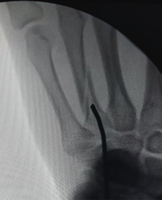
4                                                                                                            Advances in Orthopedics

               (a)                               (b)                             (c)                                 (d)

Figure 3: A 17-year-old male patient whose X-rays of anteroposterior and oblique views of the hand show a displaced diaphyseal fracture of
the second metacarpal. Fluoroscopic photos show the final fixation of the displaced second metacarpal fracture with the three-point fixation
of the intramedullary K-wire.

                                           (a)                                               (b)

                       Figure 4: X-ray showing the 6-month follow-up of the patient, prior to K-wire removal.

    Three patients (13%) developed stiffness of the inter-              4. Discussion
phalangeal joint due to not completing hand exercises at
home. These patients received physical therapy and im-                 The study shows that a single antegrade K-wire can be used
proved by the end of the follow-up.                                    to treat an unstable metacarpal shaft fracture, with excellent
    None of our patients developed malrotation or wound                functional outcomes and a low complication rate.
infection. There were two patients (8.6%) who developed                    Although plate fixation is an attractive option in the
joint penetration of the metacarpophalangeal joint during              treatment of metacarpal shaft fractures due to its stable
follow-up, although this did not affect the outcome (Table 3).          fixation and biomechanical stability [21], it has a relatively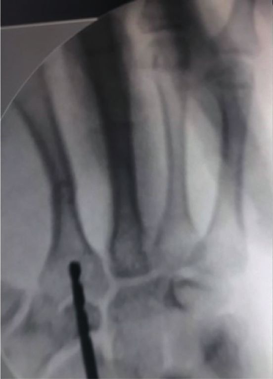
Advances in Orthopedics 5
(a) (b)
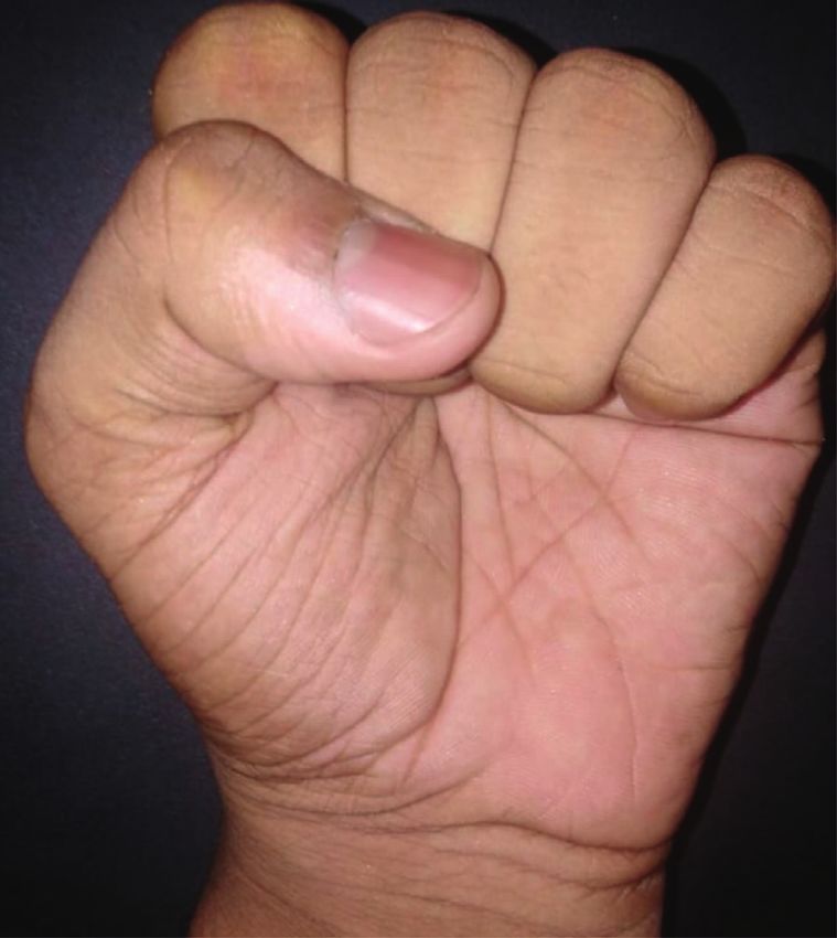
Figure 5: Functional outcome after K-wire removal with full range of motion and excellent functional outcome.
high complication rate of up to one-third of cases [22]. In a
Table 1: Demographic data. study, plate fixation resulted in functional impairments that
Characteristics Value required secondary surgery in 17% of cases [23]. Even in a
Age (years) study using modern, low-profile plates, various complica-
Minimum 16 tions led to plate removal in 40% of cases within 9.6 months
Maximum 45 after surgery [24].
Mean (SD) 28.4 8.5 To the best of our knowledge, this is the first study to
Gender describe and investigate outcomes of fixation of midshaft
Male 19 metacarpal fractures using a single, buried K-wire. To reduce
Female 6 the confounders, multiple metacarpal fractures, metacarpal
Involved bone neck fractures, and highly comminuted fractures were ex-
Second metacarpal 8 cluded from the study. In 23 cases with strict inclusion
Third metacarpal 4 criteria, the functional outcomes were excellent. The mean
Fourth metacarpal 3 modified DASH score was 2.6 ± 0.26 at 12 months post-
Fifth metacarpal 8 operatively, and the mean handgrip strength was
68% ± 12.8% after 6 months and 92.7% ± 6.9% after 12
Table 2: Results. months. These outcomes are comparable to those of most
other studies treating such fractures.
Characteristics Value The union rate was excellent (95.7%). The one case of
Time to union (weeks) nonunion was due to the use of a thin K-wire (1.2 mm), so we
Minimum 5 recommend using 1.8–2 mm K-wires. No cases of clinical
Maximum 11 malrotation were reported in our study. This indicates that
Mean (SD) 7.3 ± 1.6
insertion of a single intramedullary K-wire with the use of a
Union rate 22/23 (95.7%)
Handgrip strength (6 m) 19 splint for 2 weeks can maintain rotational stability in
Minimum 6 fracture types 77. 3.2A and 77. 3.2B.
Maximum
In a recent study using CT to measure the diameter of the
Mean (SD) 68 ± 12.8% nonthumb metacarpal shaft, the narrowest point of the
Handgrip strength (12 m)
medullary canal was found to be between 2.6 and 3.7 mm
Minimum 3 [25], supporting the observations from our study that the use
Maximum 8 of a single intramedullary K-wire with a diameter up to
Mean (SD) 92.7 ± 6.9% 2 mm gives very good stability.
DASH score (12 m) No cases of infection were detected in this study of the
Minimum 0 buried K-wire technique, although with an exposed K-wire,
Maximum 5.8 the infection rate is about 6% [26]. This is in line with results
Mean (SD) 2.6 ± 0.26 published by Ridley and colleagues [27] showing that the risk6 Advances in Orthopedics
Table 3: Complications. (institutional and national) and with the Helsinki Decla-
Nonunion Stiffness MPJ penetration
ration of 1975, as revised in 2008. This study was approved
by our institutional review board.
1 (4.3%) 3 (13%) 2 (8.6%)
of infection is higher in exposed K-wires than in buried Consent
K-wires, especially in the treatment of metacarpal fractures.
The percutaneous antegrade intramedullary fixation has Written consent has been taken from all patients to par-
been described by Landi et al. [18]. The use of the blunt tip of ticipate in the study without sharing their personal infor-
the K-wire has been described previously by Rocchi et al. mation, signed in the Arabic language, and inserted in their
[28]. Their large sample included single and multiple medical files.
K-wires and cases with both shaft and neck fractures but
obtained excellent results with minimal complications. Conflicts of Interest
However, both techniques used unburied K-wires without
focusing on the use of a single K-wire. The authors declare that they have no conflicts of interest.
Although two cases of metacarpophalangeal joint pen-
etration were observed during follow-up, the final functional
outcome was not affected.
References
Despite the short immobilization time in our study (2 [1] J. W. Karl, P. R. Olson, and M. P. Rosenwasser, “The epi-
weeks), three patients reported stiffness in the corresponding demiology of upper extremity fractures in the United States,
interphalangeal joint during follow-up. These symptoms were 2009,” Journal of Orthopaedic Trauma, vol. 29, no. 8,
improved by physiotherapy, and the patients had no limitation pp. e242–e244, 2015.
of motion at the final follow-up. Note that these cases of stiffness [2] M. Ameri, K. Aghakhani, E. Ameri, S. Mehrpisheh, and
and the joint penetration cases were in different patients. A. Memarian, “Epidemiology of the upper extremity trauma
in a traumatic center in Iran,” Global Journal of Health Sci-
Various retrograde and antegrade techniques have been ence, vol. 9, no. 4, pp. 97–105, 2017.
described over 70 years for intramedullary K-wire fixation of [3] R. J. Strauch, M. P. Rosenwasser, and J. G. Lunt, “Metacarpal
metacarpal fractures, but no technique has been proven to be shaft fractures: the effect of shortening on the extensor tendon
definitively superior [29]. A biomechanical study concluded mechanism,” The Journal of Hand Surgery, vol. 23, no. 3,
that using a single 1.6 mm K-wire results in significantly pp. 519–523, 1998.
more stiffness than three 0.8 K-wires [30]. Smooth and [4] C. K. Low, H. C. Wong, Y. P. Low, and H. P. Wong, “A
unlocked fixation devices are not out of date, but they should cadaver study of the effects of dorsal angulation and short-
be used in the right way. The recent literature continues to ening of the metacarpal shaft on the extension and flexion
prove it. The three-point intramedullary fixation system force ratios of the index and little fingers,” Journal of Hand
Surgery, vol. 20, no. 5, pp. 609–613, 1995.
could be superior to the rigid interfragmentary fixation and
[5] M. S. Birndorf, R. Daley, and D. P. Greenwald, “Metacarpal
it does not hinder the movement [19, 20]. fracture angulation decreases flexor mechanical efficiency in
However, the study has some limitations. First, it lacks a human hands,” Plastic and Reconstructive Surgery, vol. 99,
comparison group using other techniques. Second, it re- no. 4, pp. 1079–1083, 1997.
quired a second procedure to remove the K-wire, although [6] W. A. Eglseder, “Metacarpal fractures,” in Atlas of Upper
most of the patients did not report major complaints during Extremity Trauma, W. A. Eglseder, Ed., Springer International
follow-up. Finally, all patients in the study were young and Publishing, Berlin, Germany, 2018.
healthy, and the validity of this technique needs to be tested [7] A. Khan and G. Giddins, “The outcome of conservative
treatment of spiral metacarpal fractures and the role of the
in an older age group and those with osteoporosis.
deep transverse metacarpal ligaments in stabilizing these
In conclusion, the use of a single 1.8–2.0 mm K-wire and
injuries,” Journal of Hand Surgery (European Volume), vol. 40,
immobilization for 2 weeks to treat a displaced metacarpal no. 1, pp. 59–62, 2015.
shaft fracture results in excellent functional outcomes and an [8] V. W. Wong and J. P. Higgins, “Evidence-Based medicine,”
excellent union rate without significant complications. The Plastic and Reconstructive Surgery, vol. 140, no. 1, pp. 140e–
technique should be further validated in cases of multiple 151e, 2017.
fractures or open fractures but should be used with caution [9] M. H. Henry, “Fractures of the proximal phalanx and
in cases of osteoporotic fractures. metacarpals in the hand: preferred methods of stabilization,”
Journal of the American Academy of Orthopaedic Surgeons,
vol. 16, no. 10, pp. 586–595, 2008.
Data Availability [10] J. B. Friedrich and N. B. Vedder, “An evidence-based ap-
proach to metacarpal fractures,” Plastic and Reconstructive
The datasets used and analyzed during the current study are
Surgery, vol. 126, no. 6, pp. 2205–2209, 2010.
available from the corresponding author on request. [11] S. A. A. L. F. H. VOM, “Intramedullary fixation in fractures of
the hand and fingers,” Journal of Bone & Joint Surgery, vol. 35-
Ethical Approval A, no. 1, 1953.
[12] G. Foucher, ““Bouquet” osteosynthesis in metacarpal neck
All procedures followed were following the ethical standards fractures: a series of 66 patients,” The Journal of Hand Surgery,
of the responsible committee on human experimentation vol. 20, no. 3, pp. S86–S90, 1995.Advances in Orthopedics 7
[13] A. A. Faraj and T. R. C. Davis, “Percutaneous intramedullary [28] L. Rocchi, G. Merendi, L. Mingarelli, and F. Fanfani,
fixation of metacarpal shaft fractures,” Journal of Hand “Antegrade percutaneous intramedullary fixation technique
Surgery, vol. 24, no. 1, pp. 76–79, 1999. for metacarpal fractures: prospective study on 150 cases,”
[14] B. J. Mockford, N. S. Thompson, P. C. Nolan, and Techniques in Hand & Upper Extremity Surgery, vol. 22, no. 3,
J. W. Calderwood, “Antegrade intramedullary fixation of pp. 104–109, 2018.
displaced metacarpal fractures: a new technique,” Plastic and [29] J. P. Corkum, P. G. Davison, and D. H. Lalonde, “Systematic
Reconstructive Surgery, vol. 111, no. 1, pp. 351–354, 2003. review of the best evidence in intramedullary fixation for
[15] N. D. Downing and T. R. C. Davis, “Intramedullary fixation of metacarpal fractures,” Hand, vol. 8, no. 3, pp. 253–260, 2013.
unstable metacarpal fractures,” Hand Clinics, vol. 22, no. 3, [30] S. V. Hiatt, M. T. Begonia, G. Thiagarajan, and
pp. 269–277, 2006. R. L. Hutchison, “Biomechanical comparison of 2 methods of
[16] B. J. Zirgibel and W. S. Macksoud, “Self-correcting intra- intramedullary K-wire fixation of transverse metacarpal shaft
medullary Kirschner wire fixation of metacarpal shaft frac- fractures,” The Journal of Hand Surgery, vol. 40, no. 8,
tures,” Techniques in Hand & Upper Extremity Surgery, vol. 17, pp. 1586–1590, 2015.
no. 2, pp. 87–90, 2013.
[17] E. M. Van Bussel, R. M. Houwert, T. J. M. Kootstra et al.,
“Antegrade intramedullary Kirschner-wire fixation of dis-
placed metacarpal shaft fractures,” European Journal of
Trauma and Emergency Surgery, vol. 45, no. 1, pp. 65–71, 2019.
[18] A. Landi, R. Luchetti, F. Catalano, and M. Altissimi, Trattato
di Chirurgia della mano, Verduci, Rome, Italy, chapter 18,
2007.
[19] R. De Vitis, M. Passiatore, V. Cilli, J. Maffeis, G. Milano, and
G. Taccardo, “Intramedullary nailing for treatment of forearm
non-union: is it useful?-a case series,” Journal of Orthopaedics,
vol. 20, pp. 97–104, 2020.
[20] M. Passiatore, R. De Vitis, A. Perna, M. D’Orio, V. Cilli, and
G. Taccardo, “Extraphyseal distal radius fracture in children:
is the cast always needed? A retrospective analysis comparing
Epibloc system and K-wire pinning,” European Journal of
Orthopaedic Surgery & Traumatology, vol. 30, no. 7,
pp. 1243–1250, 2020.
[21] B. D. Curtis, O. Fajolu, M. E. Ruff, and A. S. Litsky, “Fixation
of metacarpal shaft fractures: biomechanical comparison of
intramedullary nail crossed K-wires and plate-screw con-
structs,” Orthopaedic Surgery, vol. 7, no. 3, pp. 256–260, 2015.
[22] C. Fusetti, H. Meyer, N. Borisch, R. Stern, D. D. Santa, and
M. Papaloı̈zos, “Complications of plate fixation in metacarpal
fractures,” The Journal of Trauma: Injury, Infection, and
Critical Care, vol. 52, no. 3, pp. 535–539, 2002.
[23] A. P. A. Greeven, S. Bezstarosti, P. Krijnen, and I. B. Schipper,
“Open reduction and internal fixation versus percutaneous
transverse Kirschner wire fixation for single, closed second to
fifth metacarpal shaft fractures: a systematic review,” Euro-
pean Journal of Trauma and Emergency Surgery, vol. 42, no. 2,
pp. 169–175, 2016.
[24] S. M. Cha, H. D. Shin, and Y. K. Kim, “Comparison of low-
profile locking plate fixation versus antegrade intramedullary
nailing for unstable metacarpal shaft fractures--A prospective
comparative study,” Injury, vol. 50, no. 12, pp. 2252–2258,
2019.
[25] M. L. Dunleavy, X. Candela, and M. Darowish, “Morpho-
logical analysis of metacarpal shafts with respect to retrograde
intramedullary headless screw fixation,” Hand, Article ID
1558944720937362, 2020.
[26] L. P. Hsu, E. G. Schwartz, D. M. Kalainov, F. Chen, and
R. L. Makowiec, “Complications of K-wire fixation in pro-
cedures involving the hand and wrist,” The Journal of Hand
Surgery, vol. 36, no. 4, pp. 610–616, 2011.
[27] T. J. Ridley, W. Freking, L. O. Erickson, and C. M. Ward,
“Incidence of treatment for infection of buried versus exposed
kirschner wires in phalangeal, metacarpal, and distal radial
fractures,” The Journal of Hand Surgery, vol. 42, no. 7,
pp. 525–531, 2017.You can also read