Update to the difficulties in diagnostic and therapeutic management of the rare carpometacarpal joint II-V dislocation injuries: Case presentation ...
←
→
Page content transcription
If your browser does not render page correctly, please read the page content below
Trauma and Emergency Care
Case Report ISSN: 2398-3345
Update to the difficulties in diagnostic and therapeutic
management of the rare carpometacarpal joint II-V
dislocation injuries: Case presentation and review of
literature
Ingo Schmidt*
Medical Centre Wutha-Farnroda GmbH (Department of Orthopaedics, Traumatology and Hand Surgery), Ringstr. 20, 99848 Wutha-Farnroda, Germany
Abstract
This case report describes specific features and difficulties about the rare carpometacarpal joint dislocation injury. The carpometacarpal joint IV seems to be the main
problem in radiographic assessment because it is partially overlapped by the hook of the hamate bone. Therefore, computed tomography scans should be done in every
instance especially after a closed reduction procedure. When joint transfixations become necessary, then the K-wires should not cross the midcarpal joint in order to
obtain early wrist motion.
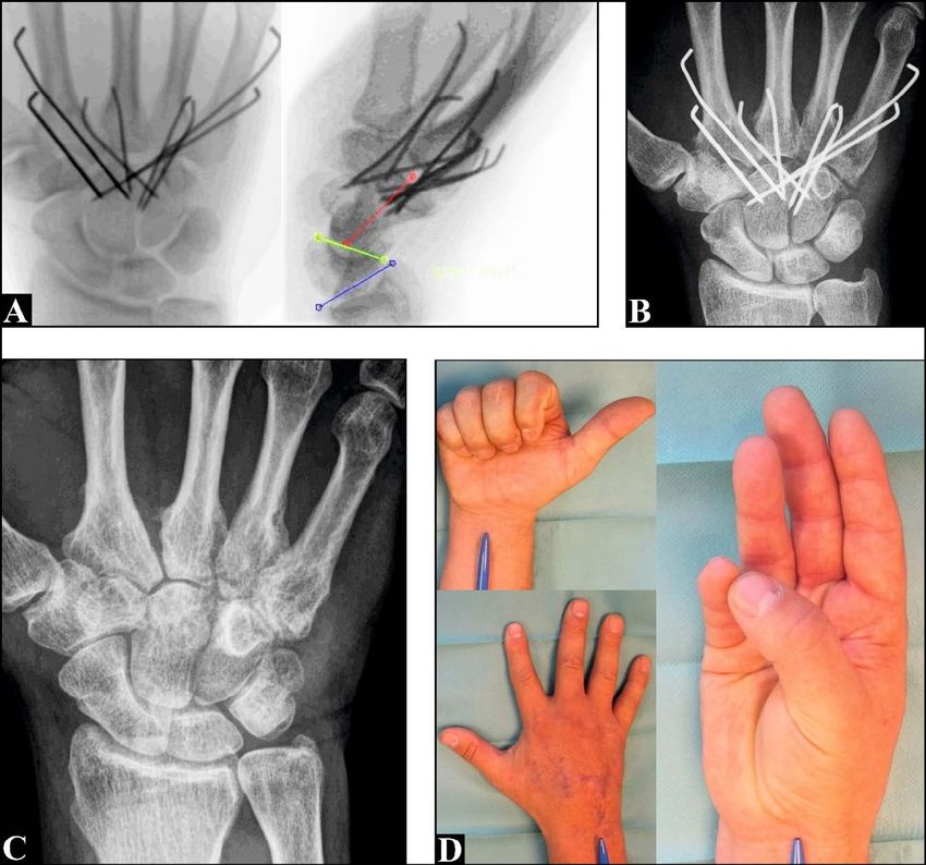
Abbreviations: CMCJ: Carpometacarpal joint; MCPJ: Metacarpo- that revealed persistent or recurrent subdislocation of the CMCJ III
phalangeal joints; K-wires: Kirschner-wires; CT: Computed tomogra- [Figure 2D], and nearly complete dislocation of the CMCJ IV [Figure
phy; ORIF: Open reduction and internal fixation 2E]. Moreover, CT scan demonstrated impressively that the CMCJ
IV could not sufficiently assessed in the postoperative radiograph
Introduction because this joint was ulnar-sided completely overlapped by the hook
Dislocation involving one or more carpometacarpal as well of the hamate bone [Figure 2E]. An open reduction became necessary
as metacarpophalangeal joint(s) (CMCJ, MCPJ) with or without secondarily with joint transfixations utilizing two K-wires for each
concomitant fractures (i.e. fracture-dislocation injury) is a rare joint which did not cross the midcarpal joint that allows an early active
high-energy injury with various pattern, it can be associated with a movement for the wrist too [Figures 3A-B]. The hand was immobilized
polytrauma after traffic accidents, and it is observed of both in dorsal with a plaster splint for two weeks, after that, movement of the wrist
and in volar direction [1-4]. Normally, the CMCJ space of an uninjured and all finger joints were freed without load for another four weeks.
hand is clearly visible and limited by the parallel running articular The K-wires were completely removed three months after second
surfaces. Typically for a dislocation, the joint space is not visible due surgery. At the 1-year follow-up, there were unchanged stable and
to an overlapping of the carpal articular surface by the base of the well-aligned CMCJs II-V but with pronounced signs of post-traumatic
opposing metacarpal bone in the posterior-anterior (PA) radiograph, osteoarthritis of the CMCJ IV and V radiographically [Figure 3C], and
however, this sign can not often easily assessed potentially leading to a function of all finger joints had been completely restored [Figure 3D].
missed diagnosis of a primary injury [5,6], or a persistent or recurrent Discussion
(sub)dislocation after closed reduction such as presented with our case.
Main causes for polytraumata are traffic accidents (cars,
Case presentation motorcycles, bicycles, pedestrians) with a relative portion ranging
A 46-year-old male sustained a polytrauma (Injury Severity Score from 65-88%, and followed by accidental and suicidal falls [7,8]. Most
5, scale 1-6) due to a car accident associated with multiple fractures frequently observed in polytraumatized patients are fractures of the long
of the ribs that led to a right pneumothorax, the left clavicle, the right bones with a relative portion of 86% (most frequently fractures at the
distal femur, the left lower leg, the left radius shaft, the left hand, and lower extremities with a male predominance), whereas injuries at the
CMCJ II-V dislocations at his right hand [Figures 1A-E, Figure 2A].
Primarily, a closed reduction with transfixation of the CMCJ II and V
*Correspondence to: Ingo Schmidt, Medical Centre Wutha-Farnroda GmbH
(i.e. border digit rays) utilizing two percutaneously drilled K-wires was
(Department of Orthopaedics, Traumatology and Hand Surgery), Ringstr. 20,
done and intraoperative fluoroscopy showed well-aligned CMCJs II-V 99848 Wutha-Farnroda, Germany, E-mail: schmidtingo62@googlemail.com
as suggested by us [Figure 2B], noted that persistent (sub)dislocations
as well as joint instabilities could not observed in the lateral and oblique Key words: polytrauma, carpometacarpal joint, dislocation injury, anatomical
plane with dynamic fluoroscopy. However, the PA radiograph one day landmarks, computed tomography, reduction
after surgery did not allow an exact assessment of the CMCJ IV [Figure Received: March 09, 2021; Accepted: March 19, 2021; Published: March 26,
2C]. Therefore, a computed tomography (CT) scan was detected by us 2021
Trauma Emerg Care, 2021 doi: 10.15761/TEC.1000209 Volume 6: 1-6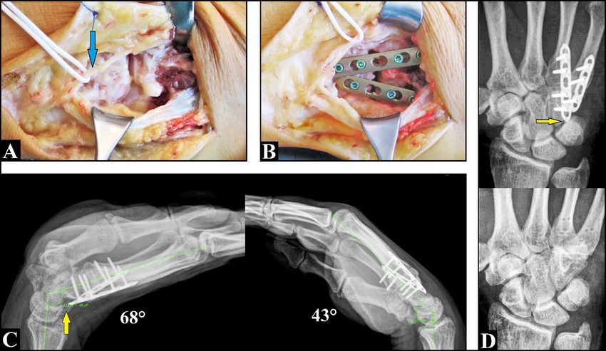
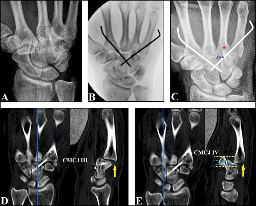
Schmidt I (2021) Update to the difficulties in diagnostic and therapeutic management of the rare carpometacarpal joint II-V dislocation injuries: Case presentation
and review of literature

Figure 1 (Case presentation). (A) The radiograph of the chest showing the intratracheal tubulus for the the required arteficial respiration, the right intrathoracic drain for treatment of the
pneumothrax, and the fracture of the left clavicle (arrows). The fracture of the clavicle was treated by ORIF. (B) The highly comminuted right distal femoral fracture was definitively treated
by ORIF after initial external fixation. (C) The highly comminuted fracture of the left lower leg was definitively treated by ORIF after initial external fixation. (D) The left radius shaft
fracture was treated by ORIF. (E) The multiple fractures of the left hand were treated by ORIF

Figure 2 (Case presentation). (A) Initial PA radiograph of the right hand demonstrating the typical overlapping in the CMCJs II-V. (B) Intraoperative fluoroscopy after closed reduction
showing the K-wire transfixation of the border digit rays. Note that the CMCJ II-V joint spaces are well visible suggesting correctly performed reductions. (C) Postoperative PA radiograph
after closed reduction confirmed the intraoperative findings, however, the CMCJ IV could not clearly assessed. The red dot marks the distal shadow of the apex of the hook of the hamate
bone which is more distally located than the joint space (blue dotted line). (D) Sagittal CT scan after closed reduction showing the persistent or recurrent subdislocation of the third
metacarpal bone against the capitate bone in dorsal direction (arrow). (E) Sagittal CT scan after closed reduction showing the persistent or recurrent nearly complete dislocation of the fourth
metacarpal bone against the hamate bone in dorsal direction (arrow). Note that the articular space of the hamato-metacarpal joint (light blue dotted lines) is ulnar-sided completely overlapped
by the hook of the hamate bone (yellow dotted lines)

Trauma Emerg Care, 2021              doi: 10.15761/TEC.1000209                                                                                                               Volume 6: 2-6
Schmidt I (2021) Update to the difficulties in diagnostic and therapeutic management of the rare carpometacarpal joint II-V dislocation injuries: Case presentation
and review of literature

Figure 3 (Case presentation). (A) Intraoperative fluoroscopy after open reduction and K-wire transfixation without crossing the midcarpal joint showing well-aligned CMCJs II-V. The
three colored lines on the lateral view demonstrating the relative portion of both the radiocarpal and the midcarpal joint at terminal range of extension in the absence of persisting CMCJs
instabilities (i.e. dynamic fluoroscopy). (B) PA radiograph after open reduction demonstrating well-aligned CMCJs II-V too. (C) PA radiograph at the 1-year follow-up demonstrating
unchanged well-aligned CMCJs II-V, but there are pronounced signs of post-traumatic osteoarthritis at the CMCJ IV and V. All K-wires were removed three months after open reduction.
(D) Clinical photographs at the 1-year follow-up showing the complete functional restoration of all finger joints

Figure 4 (57-year-old female, after a fall downstairs). PA radiograph of the left hand showing the isolated CMCJ V dislocation injury. Note the elapsed Gilula’s third arc between the
fifth metacarpal and the hamate bone, and the interrupted broad letter „M“ configuration in the CMCJ V due to the dislocation of the metacarpal bone in dorsal and ulnar direction (circle)

Trauma Emerg Care, 2021             doi: 10.15761/TEC.1000209                                                                                                            Volume 6: 3-6
Schmidt I (2021) Update to the difficulties in diagnostic and therapeutic management of the rare carpometacarpal joint II-V dislocation injuries: Case presentation
and review of literature
hand with a relative portion in up to 3.3% are clearly underrepresented of at least the intermetacarpal ligament, and complete dislocation the
[1,9,10]. The risk of getting hand injuries in polytraumatized patients is rupture of both palmar and dorsal ligaments [20].
reported to be in up to 20% (excluding distal radius fractures), and in
Divergent multiple CMCJ dislocations are possible as well. It
75% of all cases carpal or metacarpal fractures were found [11]. CMCJ
is defined as volar dislocation of ≥ 1 joint with concomitant dorsal
(fracture-)dislocation injuries represent less than 1% of all hand and
wrist injuries, and it is mainly caused in 54% by punches, followed by dislocation of ≥ 1 joint, and this variant of injury is caused of either
traffic accidents in 23% and by falls from height in 14% of cases [12-14]. by a twisting mechanism represented by supination of the metacarpal
arc around an axis passing between the third and fourth metacarpals,
The CMCJs II-V are saddle joints (comparable with the CMCJ or by a direct impact immediately followed by a rotatory force [22,23].
I) that are stabilized like an amphiarthrosis by the volar and more Noted that CMCJ dislocations can be associated with concomitant
stronger dorsal ligaments, intermetacarpal and transverse ligaments, MCPJ (fracture-)dislocation injury as well [24,25]. Moreover, Gaheer
the long flexor and extensor tendons, the intrinsic muscles of the hand; and Ferdinand [26] reported about one case with a third metacarpal
and the ulnar-sided CMCJs (II, III) are more mobile than the radial- base fracture associated with primarily missed dislocations of the
sided CMCJs (IV, V) [15,16]. In direction to the proximal carpal row CMCJ IV and V, Brinkman et al. [27] observed one case with CMCJ
the opposing joint surfaces of the capitate and hamate bone form up II-V dislocations associated with a concurrent fracture of the hamate
to the concave to distally third Gilula’s arc [17]. In distal direction, the bone, and Feder et al. [28] reported about one case with a 180° in situ
uninjured CMCJs showing parallel running articular surfaces to the dislocation of the trapezoid bone associated with concomitant CMCJ
opposing metacarpal bones with a distance of 1.0-2.0 mm and forming II and III dislocations. Noted was well that a newest study including
a broad letter „M“ configuration [12]. The CMCJ III is more proximally 139,931 polytraumatized patients revealed that the relative portion
located than the other joints producing the keystone phenomenon for of primarily missed hand injuries is 6.6% (excluding distal radius
load transfer through the wrist [18]. CMCJ dislocation injuries can be fractures), and 11.2% of these patients sustained carpal fractures/
caused by the apply of either direct or indirect forces. Direct forces dislocations [29].
can occur by the abrupt reflexive clasping of the handlebars (cars or
motorcycles) during the sudden impact such as suggested by us with In diagnostic management PA, lateral and 45° oblique radiographs
our case, and indirect (i.e. axial) forces especially by an intercepting are recommended, and intraoperative dynamic stress fluoroscopy can
reaction of the hand in the event of a fall or by punches [11,14,19, 20]. be helpful as well [21,26,30]. However, in up to 70% of cases (sub)
The direction of dislocation depends on the position of the wrist at the dislocations can be overlooked [5]. Therefore, CT is recommended
time of the impact (in dorsal direction when the wrist is held in flexion primarily if a (sub)dislocation injury is clinically suggested but not
and vice versa). Dislocations in dorsal directions are more common correctly assessed by radiographs. With our case, there were persistent
than in volar direction, and dislocations of the CMCJ IV and V are more or recurrent (sub)dislocations after closed reduction that could not
common than the CMCJ II and III due to its anatomical pre-existing correctly assessed by us of both in intraoperative fluoroscopy and
higher mobility [Figure 4] [16,21]. Subdislocation implies the rupture postoperative radiographs, and the CMCJ IV seems to be the main
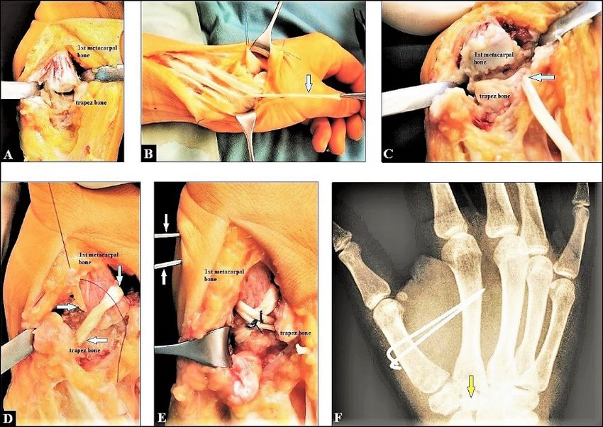
Figure 5 (A 32-year-old female presented with a right CMCJ I dislocation injury after a fall while alpine skiing, intraoperative clinical photographs and postoperative PA
radiograph). (A) The dorsal capsule and ligaments were completely disrupted. (B) Dissection of a distally pedicled abductor pollis longus tendon slip. (C) Passing the tendon slip through
a bony tunnel of the trapez bone. (D) Passing the tendon slip through a bony tunnel of the opposing metacarpal bone. (E) Suturing of the tendon slip with itself. (F) The CMCJ I was
immobilized for six weeks postoperatively utilizing transmetacarpal K-wire transfixation
Trauma Emerg Care, 2021 doi: 10.15761/TEC.1000209 Volume 6: 4-6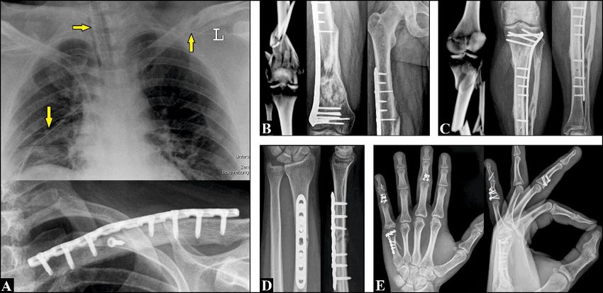
Schmidt I (2021) Update to the difficulties in diagnostic and therapeutic management of the rare carpometacarpal joint II-V dislocation injuries: Case presentation
and review of literature

Figure 6 (26-year-old male, chronic painful instabilities of the right CMCJ IV and V after a fall from 2 m height one year previously, primarily diagnosed as joint sprain and
treated conservatively by splinting). (A) Intraoperative clinical photograph showing the ligamentous instabilities at the CMCJ IV and V. The dorsal cutaneous branch of the ulnar nerve
was carefully dissected and obtained (arrow). (B) Intraoperative clinical photograph showing the fusions of both CMCJs utilizing two 2,0 mm titanium locking plates. After resection of the
corresponding articular surfaces the joint spaces were filled off with cancellous iliac crest bone grafts. (C) Lateral radiographs one year after surgery demonstrating an excellent functional
outcome for the wrist, but noted that there was a painful impingement between the radial-sided titanium plate and the pisiforme bone at terminal range of active extension (arrow). (D)
PA radiographs one year after surgery (before and after removal of both plates) showing complete bony fusions of both CMCJs. Note that the radial-sided titanium plate compromised the
pisiforme bone (arrow)

problem due to its ulnar-sided complete overlapping by the hook of                                Conclusion
the hamate bone. Thus, a postoperative CT after a closed reduction
procedure must be recommended by us in every instance. In the                                         Concerning our presented case there are five keystones: (1) it is
literature there are only a few case reports about persistent instability                         unclear if there was a persistent or a recurrent (sub)dislocation within
after closed reduction and it was stated that only an open reduction is                           one day after the closed reduction procedure with our patient, (2) the
able to guarantee anatomical reduction [31].                                                      CMCJ IV seems to be the main problem in radiographic assessment
                                                                                                  that is based on its partial overlapping by the hook of the hamate bone,
     The therapeutic approach involves both closed and open reduction                             (3) CT scans should be done in every instance after closed reduction
procedures with or without CMCJ transfixation using K-wires. For                                  with or without K-wire transfixation, or a primary injury of a CMCJ
the recovery of polytraumatized patients it is absolutely necessary to                            is suggested clinically and the radiographs do not allow an exact
achieve the earliest possible mobilization involving their independence                           assessment, (4) transfixation of each CMCJ utilizing two K-wires
in daily life for example to perform body hygiene that requires                                   without crossing the midcarpal joint allows an early active motion of
sufficient wrist motion [1]. The extension-flexion motion arc is realized                         both for the wrist and all finger joints which is an important factor for a
with a relative portion of approximately 50% of both in radiocarpal                               required early mobilization and recovery of a polytraumatized patient,
and midcarpal joint, and for radial-ulnarduction the opposite shift of                            and (5) a CMCJ dislocation injury must be considered as a predicting
                                                                                                  factor for development of post-traumatic osteoarthritis.
the proximal carpal row is needed [32]. Therefore, it is recommended
that the K-wires should not cross the midcarpal joint, and additionally                           Declaration of conflicting interests
in order to avoid a breakage of the K-wires if wrist motion is freed. The
difference in the therapeutic approach to the CMCJ I dislocation injury                               The author declares no potential conflicts of interest with respect to
                                                                                                  the research, authorship, and/or publication of this article.
is that here an open reduction combined with a ligamentous repair
should be recommended in every instance in order to obtain a stable                               Funding
thumb’s circumduction [Figures 5A-F] [33].
                                                                                                       None.
    Delayed diagnosis and/or reduction usually result in undesirable
                                                                                                  References
outcome of pain, reduced grip strength, and degenrative arthritis that
is comparable to those after MCPJ dislocation injuries [30,34,35].                                1. Schmidt I, Markgraf E, Friedel R (1997) Die Handverletzung beim Polytrauma. OP-
                                                                                                     JOURNAL 13: 146-150 (Thieme, Stuttgart-New York).
However, despite sufficient management of CMCJ dislocation injuries
43% of patients reported residual pain and impaired function, and                                 2. Mueller JJ (1986) Carpometacarpal dislocations: report of five cases and review of the
                                                                                                     literature. J Hand Surg Am 11: 184-188. [Crossref]
13% of patients can not return to full work and sporting activities [36].
For persisting chronic and painful CMCJ instability or symptomatic                                3. Fotiadis E, Svarnas T, Lyrtzis C, Papadopoulos A, Akritopoulos P, et al. (2010) Isolated
                                                                                                     thumb carpometacarpal joint dislocation: a case report and review of the literature. J
arthritis CMCJ fusion will be inevitable [Figures 6A-D] [33,37].                                     Orthop Surg Res 5: 16. [Crossref]

Trauma Emerg Care, 2021             doi: 10.15761/TEC.1000209                                                                                                               Volume 6: 5-6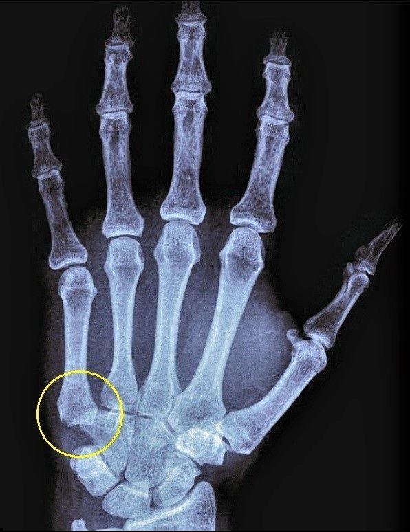
Schmidt I (2021) Update to the difficulties in diagnostic and therapeutic management of the rare carpometacarpal joint II-V dislocation injuries: Case presentation
and review of literature

4. Beylich T, Schmidt I (2021) Open Dislocation Injury of the Metacarpophalangeal               22. Kumar R, Malhotra R (2001) Divergent fracture-dislocation of the second
   Joints III-V: Case Report. Plast Reconstr Surg Glob Open 9: e3424.                               carpometacarpal joint and the three ulnar carpometacarpal joints. J Hand Surg Am 26:
                                                                                                    123-129.
5. Henderson JJ, Arafa MA (1987) Carpometacarpal dislocation. An easily missed
   diagnosis. J Bone Joint Surg Br 69: 212-214. [Crossref]                                      23. Chikate A, Mthethwa J (2017) A Review of the Diagnosis and Management of Index
                                                                                                    through Fifth Carpometacarpal Dislocations. MOJ Orthop Rheumatol 9: 00534.
6. Jameel J, Zahid M, Abbas M, Khan AQ (2013) Volar dislocation of the second, third,
   and fourth carpometacarpal joints: a rare and easily missed diagnosis. J Orthop              24. Jari S, Waseem M, Srinivasan MS (2000) Simultaneous Bennett’s fracture and
   Traumatol 14: 67-70. [Crossref]                                                                  metacarpophalangeal dislocation of the same thumb in a soccer player. Br J Sports Med
7. Regel G, Lobenhoffer P, Lehmann U, Pape HC, Pohleman T, Tscherne H (1993)                        34: 463-464. [Crossref]
   Results of treatment of polytraumatized patients. A comparative analysis of 3,406 cases
                                                                                                25. Kocazeybek E, Demirel M, Arzu U, Ergin ON (2018) Simultaneous metacarpophalangeal
   between between 1972 and 1991. Unfallchirurg 96: 350-362.
                                                                                                    dislocation and carpometacarpal fracture-dislocation of the ring finger: a case report. J
8. Ruchholtz S, Nast-Kolb D, Waydhas C, Schweiberer L (1996) The injury pattern                     Orthop Case Rep 8: 68-70. [Crossref]
   in polytrauma. Value of information regarding accident process in clinical acute
   management. Unfallchirurg 99: 633-641.                                                       26. Gaheer RS and Ferdinand RD (2011) Fracture dislocation of carpometacacarpal joints:
                                                                                                    a missed injury. Orthopedics 34: 399.
9. Welkerling H, Wening JV, Langemdorff HU, Jungbluth KH (1991) Computer-assisted
   data analysis of injuries of the skeletal system in polytrauma patients. Zentralbl Chir      27. Brinkman JN, Hartholt KA, de Vries MR (2016) Multiple carpometacarpal dislocations
   116: 1263-1272.                                                                                  and an associated fracture of the hamate: an uncommon injury. BMJ Case Rep 2016:
                                                                                                    bcr2015213106.
10. Reynolds BM, Balsano NA, Reynolds FX (1971) Falls from height: a surgical
    experience of 200 consecutive cases. Ann Surg 174: 304-308. [Crossref]                      28. Feder OI, Letzelter JP, Haquebord JH (2021) Dorsal Dislocation oft he Trapezoid with
                                                                                                    Metacarpal Instability. J Wrist Surg DOI: 10.1055/s-0040-1715801 (eFirst).
11. Schaller P, Geldmacher J (1994) Hand injury in polytrauma. A retrospective study of
    782 cases. Handchir Mikrochir Plast Chir 26: 307-312. [Crossref]                            29. Fitschen-Oestern S, Lippross S, Lefering R et al. (2020) Missed hand and forearm
                                                                                                    injuries in multiple trauma patients: An analysis from the TraumaRegister DGU®.
12. Gurland M (1992) Carpometacarpal joint injuries of the fingers. Hand Clin 8: 733-744.           Injury 51: 1608-1617.
    [Crossref]
                                                                                                30. Woon CYL, Chong KC, Low CO (2006) Carpometacarpal joint dislocations of the
13. Sharma AK, John JT (2005) Unusual Case of Carpometacarpal Dislocation of All Four
                                                                                                    index to small finger: Three cases and a review of literature. Injury Extra 37: 466-472.
    Fingers of Ulnar Side of Hand. Med J Armed Forces India 61: 188-189. [Crossref]
14. Yoshida R, Shah MA, Patterson RM, Buford Jr WL, Knighten J, Viegas SF (2003)                31. Kimura H, Toga A, Suzuki T, Iwamato T (2021) Open Reduction for Dorsal Dislocation
    Anatomy and pathomechanics of ring and small finger carpometacarpal joint injuries. J           of Second to Fifth Carpometacarpal Joints: A Case Report. J Wrist Surg DOI: 10.1005/
    Hand Surg Am 28: 1035-1043. [Crossref]                                                          s-0040-1715802 (eFirst).

15. El-Shennawy M, Nakamura K, Patterson RM, Viegas SF (2001) Three-dimensional                 32. Schmidt I (2019) Functional Outcomes After Salvage Procedures for Wrist Trauma and
    kinematic analysis of the second through fifth carpometacarpal joints. J Hand Surg Am           Arthritis (Four-Corner Fusion, Proximal Row Carpectomy, Total Wrist Arthroplasty,
    26: 1030-1035. [Crossref]                                                                       Total Wrist Fusion, Wrist Denervation): A Review of Literature. Open Orthop J 13:
                                                                                                    217-231.
16. Pundkare GT, Patil AM (2015) Carpometacarpal Joint Fracture Dislocation of Second
    to Fifth Finger. Clin Orthop Surg 7: 430-435. [Crossref]                                    33. Schmidt I (2018) The various patterns of dislocation injuries at the carpometacarpal
                                                                                                    joints: A case series and brief review of literature with regards to the main topics.
17. Metz VM, Wunderbaldinger P, Gilula LA (1996) Update on imaging techniques of the                Trauma Emerg Care 3.
    wrist and hand. Clin Plast Surg 23: 369-384. [Crossref]
                                                                                                34. Imbriglia JA (1979) Chronic dorsal carpometacarpal dislocation of the index, middle,
18. Hartwig RH, Louis DS (1979) Multiple carpometacarpal dislocations: a review of four
                                                                                                    ring, and little fingers: a case report. J Hand Surg Am 4: 343-345. [Crossref]
    cases. J Bone Joint Surg Am 61: 906-908. [Crossref]
                                                                                                35. Dinh P, Franklin A, Hutchinson B, Schnall SB, Fassola I (2009) Metacarpophalangeal
19. Hems TE, Simpson H (1992) Prevention of hand injuries in cycle accidents. J Trauma
    32: 683-685.                                                                                    joint dislocation. J Am Acad Orthop Surg 17: 318-324. [Crossref]

20. Chardel P (2000) Dislocations and Fracture Dislocations of the Carpometacarpal Joints       36. Lawlis 3rd JF, Gunther SF (1991) Carpometarpal dislocations. Long-term follow-up. J
    of the Fingers and Thumb. Emerg Surg Hand 6: 829.                                               Bone Joint Surg Am 73: 52-59. [Crossref]

21. Kural C, Başaran SH, Ercin E, Bayrak A, Bilgili MG, Baca E (2014) Fourth and fifth          37. Büren C, Lögters T, Gehrmann S, Windolf J (2016) Carpometacarpal fracture
    carpometacarpal fracture dislocations. Acta Orthop Traumatol Turc 48: 655-660. [Crossref]       dislocations. OUP 2: 088-093.

Copyright: ©2021 Schmidt I. This is an open-access article distributed under the terms of the Creative Commons Attribution License, which permits unrestricted
use, distribution, and reproduction in any medium, provided the original author and source are credited.

Trauma Emerg Care, 2021              doi: 10.15761/TEC.1000209                                                                                                            Volume 6: 6-6
You can also read