ST-segment elevation myocardial infarction in post-COVID-19 patients: A case series
←
→
Page content transcription
If your browser does not render page correctly, please read the page content below
Ann Acad Med Singap 2021;50:425-30
COMMENTARY                                                                                      https://doi.org/10.47102/annals-acadmedsg.202175

ST-segment elevation myocardial infarction in post-COVID-19 patients:
A case series
Shiun Woei Wong 1,3MRCP, Bingwen Eugene Fan 2,3MRCP, Wenjie Huang 1,3 MRCP, Yew Woon Chia 1,3FRCPEd

                ABSTRACT
                Coronavirus disease 2019 (COVID-19) is associated with an increased risk of thromboembolic events in the
                acute setting. However, the abnormal thrombotic diathesis is not known to persist into the recovery phase of
                COVID-19 infection.
                   We described 3 cases of ST-segment elevation myocardial infarction in healthy male patients who
                recovered from COVID-19 with no prior cardiovascular risk factors. They shared features of elevated
                von Willebrand factor antigen, factor VIII and D-dimer level. One patient had a borderline positive lupus
                anticoagulant. Intravascular ultrasound of culprit vessels revealed predominantly fibrotic plaque with
                minimal necrotic core. Clot waveform analysis showed parameters of hypercoagulability. They were
                treated with dual antiplatelet therapy, angiotensin-converting-enzyme inhibitor, beta blocker and statin.
                These cases highlight the strong thrombogenic nature of COVID-19 that persisted among patients who
                recovered from infection. Several suspected mechanisms could explain the association between vascular
                thrombosis in the convalescent period (endothelial dysfunction, hypercoagulability, systemic inflammatory
                response and vasculopathy). Additional studies on “long COVID” are essential for identifying endotheliopathy
                and thrombotic sequalae.

                                                                                       Ann Acad Med Singap 2021;50:425-30

                Keywords: Coronary artery disease, hypercoagulability, pandemic, thrombosis

Coronavirus disease 2019 (COVID-19) has been                                (Table 1). Prior to their AMI presentation, the cases
associated with thromboembolic phenomenon in the                            were asymptomatic and belonged to a low-risk group.
early phase of disease. Growing evidence suggests                           They did not require hospital admission nor
a hypercoagulable state as well as abnormal platelet                        thromboprophylaxis and were quarantined at isolation
activation, impaired fibrinolysis, and endothelial                          facilities.6
dysfunction in COVID-19 patients, resulting in                                Case 1. A 38-year-old man, with no significant
thrombosis.1-3 The lungs are thought to be the epicentre                    cardiovascular risk factors, was admitted to the
of thrombosis, where thrombosis may manifest as in                          emergency department with an out-of-hospital
situ pulmonary thrombosis, as well as systemic micro-                       ventricular fibrillation (VF) cardiac arrest. He presented
and macrovascular thrombosis. Little is known about                         80 days after his positive SARS-CoV-2 antibody serology
post-COVID-19 thrombotic complications.4                                    on 9 July 2020. He was successfully resuscitated, and
  Singapore adopted a mass screening strategy for its                       a subsequent electrocardiogram (ECG) showed ST-
foreign workers after COVID-19 clusters were discovered                     segment elevation in anterior leads. He was intubated
in their dormitories.5 We report 3 cases of healthy                         for airway protection and started on vasopressor support
male foreign workers who presented with massive                             for cardiogenic shock. Urgent coronary angiogram
acute myocardial infarction (AMI) after recovery from                       showed single vessel coronary artery disease with a
COVID-19. All 3 had raised immunoglobulin G levels                          complete occlusion of the left anterior descending
for severe acute respiratory syndrome coronavirus-2                         artery (LAD) (Fig. 1A). Intravascular ultrasound (IVUS)
(SARS-CoV-2) that is indicative of seroconversion                           with Eagle Eye IVUS Catheter (Philips Healthcare,

1
  Department of Cardiology, Tan Tock Seng Hospital, Singapore
2
  Department of Haematology, Tan Tock Seng Hospital, Singapore
3
  Lee Kong Chian School of Medicine, Nanyang Technological University, Singapore
Correspondence: Dr Shiun Woei Wong, Department of Cardiology, Tan Tock Seng Hospital, 11 Jalan Tan Tock Seng, Singapore 308433.
Email: shiun_woei_wong@ttsh.com.sg

                                           Ann Acad Med Singap Vol 50 No 5 May 2021 | annals.edu.sg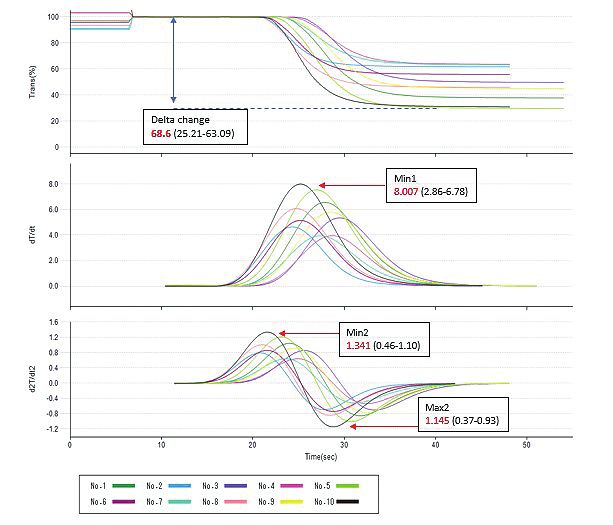
426                                         Post-COVID-19 ST-segment elevation MI—Shiun Woei Wong et al.

 Table 1. Characteristics and investigation of 3 cases of post-COVID-19 acute myocardial infarction

                                             Case 1                             Case 2                                 Case 3

 Age, years                                  38                                 50                                     36

 Sex                                         Male                               Male                                   Male

 Cardiovascular risk factors                 None                               None                                   None

 SARS-CoV-2 RT-PCR results and dates         Negative                           Positive                               Negative
                                             24 Jun, 27 Sep,                    4 Jun 2020                             6 Jun, 14 Jul, 30 Nov 2020
                                             28 Sep, 3 Oct 2020
                                                                                Negative
                                                                                29 Nov, 30 Nov 2020

 SARS-CoV-2 total antibody result and        Positive                           Positive                               Positive
 date                                        9 Jul 2020                         8 Jun 2020                             18 Jun 2020

 Arterial event                              27 Sep 2020                        29 Nov 2020                            30 Nov 2020

                                             VF arrest, myocardial              VF arrest, myocardial infarction,      Myocardial infarction, occlusion
                                             infarction, occlusion of LAD       occlusion of proximal LAD              of LAD

 No. of days from positive serology to       80                                 174                                    165
 thrombosis

 D-dimer, μg/mL                              >4                                 1.02                                   0.43
 (reference range
Post-COVID-19 ST-segment elevation MI—Shiun Woei Wong et al. 427
A B
C D
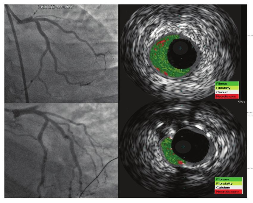
Fig. 1. (A) Coronary angiogram of Case 1 revealed acute occlusion of the ostial left anterior
descending artery. (B) Virtual histology showed focal, heavily fibrous plaque burden of 59%
with minimal necrotic core. (C) Coronary angiogram of Case 2 showed acute occlusion of the
left anterior descending artery. (D) Subsequent virtual histology showed predominantly fibrous
plaque burden of 31%.
positive lupus anticoagulant. A further workup did the ostial segment of LAD. Virtual histology (via
not reveal any evidence of hyperlipidaemia, diabetes IVUS) revealed a predominantly fibrous plaque
mellitus, thyroid disorder or metabolic syndrome. (31% burden) that was successfully treated through
The patient’s hospitalisation was complicated by thrombectomy, followed by stent implantation
lower limb compartment syndrome, which was treated (Figs. 1C and 1D). Given the thrombus burden, he was
with fasciotomy; acute kidney injury; and critical illness treated with glycoprotein IIb/IIIa inhibitor (eptifibatide)
neuromyopathy. Tracheostomy was performed in view infusion. TTE showed mild left ventricular systolic
of prolonged mechanical ventilation and myopathy. He dysfunction (left ventricular ejection fraction 45%)
is currently undergoing intensive rehabilitation in our with hypokinesia in the apex and anterior wall.
institution. The patient had a high fibrinogen level of 5.7g/L
Case 2. A 50-year-old man presented to the emergency (1.8–4.5g/L); elevated VWF level of 215% (56–160%);
department for worsening chest pain. Initial physical factor VIII level of 338%; and a raised D-dimer level
examination revealed his blood pressure as 116/93mmHg; of 1.02μg/mL (428 Post-COVID-19 ST-segment elevation MI—Shiun Woei Wong et al.
Fig. 2. Clot waveform analysis of Case 2. Patient’s tracing is in black; healthy controls (for reference) in multicolour. CWA showed markedly
elevated median Min1 of 8.007%/s, elevated median Min2 of 1.341 and high median delta change of 68.6%/s.
Min1: maximum velocity; Min2: maximum acceleration; Max2: maximum deceleration
Case 3. A 36-year-old man was admitted to our during the convalescent period.7,8 We described 3 cases
hospital with a persistent chest pain of 3 hours. On of COVID-19 recovered patients who presented with
admission, his blood pressure and heart rate were AMI. The remarkable characteristics of these cases
120/88mmHg and 88 beats per minute, respectively. included their relatively young age without preexisting
Oxygen saturation was 100% on room air with normal cardiovascular risk factors, preceding asymptomatic
lung auscultation. ECG showed hyperacute T waves SARS-CoV-2 infection, and the presence of a long
in the anterior leads and his RT-PCR results for latency period between initial positive SARS-CoV-2
COVID-19 was negative. He presented 165 days after serology and their AMI.
his positive SARS-CoV-2 antibody serology on 18 June Thrombosis has been classically associated with
2020. He demonstrated a raised factor VIII level of Virchow’s triad of blood stasis, endothelial activation,
162% and a VWF level of 92%. Coronary angiogram and hypercoagulable state. However, in the convalescent
revealed an acute mid-LAD occlusion, for which he phase after a COVID-19 infection, studies have
underwent angioplasty with successful implantation described a waning hypercoagulable state with possible
of a drug-eluting stent (Fig. 3). After percutaneous persistence of endothelial dysfunction in patients.
coronary intervention, the chest pain and ST-segment This has been well described in children during their
deviation resolved. The patient was discharged well on recovery from COVID-19.
day 6 of his hospitalisation.
Multisystem inflammatory syndrome in children
While it is known that approximately 30% of (MIS-C) is a newly defined post-viral myocarditis
myocardial infarctions are preceded by an upper and inflammatory vasculopathy of children following
respiratory infection, in particular influenza; less is COVID-19 infection. MIS-C is likely due to viral
known about the thrombotic sequelae in COVID-19 trophism of myocardial and endothelial cells by the
Ann Acad Med Singap Vol 50 No 5 May 2021 | annals.edu.sgPost-COVID-19 ST-segment elevation MI—Shiun Woei Wong et al. 429
Fig. 3. (A) Coronary angiogram of Case 3 showed an occlusion of mid left anterior descending artery. (B) Re-establishment
of blood flow after implantation of drug eluting stent.
coronavirus. The best evidence supporting MIS-C have “thrombin burst”; (2) peak of second derivative curve;
been demonstrated in paediatric cases presenting with (3) clot maximum deceleration; and (4) delta change
self-limited, chilblain-like acral purpuric lesion. 9 (decreased light transmission reflective of increased
The children remain otherwise asymptomatic, and clot thickness) as demonstrated by Fan et al. 14 In
interestingly, often test negative for SARS-CoV-2 in addition, autopsy series demonstrated the presence
nasopharyngeal samples. Some acute viral infections are of cardiac microemboli despite the absence of viral
associated with transiently elevated lupus anticoagulant, particles in the myocardium.15
but they can persist and lead to thromboembolic Our case series suggest that life-threatening
complications by various mechanisms, including the myocardial infarction can occur unexpectedly in
release of microparticles and exposure of prothrombotic otherwise healthy patients with asymptomatic
phospholipids. 10 Although the significance of these COVID-19 infection. Physicians should have a high
antibodies is not well established yet, COVID-19- index of suspicion in managing patients in the
induced lupus anticoagulant could favour the occurrence convalescent phase. Screening for and strict management
of thromboembolic events in children populations and of cardiovascular risk factors are of utmost importance
hence should be systematically tested for. post COVID-19. Further longitudinal studies in patients
Virtual histology via IVUS revealed heavily fibrous with “long COVID” should be performed to look
plaques in the coronary arteries of the cases described. for post-COVID-19 associated endotheliopathy and
This is unusual as fibrotic lesions are usually deprived thrombotic sequelae, where there may be a role for
of lipid and inflammatory cells, and hence less likely thromboprophylaxis in high-risk groups.
to rupture and generate thrombosis.11 A fibrotic plaque
consists mainly of fibrous tissue without a necrotic core
or calcium.12,13 This type of plaque is mostly indolent REFERENCES
and stable in comparison with thin cap fibroatheroma, 1. Gu SX, Tyagi T, Jain K, et al. Thrombocytopathy and endotheliopathy:
the main culprit in acute coronary syndrome. crucial contributors to COVID-19 thromboinflammation. Nat Rev
Interestingly, in the current cases described, delayed Cardiol 2021;18:194-209.
thrombotic arterial events occurred 80–174 days from 2. Tang N, Li D, Wang X, et al. Abnormal coagulation parameters are
the onset of positive SARS-CoV-2 serology. Laboratory associated with poor prognosis in patients with novel coronavirus
pneumonia. J Thromb Haemost 2020;18:844-7.
evaluation of the haemostatic profiles with raised
3. Lillicrap D. Disseminated intravascular coagulation in patients with
factor VIII, VWF and D-dimer supported an ongoing 2019-nCoV pneumonia. J Thromb Haemostat 2020;18:786-7.
vasculopathy. The CWA, a global haemostatic test that
4. Fan BE, Umapathi T, Chua K, et al. Delayed catastrophic thrombotic
was performed on case 2, demonstrated parameters of events in young and asymptomatic post COVID-19 patients.
hypercoagulability. There were increased (1) clot Min1 J Thromb Thrombolysis 2021;51:971-7.
Ann Acad Med Singap Vol 50 No 5 May 2021 | annals.edu.sg430 Post-COVID-19 ST-segment elevation MI—Shiun Woei Wong et al.
5. Chen JI, Yap JCH, Hsu LY, et al. COVID-19 and Singapore: From infection: a systematic review and meta-analysis. Lupus 2018;
early response to circuit breaker. Ann Acad Med Singap 2020; 27:572-83.
49:561-72. 11. Bharadwaj AS, Vengrenyuk Y, Yoshimura T, et al. Multimodality
6. Tan THY, Toh MPHS, Vasoo S, et al. Coronarvirus disease 2019 intravascular imaging to evaluate sex difference in plaque
(COVID-19): The Singapore experience. A review of the first eight morphology in stable CAD. JACC Cardiovasc Imaging 2016;9:400-7.
months. Ann Acad Med Singap 2020;49:764-78. 12. Honda S, Kataoka Y, Kanaya T, et al. Characterization of coronary
7. Spodick DH, Flessas AP, Johnson MM. Association of acute atherosclerosis by intravascular imaging modalities. Cardiovasc
respiratory symptoms with onset of acute myocardial infarction: Diagn Ther 2016;6:368-81.
prospective investigation of 150 consecutive patients and matched 13. Bentzon JF, Otsuka F, Virmani R, et al. Mechanisms of plaque
control patients. Am J Cardiol 1984;53:481-2. formation and rupture. Circ Res 2014;114:1852-66.
8. Bainton D, Jones GR, Hole D. Influenza and ischaemic heart disease: 14. Fan BE, Ng J, Seok SSW, et al. COVID-19 associated coagulopathy
a possible trigger for acute myocardial infarction? Int J Epidemiol in critically ill patients: A hypercoagulable state demonstrated by
1978;7:231-9. parameters of haemostasis and clot waveform analysis. J Thromb
9. Hernandez C, Bruckner AL. Focus on “COVID Toes”. JAMA Thrombolysis 2021;51:663-74.
Dermatol 2020;156:1003. 15. Guagliumi G, Sonzogni A, Pescetelli I, et al. Microthrombi and
10. Abdel-Wahab N, Talathi S, Lopez-Olivo MA, et al. Risk ST-segment elevation myocardial infarction in COVID-19.
of developing antiphospholipid antibodies following viral Circulation 2020;142:804-9.
Ann Acad Med Singap Vol 50 No 5 May 2021 | annals.edu.sgYou can also read