Corticospinal Tract (CST) reconstruction based on fiber orientation distributions (FODs) tractography
←
→
Page content transcription
If your browser does not render page correctly, please read the page content below
Corticospinal Tract (CST) reconstruction based on fiber orientation distributions
                                                                            (FODs) tractography

                                                                                                   Youshan Zhang
                                                                                    Computer Science and Engineering Department
                                                                                                  Lehigh University
                                                                                                   Bethlehem, PA
                                                                                                 yoz217@lehigh.edu
arXiv:1904.11136v1 [q-bio.NC] 23 Apr 2019

                                               Abstract—The Corticospinal Tract (CST) is a part of pyra-        About 75%-90% of the CST fibers descend to the lateral
                                            midal tract (PT) and it can innervate the voluntary movement        corticospinal tract by decussation of the pyramid, but a few
                                            of skeletal muscle through spinal interneurons (the 4th layer of    fibers form the anterior corticospinal tract by not crossing
                                            the Rexed gray board layers), and anterior horn motorneurons
                                            (which control trunk and proximal limb muscles). Spinal cord        the pyramid [9], [10]. The fibers, which first passed through
                                            injury (SCI) is a highly disabling disease often caused by          the medial posterior spinocerebellar tract then went into
                                            traffic accidents. The recovery of CST and the functional           the lumbosacral spinocerebellar tract, were not yet appeared
                                            reconstruction of spinal anterior horn motor neurons play an        [11], [12]. The main function of the CST is innervating
                                            essential role in the treatment of SCI. However, the localization   the voluntary movement of skeletal muscle through spinal
                                            and reconstruction of CST are still challenging issues, the
                                            accuracy of the geometric reconstruction can directly affect        interneurons (the 4th layer of the Rexed gray board layers),
                                            the results of the surgery. The main contribution of this paper     and anterior horn motorneurons (which control trunk and
                                            is the reconstruction of the CST based on the fiber orientation     proximal limb muscles), then the CST terminates in the spinal
                                            distributions (FODs) tractography. Differing from tensor-based      motor cells (which control the fine motor of small muscle
                                            tractography in which the primary direction is a determined         in extremities) [13]. Spinal cord injury (SCI) is a highly
                                            orientation, the direction of FODs tractography is determined
                                            by the probability. The spherical harmonics (SPHARM) can            disabling disease in clinical practice. With the popularization
                                            be used to approximate the efficiency of FODs tractography.         of modern transport vehicles and the acceleration of the
                                            We manually delineate the three ROIs (the posterior limb of         process of industrialization, the number of patients with SCI
                                            the internal capsule, the cerebral peduncle, and the anterior       is increasing day by day, many people were injured in traffic
                                            pontine area) by the ITK-SNAP software, and use the pipeline        accidents. If the CST is damaged, it can seriously affect the
                                            software to reconstruct both the left and right sides of the CST
                                            fibers. Our results demonstrate that FOD-based tractography         quality of life of patients and even lead to paralysis. Patients
                                            can show more and correct anatomical CST fiber bundles.             with rigid muscle, spasm, paralysis and other pathologies
                                                                                                                are usually considered to have CST injury [14]. When the
                                               Keywords-Corticospinal Tract reconstruction; fiber orienta-
                                            tion distributions; tractography;                                   CST is damaged in the SCI, the motor pathways are affected,
                                                                                                                and these surviving spinal cord nerve cells, which are below
                                                                                                                the injury surface, lose neural innervation of the brain, and
                                                                  I. I NTRODUCTION
                                                                                                                are unable to perform the random functional activity [15].
                                               The Corticospinal Tract (CST) is a part of pyramidal tract       In addition, the repair of the CST after the SCI is still a
                                            (PT), and many of its fibers originate from the primary motor       vital research topic in neuroscience, and the recovery of
                                            cortex (the precentral gyrus) then terminate in the spinal cord     limb motor function can be promoted by the CST repair or
                                            [1], [2]. The CST is thought to originate from the premotor         functional remodeling. Although many types of experimental
                                            area, the supplementary motor area, and the somatosensory           research have made great progress, there has been no report
                                            cortex, and then terminate at the thoracic levels (20%),            of successful clinical application [16]. Therefore, the recovery
                                            lumbosacral levels (25%) and cervical levels (55%) [3], [4].        of CST and the functional reconstruction of spinal anterior
                                            Evidence shows that the CST serves as the major downstream          horn motor neurons play an essential role in the treatment
                                            motor tract in the mammalian spinal cord, and the fibers            of SCI [17]. However, the localization and reconstruction
                                            control the voluntary movement of skeletal muscle by directly       of CST are still challenging issues, and the accuracy of the
                                            or indirectly innervating the neurons in the anterior horn [5].     geometric reconstruction can directly affect the results of
                                            However, the structural and functional properties of the CST        the surgery. Furthermore, there are few methods that can be
                                            are far more complex than researchers previously thought,           used to reconstruct the CST and the validation of clinical
                                            and we continue to have a new understanding about the CST           application is still uncertain [18].
                                            with the advancement of technology, thus the CST is still              Diffusion tensor imaging (DTI) is a non-invasive technique
                                            one of the hotspots in neuroscience research [6], [7], [8].         that can estimate the integrity of the white matter tracts
Figure 1: The four different images are used in the reconstruction of the CST fibers. (a): FOD images, which are the FODs
tractography files, (b): FA images which can be used to draw the ROIs using ITK-SNAP, (c): The CortexLabel images that
contain the labels of all the cortex; the labels of the left and right precentral gyrus are 49 and 50. (d): The mask of the
tractography image. In each image, the left is the coronal slice, the middle is the sagittal slice and the right is the axial slice
(A: anterior, I: inferior, R: right, L: left).

Figure 2: The seed region and three ROIs in the reconstruction of the CST fibers. (a): the coronal slice of the seed region
precentral gyrus, a1 is the left precentral gyrus and a2 is the right precentral gyrus. From (b) to (d) are the axial slice the
three ROIs (the posterior limb of the internal capsule, the cerebral peduncle, and the anterior pontine area). These three ROIs
are manually delineated by the ITK-SNAP. 1-6 are six labels, 1, 3 and 5 are the left ROIs, 2, 4, and 6 are the right ROIs (A:
anterior, P: posterior, R: right, L: left).

by using the diffusion of water molecules [19], [20], [21].        tractography is fiber orientation distribution (the direction
Diffusion tensor tractography (DTT), a three-dimensional           is determined by the probability), the spherical harmonics
visualized version of DTI, has been widely used to reconstruct     (SPHARM) can be used to approximate the efficiency of FOD
the CST [8], [22], [23]. However, there are several challenges     [24]. In this paper, we manually delineate the ROIs by the
in these papers. Firstly, the seed regions or the regions of       ITK-SNAP software[27], and use the Lab of NeuroImaging
interest (ROIs) are different in these papers, the models of       (LONI) pipeline software[28] to reconstruct both the left and
these papers did not cover all the anatomic areas of the CST,      right sides of the CST fibers. Our results demonstrate that
and the results did not reflect the complexity of the CST          FOD-based tractography can show correct anatomical CST
fibers. Secondly, the validation of these models in the clinical   fiber bundles.
application still not fully clarified.
                                                                                            II. M ETHOD
   The main contribution of this paper is the reconstruction of
the CST based on the fiber orientation distributions (FODs)        A. Data
tractography [24], [25]. The major difference between the             In this paper, the data are from Human Connectome Project
FODs tractography and the tensor-based tractography is the         (HCP) [29], which can be found on LONI IDA (https://ida.
definition of the tracts direction. Tensor-based tractography      loni.usc.edu/services/NewUser.jsp) or ConnectomeDB (https:
relies on the three non-negative eigenvalues and eigenvectors,     //db.humanconnectome.org/app/template/Login.vm) . Figure 1
the main direction is a determined orientation [26]. The FOD       shows the four images ( fiber orientation distributions (FOD),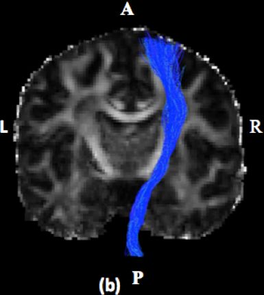
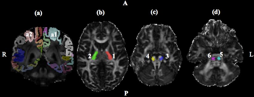
Figure 3: The workflow of CST reconstruction method from Pipeline software, which is used to generate the reconstruction
CST fibers based on the FODs tractography. The input files are the gray circle buttons (Mask, FOD, Num track, Radius,
CortexLabel, and the CST ROIs), the output files are the gray triangular button, which is the fibers of the CST. The middle
FSL Maths modules can add more images in one module, the FSLChangeFiletype module can change the file type of the
image, and the streamtrack module can compute the fibers of the CST based on the FODs.

fractional anisotropy(FA), CortexLabel and Mask), which are       we exclude this area in our study. In this paper, the seed
used to reconstruct the CST fibers.                               region is the precentral gyrus, the ROIs are the PLIC, CP
                                                                  and the aiP. Figure 2 shows the seed region and ROIs in the
B. ROIs                                                           CST reconstruction.
   The CST primarily originates from the motor cortex (the
precentral gyrus), then descends to the forebrain, pons,          C. Workflow of the Pipeline
medulla, and pyramidal decussation and terminates at thoracic        We use Pipeline software [28] to reconstruct the CST fibers
levels, lumbosacral levels, and cervical levels. Therefore, the   after we choose the seed region and the ROIs. Figure 3 shows
ROIs should be in these different areas, but the proper ROIs      the reverent workflow of the Pipeline when we compute the
are still hard to decide. Many papers used the precentral         fibers of the CST.
gyrus as the starting point since precentral gyrus is the major
area controls the movement of the arm, leg and the trunk          D. Parameters
[30], [8], [31]. However, the organization of the fibers can         The input files Mask, FOD, and CortexLabel are the three
be distorted in patients with brain tumors [8], so more ROIs      images which are mentioned in section A (data part), the
along the CST should be included. Another two ROIs (the           CST ROIs are the images which are generated by manual
posterior limb of the internal capsule (PLIC) and anterior        delineation by the ITK-SNAP. The number of the tracks
pontine area (aiP)) were used in Carolin et al., 2015. The        set as 1000, the radius is the 1.5, the cortex label of left
internal capsule lies in the forebrain, the pons is a part of     precentral gyrus is 49 and the right precentral gyrus is 50.
the brainstem, both of these two regions are involved in          Besides, the left labels of the three ROIs are 1, 3 and 5, and
the movement pathways, they can affect the movement of            the right labels are the 2, 4, and 6. The left and the right
the leg, trunk, and arm. Therefore, the PLIC and aiP are          CST fibers are computed separately, the cortex label and the
two essential ROIs. Several papers showed that the cerebral       ROIs labels should be consistent with the above parameters.
peduncle (CP) of the midbrain is another important ROI of
the CST reconstruction, since it also involved in innervating     E. Outlier tracts remove
the movement pathways [30], [32]. Although Venkateswaran            The result of the workflow is a tck file, we convert it into
and Erik, 2017 used centrum semiovale at top of lateral           the trk file by Matlab since the BrainSuite software [33]
ventricle (CSoLV) as additional ROIs[6], the CSoLV is not         cannot recognize the tck files. In addition, some fibers are
significant comparing with the other three ROIs, therefore,       not normal and may be too long and project to other areas.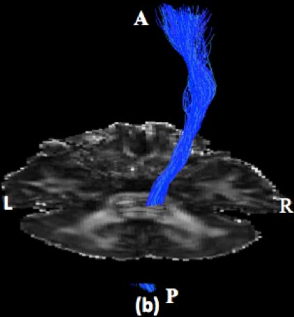
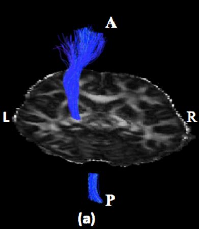
Figure 4: The coronal slice of the reconstructed CST fibers. It overlays the fibers with the cortex label image to show the
projection of the fiber tracts into the precentral gyrus. (a): the left CST fibers, (b): the right CST fibers, (c): both the left and
right CST fibers (A: anterior, P: Posterior, R: right, L: left).
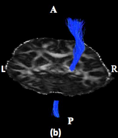
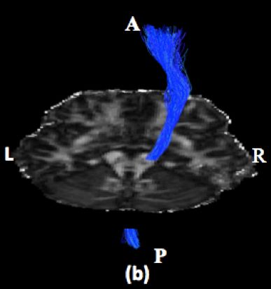
Figure 5: The coronal slice of the reconstructed CST fibers. It Figure 6: The axial slice of the reconstructed CST fibers. It
overlays the fibers with the FA image to show the projection overlays the fiber bundle with the FA image that shows the
of the fiber tracts into the precentral gyrus. (a): the left CST internal capsule and the thalamus. (a): the left CST fibers,
fibers, (b): the right CST fibers (A: anterior, P: Posterior, R: (b): the right CST fibers (A: anterior, P: Posterior, R: right,
right, L: left). L: left).
Firstly, we filter the outlier tracts, and then some obvious prove that the FOD-based tractography can provide a smooth,
abnormal tracts are removed using the BrainSuite 16 a1 track clear and reliable reconstruction of the CST fibers.
filtering options. IV. D ISCUSSION
III. R ESULTS Comparing our results with [34], [35], our reconstruction
results show more fibers, which shows the high accuracy of
The reconstructed CST fibers are visualized by the our methods. One limitation of the paper is that we only
BrainSuite 16 a1, to show the anatomical validity of the choose three ROIs (the posterior limb of the internal capsule,
reconstruction results, we overlay the CST fiber bundles the cerebral peduncle, and the anterior pontine area); there
with the FA image. Fig. 4 and 5 show the projection to the are more regions which can be explored, such as top of
precentral gyrus by overlaying the fiber tracts with the cortex the lateral ventricle (CSoLV) though it might not be as
label and FA images. Fig. 6 shows the results of the CST important as above three ROIs, it still can be an interesting
fibers through the posterior of the internal capsule and the direction. Our study also had a limited sample size. However,
thalamus. Fig. 7 shows the results of the cerebral peduncle Our tractography findings suggest the FODs reconstruction
by overlaying the fiber bundles with the FA image. And the method can accurately reconstruct the CST fibers; thus, the
Fig. 8 overlays the fiber bundle with the FA image to the power of our study is sufficient. For future work, we should
anterior pontine area. These results demonstrate that the fiber explore our reconstruction of the CST in recovery from the
bundles are anatomically correct. Furthermore, these results SCI injury.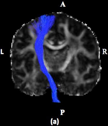
differences for motor specialization? Muscle & Nerve: Official
                                                                             Journal of the American Association of Electrodiagnostic
                                                                             Medicine, 32(3):261–279, 2005.

                                                                         [2] Sung Ho Jang. The corticospinal tract from the viewpoint
                                                                             of brain rehabilitation. Journal of rehabilitation medicine,
                                                                             46(3):193–199, 2014.

                                                                         [3] Roger N Lemon. Descending pathways in motor control. Annu.
                                                                             Rev. Neurosci., 31:195–218, 2008.

Figure 7: The axial slice of the reconstructed CST fibers. It            [4] Joseph C Masdeu, José Biller, et al. Localization in clinical
overlays the fibers with the FA image to show the middle                     neurology. Lippincott Williams & Wilkins, 2011.
brain (mainly the cerebral peduncle). (a): the left CST fibers,          [5] Patrick Freund. Anti-Nogo-A antibody treatment enhances
(b): the right CST fibers (A: anterior, P: Posterior, R: right,              functional recovery and sprouting of the corticospinal tract
L: left).                                                                    after spinal cord injury in adult primates. PhD thesis,
                                                                             Université de Fribourg, 2008.

                                                                         [6] Erin D Bigler. Incorporating neuroimaging into cognitive as-
                                                                             sessment. The Role of Technology in Clinical Neuropsychology,
                                                                             page 377, 2017.

                                                                         [7] Aleksandr Aleksandrovich Potapov, Aleksandr Nikolae-
                                                                             vich Konovalov, Valerii Nikolaevich Kornienko, Alek-
                                                                             sandr Dmitrievich Kravchuk, Leonid Boleslavovich Likhter-
                                                                             man, Evgeniya Vladimirovna Aleksandrova, Anton Grigore-
                                                                             vich Gavrilov, Sergei Alekseevich Goryainov, Gleb Valerevich
                                                                             Danilov, et al. Current technologies and basic research in
                                                                             neurosurgery. Herald of the Russian Academy of Sciences,
Figure 8: The axial slice of the reconstructed CST fibers. It                85(2):112–121, 2015.
overlays the fibers with the FA image to show the anterior
pontine area. (a): the left CST fibers, (b): the right CST fibers        [8] Carolin Weiss, Irada Tursunova, Volker Neuschmelting,
                                                                             Hannah Lockau, Charlotte Nettekoven, Ana-Maria Oros-
(A: anterior, P: Posterior, R: right, L: left).                              Peusquens, Gabriele Stoffels, Anne K Rehme, Andrea Maria
                                                                             Faymonville, N Jon Shah, et al. Improved ntms-and dti-derived
                                                                             cst tractography through anatomical roi seeding on anterior
                        V. C ONCLUSION                                       pontine level compared to internal capsule. NeuroImage:
                                                                             Clinical, 7:424–437, 2015.
   In this paper, we reconstruct the Corticospinal Tract using
fiber orientation distributions tractography. Comparing with             [9] Usha Kant Misra, Jayantee Kalita, Ruchi Srivastava, Pradeep P
determined orientation tensor-based tractography, the main                   Nair, Manoj Kumar Mishra, and Anirban Basu. A study
direction of FODs is based on probabilistic direction. We                    of cytokines in tuberculous meningitis: clinical and mri
                                                                             correlation. Neuroscience letters, 483(1):6–10, 2010.
manually delineate the three ROIs (the posterior limb of
the internal capsule, the cerebral peduncle, and the anterior           [10] Adam Fisch. Neuroanatomy: Draw it to Know it. OUP USA,
pontine area) by the ITK-SNAP software and use the pipeline                  2012.
software reconstructs both the left and right sides of the CST
fibers. Our results demonstrate that FOD-based tractography             [11] Karl-Erik Andersson and Gorm Wagner. Physiology of penile
                                                                             erection. Physiological reviews, 75(1):191–236, 1995.
can show correct anatomical CST fiber bundles. The results
of our study show that the accurate reconstruction of CST               [12] Craig Watson. Basic Human Neuroanatomy: A Clinically
can be applied in improving both diagnostics and treatment                   Oriented Atlas. Lulu. com, 2012.
of Spinal cord injury in the future.
                                                                        [13] Anders Holtz and Richard Levi. Spinal cord injury. Oxford
                      ACKNOWLEDGMENT                                         university press, 2010.

   We want to thank professor Yonggang Shi for guiding us               [14] KENNETH H ABBOTT. Abdominal rigidity: A symptom
to do research on this topic and give us valuable suggestions                of concussion of the spinal cord. Archives of Neurology &
in explaining the topic more professionally.                                 Psychiatry, 57(2):220–227, 1947.

                          R EFERENCES                                   [15] Irin C Maier and Martin E Schwab. Sprouting, regeneration
                                                                             and circuit formation in the injured spinal cord: factors and
 [1] Roger N Lemon and James Griffiths. Comparing the function               activity. Philosophical Transactions of the Royal Society of
     of the corticospinal system in different species: organizational        London B: Biological Sciences, 361(1473):1611–1634, 2006.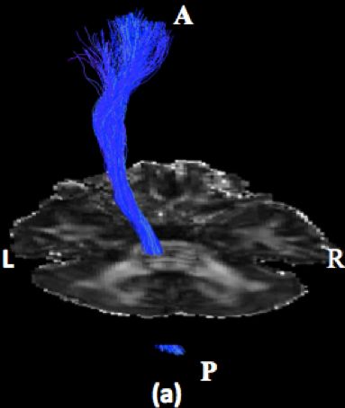
[16] James W Rowland, Gregory WJ Hawryluk, Brian Kwon, and                 [28] Ivo Dinov, Kamen Lozev, Petros Petrosyan, Zhizhong Liu, Paul
     Michael G Fehlings. Current status of acute spinal cord                    Eggert, Jonathan Pierce, Alen Zamanyan, Shruthi Chakrapani,
     injury pathophysiology and emerging therapies: promise on                  John Van Horn, D Stott Parker, et al. Neuroimaging study
     the horizon. Neurosurgical focus, 25(5):E2, 2008.                          designs, computational analyses and data provenance using
                                                                                the loni pipeline. PloS one, 5(9):e13070, 2010.
[17] Chihiro Tohda and Tomoharu Kuboyama. Current and future
     therapeutic strategies for functional repair of spinal cord injury.   [29] Qiuyun Fan, Thomas Witzel, Aapo Nummenmaa, Koene RA
     Pharmacology & therapeutics, 132(1):57–71, 2011.                           Van Dijk, John D Van Horn, Michelle K Drews, Leah H
                                                                                Somerville, Margaret A Sheridan, Rosario M Santillana, Jenna
[18] Arish A Qazi, Alireza Radmanesh, Lauren O’donnell, Gordon                  Snyder, et al. Mgh–usc human connectome project datasets
     Kindlmann, Sharon Peled, Stephen Whalen, Carl-Fredrik                      with ultra-high b-value diffusion mri. Neuroimage, 124:1108–
     Westin, and Alexandra J Golby. Resolving crossings in                      1114, 2016.
     the corticospinal tract by two-tensor streamline tractography:
     method and clinical assessment using fmri. Neuroimage,                [30] Ryan P Cabeen and David H Laidlaw. Corticospinal tract
     47:T98–T106, 2009.                                                         reconstruction with deterministic multi-fiber tractography and
                                                                                model-based processing.
[19] Susumu Mori, Barbara J Crain, Vadappuram P Chacko, and
     Peter CM Van Zijl. Three-dimensional tracking of axonal               [31] Sang Seok Yeo, Sung Ho Jang, and Su Min Son. The different
     projections in the brain by magnetic resonance imaging. Annals             maturation of the corticospinal tract and corticoreticular path-
     of Neurology: Official Journal of the American Neurological                way in normal brain development: diffusion tensor imaging
     Association and the Child Neurology Society, 45(2):265–269,                study. Frontiers in human neuroscience, 8:573, 2014.
     1999.
                                                                           [32] Judith D Schaechter, Zachary P Fricker, Katherine L Perdue,
[20] Denis Le Bihan, Jean-François Mangin, Cyril Poupon, Chris A               Karl G Helmer, Mark G Vangel, Douglas N Greve, and
     Clark, Sabina Pappata, Nicolas Molko, and Hughes Chabriat.                 Nikos Makris. Microstructural status of ipsilesional and
     Diffusion tensor imaging: concepts and applications. Journal               contralesional corticospinal tract correlates with motor skill in
     of Magnetic Resonance Imaging: An Official Journal of the                  chronic stroke patients. Human brain mapping, 30(11):3461–
     International Society for Magnetic Resonance in Medicine,                  3474, 2009.
     13(4):534–546, 2001.
                                                                           [33] David W Shattuck and Richard M Leahy. Brainsuite: an
[21] Damiano Giuseppe Barone, Theresa A Lawrie, and Michael G                   automated cortical surface identification tool. Medical image
     Hart. Image guided surgery for the resection of brain tumours.             analysis, 6(2):129–142, 2002.
     The Cochrane database of systematic reviews, (1):CD009685–
     CD009685, 2014.                                                       [34] Gert Kristo, Alexander Leemans, Beatrice de Gelder, Mathijs
                                                                                Raemaekers, Geert-Jan Rutten, and Nick Ramsey. Reliability
[22] Lidia M Nagae-Poetscher, Hangyi Jiang, Setsu Wakana, Xavier                of the corticospinal tract and arcuate fasciculus reconstructed
     Golay, Peter CM Van Zijl, and Susumu Mori. High-resolution                 with dti-based tractography: implications for clinical practice.
     diffusion tensor imaging of the brain stem at 3 t. American                European radiology, 23(1):28–36, 2013.
     Journal of Neuroradiology, 25(8):1325–1330, 2004.
                                                                           [35] Jitka Hüttlova, Zora Kikinis, Milos Kerkovsky, Sylvain Bouix,
[23] Kyung Hee Do, Sang Seok Yeo, Jun Lee, and Sung Ho                          Mai-Anh Vu, Nikos Makris, Martha Shenton, and Tomas
     Jang. Injury of the corticoreticular pathway in patients with              Kasparek. Abnormalities in myelination of the superior
     proximal weakness following cerebral infarct: diffusion tensor             cerebellar peduncle in patients with schizophrenia and deficits
     tractography study. Neuroscience letters, 546:21–25, 2013.                 in movement sequencing. The Cerebellum, 13(4):415–424,
                                                                                2014.
[24] Alexandra Kammen, Meng Law, Bosco S Tjan, Arthur W Toga,
     and Yonggang Shi. Automated retinofugal visual pathway
     reconstruction with multi-shell hardi and fod-based analysis.
     Neuroimage, 125:767–779, 2016.

[25] J Donald Tournier, Fernando Calamante, and Alan Connelly.
     Improved probabilistic streamlines tractography by 2nd order
     integration over fibre orientation distributions. In Proceedings
     of the international society for magnetic resonance in medicine,
     volume 18, page 1670, 2010.

[26] Jacques-Donald Tournier, Susumu Mori, and Alexander Lee-
     mans. Diffusion tensor imaging and beyond. Magnetic
     resonance in medicine, 65(6):1532–1556, 2011.

[27] Paul A Yushkevich, Yang Gao, and Guido Gerig. Itk-snap:
     An interactive tool for semi-automatic segmentation of multi-
     modality biomedical images. In Engineering in Medicine and
     Biology Society (EMBC), 2016 IEEE 38th Annual International
     Conference of the, pages 3342–3345. IEEE, 2016.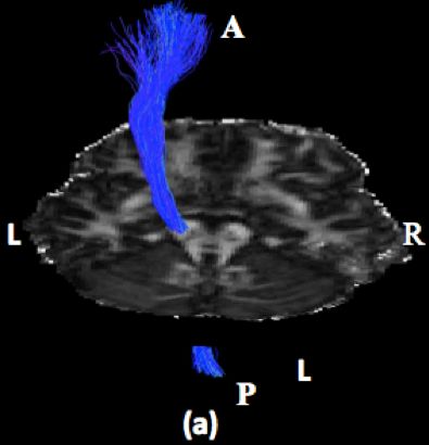
You can also read