Coxiella burnetii in Dromedary Camels (Camelus dromedarius): A Possible Threat for Humans and Livestock in North Africa and the Near and Middle ...
←
→
Page content transcription
If your browser does not render page correctly, please read the page content below
REVIEW
                                                                                                                                          published: 05 November 2020
                                                                                                                                        doi: 10.3389/fvets.2020.558481

                                             Coxiella burnetii in Dromedary
                                             Camels (Camelus dromedarius): A
                                             Possible Threat for Humans and
                                             Livestock in North Africa and the
                                             Near and Middle East?
                                             Christian A. Devaux 1,2*, Ikram Omar Osman 1,3 , Matthieu Million 1 and Didier Raoult 1
                                             1
                                              Aix-Marseille Univ, IRD, APHM, MEPHI, IHU-Méditerranée Infection, Marseille, France, 2 CNRS, Marseille, France, 3 Faculty
                                             of Sciences Ben-Ben-M’Sik, University Hassan II, Casablanca, Morocco

                                             The “One Health” concept recognizes that human health is connected to animal health
                                             and to the ecosystems. Coxiella burnetii–induced human Q fever is one of the most
                                             widespread neglected zoonosis. The main animal reservoirs responsible for C. burnetii
                                             transmission to humans are domesticated ruminants, primarily goats, sheep, and cattle.
                          Edited by:
                       Julio Alvarez,        Although studies are still too sparse to draw definitive conclusions, the most recent C.
  VISAVET Health Surveillance Centre         burnetii serosurvey studies conducted in herds and farms in Africa, North Africa, Arabian
                      (UCM), Spain
                                             Peninsula, and Asia highlighted that seroprevalence was strikingly higher in dromedary
                        Reviewed by:
                     Wendy Beauvais,         camels (Camelus dromedarius) than in other ruminants. The C. burnetii seroprevalence
      Cornell University, United States      in camel herds can reach more than 60% in Egypt, Saudi Arabia, and Sudan, and 70 to
                           Ana Afonso,
                                             80% in Algeria and Chad, respectively. The highest seroprevalence was in female camels
       University of São Paulo, Brazil
                                             with a previous history of abortion. Moreover, C. burnetii infection was reported in ticks
                  *Correspondence:
                   Christian A. Devaux       of the Hyalomma dromedarii and Hyalomma impeltatum species collected on camels.
                    christian.devaux@        Even if dromedary camels represent 5
                a section of the journal     million tons/year of both raw and pasteurized milk according to the Food and Agriculture
        Frontiers in Veterinary Science
                                             Organization) sustained by a rapid increase of population (growth rate: 2.26–3.76 per
            Received: 16 July 2020
                                             year in North Africa), dromedary camel breeding tends to increase from the Maghreb to
      Accepted: 28 September 2020
      Published: 05 November 2020            the Arabic countries. Because of possible long-term persistence of C. burnetii in camel
                              Citation:      hump adipocytes, this pathogen could represent a threat for herds and breeding farms
Devaux CA, Osman IO, Million M and           and ultimately for public health. Because this review highlights a hyperendemia of C.
   Raoult D (2020) Coxiella burnetii in
        Dromedary Camels (Camelus
                                             burnetii in dromedary camels, a proper screening of herds and breeding farms for C.
  dromedarius): A Possible Threat for        burnetii is urgently needed in countries where camel breeding is on the rise. Moreover,
Humans and Livestock in North Africa
                                             the risk of C. burnetii transmission from camel to human should be further evaluated.
      and the Near and Middle East?
            Front. Vet. Sci. 7:558481.       Keywords: Coxiella burnetii, dromedary camel (Camelus dromedarius), zoonoses awareness, epidemiology,
     doi: 10.3389/fvets.2020.558481          human—animal coexistence

Frontiers in Veterinary Science | www.frontiersin.org                               1                                        November 2020 | Volume 7 | Article 558481
Devaux et al. Coxiella burnetii-Infected Camels
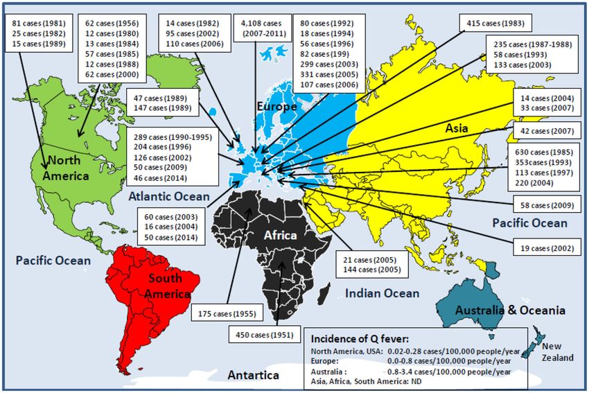
INTRODUCTION HUMAN Q FEVER IS FOUND ON SIX OF
SEVEN CONTINENTS: AN
Q fever is a neglected zoonotic disease caused by bacteria (1,
2). It is generally admitted that clones of Coxiella burnetii, EPIDEMIOLOGICAL OVERVIEW
the etiologic agent of Q fever, circulate in wildlife and infects As far back as 1950, the third World Health Assembly was
domestic ruminants. Very few bacteria are required to initiate aware of the potential danger of Q fever to public health and
the infection process (3). Usually, humans become infected passed a resolution calling for study of the C. burnetii prevalence
through the aerosol route during contact with C. burnetii– worldwide. Since then, numerous epidemiological studies have
positive domestic animals or their products (2, 4). Infection of shown that human Q fever is found almost everywhere on the
humans concerns first the farmers and other professionals that planet, with only exceptions of the Antarctica continent and
have contacts with animals (e.g., veterinarians), but epidemics New Zealand. Although seroprevalence data are available for
have been reported in other social groups. C. burnetii is a most countries, it can be considered that the true incidence of
strict intracellular Gram-negative bacterium entering different Q fever in humans is largely underestimated because (i) Q fever
cell types with progressive variation in the structure of its is a neglected infectious disease; (ii) there is a predominance of
lipopolysaccharide (LPS): a smooth LPS for the virulent phase asymptomatic forms; (iii) Q fever is rarely a notifiable disease,
I and a rough LPS for the less virulent phase II (5–7). and there is a lack of mandatory reporting (e.g., Q fever become
For symptomatic cases, human Q fever usually occurs 2 to a notifiable illness in 1977 and 1978 in Australia and the
6 weeks after bacterial exposure (8, 9). The symptomatic Netherlands, respectively, and a reportable disease in 1999 in the
primo-infection (10–60% of cases), called acute Q fever, is United States and Japan); and (iv) several local reports written in
characterized by high fever, headache, myalgia, pneumonia, and languages other than English remain ignored (Figure 1).
hepatitis (2, 10). It usually resolves spontaneously in a few On the North American continent, between 1946 and 1977,
weeks. When Q fever is suspected, confirmation is provided by a total of 1,169 human Q fever cases were reported in the
serological diagnosis based on anti–C. burnetii immunoglobulin United States (22–25). Then, from 1978 to 2016, about 200
(Ig) detection. An IgG anti–phase II Ig titer above 1:200 and cases of human Q fever were reported annually, a mean of
an IgM titer above 1:50 are considered significant for the about 0.25 cases/100,000 inhabitants/year (cases/100 kI/y), with a
diagnosis of acute Q fever. Sometimes the symptoms do not seropositivity of 3.1% in adult populations rising 22% among the
resolve (about 5% of cases) and settle in a persistent way veterinarians (26–29). In Canada, C. burnetii in humans was first
mainly in the heart valve and vascular wall but also lymph reported in 1952 (30, 31). In 1956, an outbreak with 62 human
node, and bone (11). Other disorders can be associated with cases was reported in people working in a slaughterhouse, then
persistent infections, including lung diseases, hepatitis, and B-cell individual cases in 1960 and 1966, followed by several outbreaks
lymphoma (12, 13). between 1975 and 1989 (32–34). In Central America, Q fever
Regarding domestic ruminants, C. burnetii is responsible for cases were reported in El Salvador and Mexico (35, 36). In the
epizooties with increased morbidity and mortality in livestock. South American continent, during the 2013–2014 outbreak of
It has long been considered that sheep, goats, and cattle were dengue in Brazil, C. burnetii was identified in 3.3% of patients
the main domestic source of C. burnetii worldwide among (37). Q fever cases were also reported in most South American
ecosystems in which C. burnetii clones circulate. Although C. countries (38–41). A very high incidence was also observed in the
burnetii has been classified as a notifiable animal disease by French Guiana in 2005 (150 cases/100 kI/y) (42).
the World Organization for Animal Health (14), notifications On the European continent, more than 1,000 human Q
concern only a subgroup of domestic animal species and ignore fever cases were reported among soldiers in the Balkans in the
the bacteria dynamics in different ecosystems. Among ruminant early 1940s. Large-scale outbreaks were documented over recent
species, camels are present in the countries of the Southern decades, and serosurveys suggest a seroprevalence between 2
coast of Mediterranean basin but absent from countries of the and 14% of the population (18, 43). The disease is endemic
Northern coast. The fact that some Southern countries practice in Germany with 27 to 100 Q fever annual cases (incidence is
intensive camel breeding, that a high percentage of these animals 0.08–0.14 cases/100 kI/y), and 40 Q fever outbreaks documented
are carriers of anti–C. burnetii Ig, that C. burnetii was found (44, 45). In the United Kingdom, from 1975 to 1996, between
in camels raw milk, and that camel ticks carries the bacteria 67 and 169 Q fever annual cases were reported (incidence of
must make us question our global perception of the mode 0.15–0.35 cases/100 kI/y), including eight outbreaks (46–49). In
of C. burnetii transmission in the Southern coast countries 1983, a large outbreak of 415 human Q fever cases was reported
of Mediterranean basin. This review compiles data from the in Switzerland (50). Until 2007, in the Netherlands, 5 to 16 Q
literature regarding the countries around the Mediterranean fever cases were reported annually (51, 52). In 2007–2010, a large
basin and the Arabic peninsula where camel breeding is practiced human outbreak with an estimated 44,000 people infected in 3
and highlights that the potential role of camels as a bacterial years was reported, among which were 4,108 cases of Q fever (53–
reservoir in the transmission of C. burnetii to humans should 56). In Portugal, the average frequency of Q fever is 0.1 case/100
be considered. kI/y, yet it is likely underestimated (57, 58). In the Spanish
Frontiers in Veterinary Science | www.frontiersin.org 2 November 2020 | Volume 7 | Article 558481
Devaux et al.                                                                                                                              Coxiella burnetii-Infected Camels

 FIGURE 1 | Schematic representation of human Q fever epidemiology around the world. With the exception of Antarctica and New Zealand, Q fever is a global
 zoonosis present in North America, South America, Europe, Asia, Africa, and Australia/Oceania. The clinical manifestation of Q fever in human is usually an
 undifferentiated febrile illness. Q fever was described for the first time in humans in 1937 by Burnet, who investigated several cases of Australian abattoir workers
 suffering from undifferentiated febrile illness (15, 16). During the Second World War (1941–1944), the Q fever disease was reported among German soldiers stationed
 in the Balkans, Southern Italy, Corsica, in English and American allied troops in Central Italy, and soldiers in Crimea and Ukraine. That is why the disease has had many
 synonyms: Olympus fever, Crimean fever, flu Balkan flu, Cretan pneumonia, Euboea fever, fever of the 7 days, or Derrick and Burnet’s disease (17). The causative
 agent of the disease first identified by Cox in the United States, and formerly named Rickettsia diasporica, was definitively renamed Coxiella burnetii (18–20). The figure
 illustrates the history of the major human epidemics of Q fever (outbreaks >10 linked cases) from 1950 (when the Third World Health Assembly passed a resolution
 calling for study of the prevalence of Q fever throughout the world) to the present day. Although C. burnetii infection has been classified as a notifiable animal disease
 by the World Organization for Animal Health, OIE (14), the lack of mandatory reporting of human Q fever cases in most countries, the predominance of asymptomatic
 forms, the clinical polymorphism, and the difficulty of diagnosis are likely to lead to a significant underestimation of the true incidence of the disease in humans. In
 Europe where the ECDC carries out a regular epidemiological surveillance, only 1,023 of 4,245 Q fever cases confirmed during the 2013–2017 period were reported
 by the European countries (21). It is impossible to evaluate the number of cases for the Asian and African continents. ND, not determined.

Canary Islands, a seroprevalence of infection by C. burnetii in                             On the Asian continent, 1% of patients hospitalized for
humans of 36% was reported during an outbreak of Q fever                                 infectious endocarditis and 14.6% of patients hospitalized for
(59). In France, the seroprevalence for anti–C. burnetii Ig was                          acute febrile illness/pneumonitis in India were infected by C.
estimated 5/100 kI/y (60). In Bulgaria, from 1949 to 1993, more                          burnetii (69, 70). In Iran, 4.2% of patients with febrile illness
than 20 Q fever outbreaks occurred with three major outbreaks                            and 18.1% of butchers and slaughterhouse workers carried anti–
between 1982 and 1985, and next in 1993 and 1997 (61–64). In                             C. burnetii Ig (71, 72). In China, Q fever was initially reported
the late 2010s, 139 Q fever cases were reported (incidence of                            in 1950 in a patient with pneumonia, and then in the 1960s,
0.27 cases/100 kI/y) (65). In Slovakia, a seroprevalence of 3%                           five outbreaks of Q fever occurred in abattoir workers, stockyard
was estimated for the period before 1993 (63). According to                              men, and troops (73, 74). Between 1989 and 2013, human Q
OIE, between 1996 and 2001, eight Q fever cases were reported                            fever cases were reported in people from 15 provinces in China
in Hungary, 26 cases in Ukraine, and 138 cases in Yugoslavia.                            and 4% of patients with infectious endocarditis suffered from Q
In Russia, an outbreak in Leningrad affected 48 people in 1957                           fever (75, 76). In Japan, serosurveys indicated the presence of
and between 1957 and 1995 up to 11,058 Q fever cases were                                anti–C. burnetii Ig in 16.5% of human serum samples collected
reported (66–68).                                                                        between 1978 and 1991 (77, 78). Since 1999, 7 to 46 Q fever

Frontiers in Veterinary Science | www.frontiersin.org                                3                                          November 2020 | Volume 7 | Article 558481
Devaux et al. Coxiella burnetii-Infected Camels
cases were reported annually (79–82). In the Arabian Peninsula, in Paris, in the region of Lyon and Northwestern (122). An
the presence of C. burnetii in humans was reported in 1968, and intrafamily Q fever outbreak was induced by infected pigeons
a recent serological analysis detected C. burnetii Ig in 35.2% of (18). The seroprevalence in humans can go up to 30% in the
patients with pyrexia of undetermined cause (83–85). In Qatar, Q Alps rural populations (123). In the South of France, 5 to 8% of
fever data are rare, yet a seroprevalence of 2.1% was found in US cases of endocarditis are due to C. burnetii, and a retrospective
soldiers deployed in this country (86). analysis performed on 22,496 sera showed a seroprevalence of
On the Australian/Oceanian continent, since the first 7.8% (1,754/22,496) with 323 acute Q fever (124, 125). Between
description of Q fever in 1937, the disease has continued to 1990 and 1996, three outbreaks (including 289 Q fever cases in
be endemic in Australia (87). Between 1977 and 1994, 202 to Martigues and 204 cases in Briançon) were linked to meet with
860 cases were reported annually (incidence 3.11–4.99 cases/100 infected sheep or goat, animal carcasses, and/or consumption
kI/y), despite a vaccine is recommended to farmers since 1989 of unpasteurized milk (126, 127). In 2002, an outbreak of 126
(88–91). New Zealand is considered free from Q fever. human Q fever cases possibly contaminated by ovine livestock
On the African continent, outbreaks of Q fever were reported occurred in Chamonix (128). In 2009, an outbreak of 50 human
in the early 1950s, but the disease remained neglected and cases of Q fever was reported in Cholet (129), and in 2014,
underestimated (92–95). In Rwanda, an outbreak with 450 Q an outbreak of 46 cases of Q fever occurred after people had
fever cases and 40 deaths linked to C. burnetii was reported (96). visited a sheep farm (130). In Italy, Q fever emerged in the late
In Western Africa, seroprevalence in human was found to be 1940s with epidemic outbreaks, and then it became endemic with
5% in rural Western Ivory Coast, 8% among nomads in rural sporadic occurrence (131). However, an epidemic outbreak was
Northern Burkina Faso, and 6–9% of patients hospitalized for reported in 1996 in the Vicenza region with 58 human cases
pneumonia in Cameroun (97–101). C. burnetii was incriminated after contact with infected sheep (132). In 2003, an outbreak
in 10% of children with non-malaria febrile illness (NMFI) in of 133 human Q fever was reported in Como, the prison being
Niger, 8% in Gambia, and 17% in Ghana (102–104). Q fever is mainly concerned with a prevalence of disease of 10.8% (59/547)
responsible for 2 to 9% of human hospitalization for NMFI in in prisoners, 16.5% (37/224) in guards, and 3.2% (33/1,025) in the
Middle, Central, and West Africa (105–107). In Eastern Africa, city residents (133).
C. burnetii seroprevalence was reported to be 5% in pregnant In the countries of the North coast of the Adriatic sea
women (108). A serological testing carried out in Kenya in 2016 (Slovenia, Croatia, Bosnia Herzegovina, Montenegro, Albania),
indicated that 2.5% of people were seropositive for C. burnetii several reports indicated the presence of the pathogen. A group
(109). In South Africa, a recent study reported that 38% of NMFI of 33 veterinarians contracted Q fever during a training course
patients and 61% of workers in contact with camels (farmers, in Slovenia in 2007 (134). In Croatia between 1985 and 2002,
herders, and veterinary) carried anti–C. burnetii Ig (110). 155 acute Q fever cases were hospitalized in Split, and the annual
mean incidence was 0.20–4,64 cases/100 kI/y (135, 136). In 2004,
an outbreak of 14 Q fever cases occurred in Zadar linked to
HUMAN Q FEVER EPIDEMIOLOGY contacts with infected sheep (137). During the 2008–2010 period
AROUND THE MEDITERRANEAN in Croatia, a C. burnetii seroprevalence study indicated that
27.5% (152/552) of febrile patients with prolonged cough showed
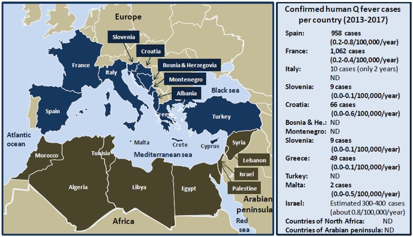
The Mediterranean is bordered by 22 riparian countries anti–C. burnetii Ig, and 5.8% developed acute Q fever (138).
(Figure 2) including the following: In the 2000s, a Q fever outbreak was reported in Albania in a
group of 115 Argentinean police officers who were exposed to
• in the North: Spain, France, Monaco principality, Italy,
contaminated dust from infected sheep during a United Nations
Slovenia, Croatia, Bosnia Herzegovina, Montenegro, Albania,
mission in Prizren in the South Kosovo, among whom 42 showed
Greece, Turkey, Malta, and Cyprus; and
clinical symptoms of Q fever (139).
• in the South: Syria, Lebanon, Israel, Palestine, Egypt, Libya,
Q fever occurred in Greece in 1946 possibly due to
Tunisia, Algeria, and Morocco.
consumption of milk from infected ovines (140). A serosurvey
On the Northern Mediterranean coast, Q fever is endemic in performed in Northern Greece in 1990 showed that 4.7%
countries of the South Europe (Spain, France, Italia). From 1981 (173/3,686) of patients with atypical pneumonia had anti–C.
to 1998, more than 600 cases of Q fever were reported in Spain, burnetii Ig. During a 2-year survey on children hospitalized
most of which sporadic, except three outbreaks in 1989 (5 cases), in Athens, acute Q fever was diagnosed in 0.67% (8/1,200) of
1990 (30 cases), and 1998 (14 cases) (112–116). Between 2000 and patients, and Q fever accounted for 2.9% of the cases with
2009, hundreds of Q fever cases were reported, most of which prolonged fever (141). In 2009, 58 cases of Q fever were reported
sporadic with an epidemic episode in the Asturias with 60 cases in Northern Greece (142). The mean rate of Q fever during
in 2003, and two outbreaks (16 and 22 cases, respectively) in 2004 to 2012 was 0.033 cases/100 kI/y. C. burnetii is endemic
Madrid (117–119). During the 2011–2015 period, 50 human Q in the island of Crete. A high seroprevalence (38.7%) of anti–
fever cases were reported in Vizcaya and among 155 subjects C. burnetii Ig was found in humans living in Crete, and 98
with febrile illness from Galicia, 25% (39/155) were diagnosed cases of Q fever were reported between 1989 and 1993 (143). In
with Q fever, and 6 patients died (120, 121). In France, Q addition, a study over a period of 6 years (1989–1995) confirmed
fever was first observed in 1948 among slaughterhouse workers that 4.6% (152/3,300) of patients suspected of infection had
in Strasbourg. Between 1949 and 1953, cases were reported anti–C. burnetii Ig (144). More recently, another serosurvey
Frontiers in Veterinary Science | www.frontiersin.org 4 November 2020 | Volume 7 | Article 558481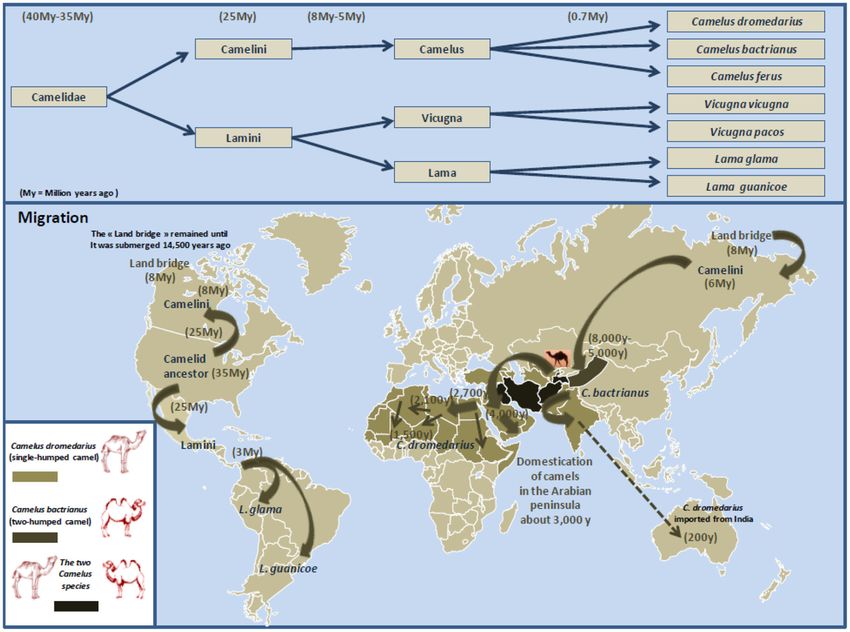
Devaux et al.                                                                                                                           Coxiella burnetii-Infected Camels

 FIGURE 2 | Schematic representation of human Q fever around the Mediterranean. Left panel: map of the Mediterranean basin. The Mediterranean Sea is bordered
 by 22 riparian countries. The countries of the Northern Mediterranean coast are represented in dark blue, and the countries of the Southern Mediterranean coast are
 represented in tanned brown. Right panel: confirmed human Q fever cases per country during the period 2013 to 2017 according to the ECDC (21). Q fever
 surveillance report, 2017. Values between brackets indicate the average number of Q fever cases per 100,000 inhabitants per year over the 5 years period. Regarding
 Italy, no data were available for the years 2013, 2014, and 2015. The number of cases of Q fever for Israel over the period 2013 to 2017 was extrapolated from the
 data published by Yarrow and colleagues (111). ND, not determined. Human Q fever occurs mostly in the form of sporadic cases. Sometimes outbreaks of Q fever
 were reported in humans. The main epidemics of Q fever described during the last 40 years in people living on the Northern coast of the Mediterranean basin are as
 follows: 2003 (60 cases), 2004 (16 cases), 2014 (50 cases) in Spain; 1992 (40 cases), 1996 (204 cases), 2002 (126 cases), 2009 (50 cases), 2014 (46 cases) in
 France; 1993 (58 cases), 2003 (133 cases) in Italy; 2007 (33 cases) in Slovenia; 2004 (14 cases) in Croatia; 2007 (42 cases) in Albania; 2009 (58 cases) in Greece; and
 2002 (19 cases) in Turkey. Human Q fever outbreaks are poorly documented concerning the countries of the Southern Mediterranean Sea coast. An epidemics of Q
 fever was described in 1955 in Algeria with 175 cases.

found a seroprevalence of 48.7% (240/493) (145). C. burnetii                           Q fever endocarditis was reported (153). A recent seroprevalence
is also present in the islands of Malta and Cyprus. In Cyprus,                         study performed in the Erzincan province in the Eastern Turkey
a serosurvey study that investigated serum samples from 547                            showed the presence of anti–C. burnetii Ig in 8.7% (32/368) of
people found that 5.3% contained anti–C. burnetii Ig, whereas a                        people (154).
more recent study using a similar number of samples indicated a                           In the Maghreb countries (Morocco, Algeria, Tunisia, Libya),
seroprevalence of 52.7% (146, 147).                                                    C. burnetii was found in the early 1950s (94). In Morocco, a
   Q fever is considered endemic in Turkey. A total of 191                             seroprevalence study conducted in 1995 reported that anti–C.
human Q fever cases were documented before 1953, most of                               burnetii Ig was present in 1% (1/300) of sera samples from
them being sporadic (148, 149). In 2002, 46 cases of febrile illness                   Casablanca and 18.3% (23/126) of samples from Fez citizens
were reported around the Black Sea in Northern Turkey, 19 with                         (155). In Algeria, the first detection of human Q fever dates back
confirmed acute Q fever (150). The search for anti–C. burnetii Ig                      to 1948 with 172 cases (156, 157). In 1955, an outbreak of Q fever
in 83 veterinarians indicated that 7–8% of them had been exposed                       concerned 175 infected soldiers from a French battalion who was
to C. burnetii. A serosurvey on blood donors in Ankara showed                          quartered in stables recently occupied by horses and sheep (158).
that anti–C. burnetii IgG was detected in 32.3% (194/601) (151).                       In 1960, several Q fever cases were reported in Eastern Algeria
In 2009, an investigation of C. burnetii prevalence in a group of                      (17). A study performed on children younger than 16 years in
407 healthy subjects living in North Turkey indicated that 8.1%                        Hoggar indicated a seroprevalence of anti–C. burnetii Ig of 20%
(33/407) of them showed evidence of contact with C. burnetii and                       (159). The follow-up of a human cohort of infective endocarditis
5.4% (22/407) were symptomatic with 17 acute Q fever and 5                             in Algiers in 2000–2003 found a C. burnetii seroprevalence of
persistent forms (152). Recently, the case of a young woman with                       3% (2/61 patients) (160). A C. burnetii seroprevalence of 15.5%

Frontiers in Veterinary Science | www.frontiersin.org                              5                                         November 2020 | Volume 7 | Article 558481
Devaux et al. Coxiella burnetii-Infected Camels
(113/729) was reported in humans in the Wilaya of Setif, an clones that are more likely to cross species barrier for infection
agropastoral region (161). In recent years, a limited number of of livestock and humans (187–190).
human cases of Q fever were reported in Algeria, and most Former epidemiological studies performed on cattle showed
cases occurred in the Northern part of the country (160, 162). that when imported into an area of endemic infection, 40% of
In Tunisia, a study of samples from a cohort of blood donors uninfected cows became C. burnetii infected within 6 months
collected in 1993 in Sousse and its rural surrounding areas (191). Although the animals can develop metritis and mastitis,
reported that 26% (130//500) of subjects had antibodies against in cattle farms the disease usually evolves subclinically (79). The
C. burnetii (163). Yet most of serosurveys performed in Tunisia different clinical manifestations of the disease can lead to late
between 1990 and 2008 suggest a seroprevalence of C. burnetii gestation abortions, fertility disorders, and premature delivery
between 1 and 3% (164–167). Information is missing regarding (192, 193). Up to 109 bacteria per gram can be contained in
the human prevalence of C. burnetii infections in Libya. The the placenta from infected ruminants (194–196). C. burnetii
serological study of foreigners (Czechoslovak citizens) returning shedding is higher in vaginal mucus and feces than milk in the
to their country after they had worked in Libya between 1984 and first 3 weeks postabortion or postpartum (197).
1988 showed an anti–C. burnetii Ig in 48 people, and about half On the Australian/Oceanian continent, Q fever is the most
of them had clinical symptoms of Q fever (168). commonly reported notifiable zoonotic disease in Australia after
In the Mashreq (Egypt, Jordan, Palestine, Lebanon, Syria), a food-borne pathogens (198). Australia became the first country
seroprevalence for anti–C. burnetii Ig ranging from 3 to 32%, to use ruminants’ vaccination. In New Zealand, in 1993, a large
was reported. An early study reported anti–C. burnetii Ig in study conducted on 2,181 cattle and 12,556 sheep concluded that
14.3% (11/77) of sera samples from Egypt (169). A C. burnetii the country was free from coxiellosis. On the North American
seroprevalence of 32% (285/883) was reported in humans living continent, a serosurvey performed in 1964 revealed that Quebec
near the Nile River Delta (170). In North Sinai in 2006, anti– had the highest seroprevalence of anti–C. burnetii Ig in bovine
C. burnetii Ig was found in 5.3% (8/150) of patients with (39.6%) (199). Sheep and goat occupy only a minor segment
pyrexia of unknown origin and 3.3% (1/30) of healthy controls of farm activities in Canada, and their seroprevalence was 6.7
(171). Another study found a seroprevalence of 16.3% (15/92) and 10.5%, respectively (19). Decades ago in the United States,
in humans who lived in agricultural districts (172). A more a seroprevalence of C. burnetii Ig study in farm animals showed
recent (2016–2017) study in El Minya Governorate reported a the highest seroprevalence among goats (41.6%), followed by
seroprevalence of anti–C. burnetii IgG of 25.7% (9/35) in farmers sheep (16.5%) and cattle (3.4%) (200). In Asia, a seroprevalence
(173). Besides a case of Q fever in a Belgian patient who developed study in Iran indicated that 13.6% (45/330) of sheep had anti–
the disease after a journey in Syria was reported (174), there C. burnetii Ig (201). The overall prevalence of anti–C. burnetii
is no information available regarding human seroprevalence of Ig in China was 15% (288/1,918) in cattle and 12% (176/1,440)
anti–C. burnetii Ig in the Palestinian, Lebanese, Jordanian, and in goats (75). A recent study of seroprevalence in goats from
Syrian populations. the Hubei province of China reported that 4.75% (55/1,157)
Q fever is endemic in Israel. Between 1981 and 1990, 758 cases of animal had anti–C. burnetii Ig (202). In Europe, the main
of Q fever were reported. A more recent series of 34 cases of sources of human infection by C. burnetii were ovine products
endocarditis allowed estimating the annual incidence of Q fever (203, 204). In 1983, a large outbreak of human Q fever was
at 3.5 cases per year, or 0.075 cases/100 kI/y, likely linked to reported in Switzerland after sheep transhumance, with 38% of
infected ruminants exposure (18, 175). In 2005, two outbreaks the animals being positive for anti–C. burnetii Ig (50). Most
of Q fever (21 cases in Haifa and 144 cases in a school in of the human epidemics reported in Germany were related to
central Israel) were reported (176, 177). A recent retrospective handling of infected sheep products (24 outbreaks), to contact
study reported 16 pediatric cases of Q fever (178). Another study with cattle (6 epidemics) or livestock (4 epidemics), or to work
investigated a cohort of patients admitted to Tel Aviv, Haifa, in slaughterhouses (2 epidemics). In 2003, 299 people were
Hadera, and Kfar Saba hospitals between 2006 and 2016 and infected when a sheep gave birth at a livestock market in Soest
confirmed 38 cases of Q fever on 205 patients (179). (205). The large Q fever epidemics reported in 2007–2010 in the
Netherlands were probably associated with the increase in goat
farming (e.g., 5,000 in 1985 and up to 375,000 in 2009) (206–
THE MAIN KNOWN RESERVOIRS OF 208), and a very high number of infected females as suggested
Coxiella burnetii: CATTLE, SHEEP, GOATS, by the frequency (20%) of abortions (209). The retrospective
WHAT ELSE? investigation of the origin of this C. burnetii outbreak in humans
revealed that C. burnetii was also found in dogs and horses, as well
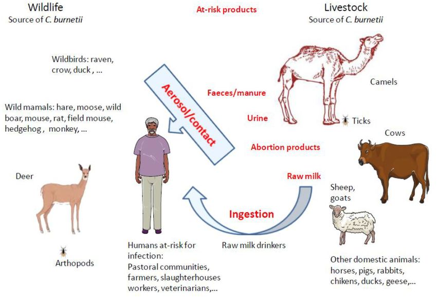
Domestic ruminants are considered the principal reservoirs for as in wild deer (210, 211). In Portugal, the frequency of exposure
Coxiella burnetii and are frequently incriminated as sources of of ovine herds at C. burnetii seems to be increasing with possible
Q fever outbreaks in humans who become infected following impact on humans (212). On the African continent, a C. burnetii
inhalation of aerosols containing particles loaded with the surveys of ovines indicated seroprevalences of 13% in Chad, 24%
bacteria or bacteria that survive in a spore-like state (95, 180, in Sudan, and 29% in Niger (105, 213, 214).
181). C. burnetii was sometimes found in other domestic animals In the countries from the Northern coast of the Mediterranean
such as poultry, cats, dogs, rabbits, and pigeons (182–186). basin, cattle, goats, and sheep are considered the major reservoir
Different C. burnetii genotypes circulate in wildlife including of C. burnetii related to human infections. Serological studies
Frontiers in Veterinary Science | www.frontiersin.org 6 November 2020 | Volume 7 | Article 558481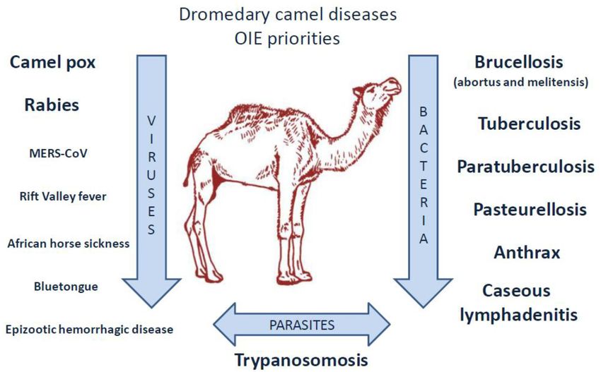
Devaux et al.                                                                                                               Coxiella burnetii-Infected Camels

performed on livestock in Madrid indicated that up to 76.6%                TABLE 1 | History of the main human Q fever epidemics in countries of the
of goats and 8.8% of cattle had anti–C. burnetii Ig (215). The             Northern coast of the Mediterranean Sea and identification of the zoonotic source
                                                                           of C. burnetii.
investigations in livestock revealed that in Northern Spain, 3%
of ovine carried C. burnetii (216). Other investigations reported          Year               Country    Probable         No. of human         References
the highest C. burnetii seroprevalence for sheep (31.5%), followed                                       origin           Q fever cases
by goat (22.4%) and cattle (5.6%), respectively (217), and 7.7%
                                                                           1987–1988          Italy      Sheep                  235                (226)
(80/1,039) of ticks (mainly Hyalomma rufipes) (218). Surveys
                                                                           1990–1995          France     Sheep                  289                (227)
carried out on 5,081 cattle abortion cases from four rural regions
                                                                           1992               France     Goat                   40                 (126)
in France between 1993 and 1996 confirmed C. burnetii infection
                                                                           1993               Italy      Sheep                  58                 (228)
in 0.5% to 3.8% of cases, while suspected for an additional 2
to 16% of cases (219). Serosurvey of C. burnetii in ruminant in            1996               France     Sheep                  29                 (227)

Sicily (Southern Italy) also showed a very high seroprevalence of          1996               France     Sheep                  204             (229, 230)

73.6% in farm sheep (220). A serosurvey in Slovenia indicated              1997               Bosnia     Sheep                  26                 (227)

that 46% of cattle, 36% (36/100) of sheep, and 2.4% (17/701)               2000               France     Goat manure            10                 (227)

of ticks (mainly Ixodes ricinus) were exposed to C. burnetii,              2000               France     Sheep                   5                 (227)
                                                                                                         manure
and ticks found positive by polymerase chain reaction (PCR)
                                                                           2002               France     Sheep                  126                (128)
were most commonly (5.09%) sampled from wild deer (221). A
                                                                           2002               Turkey     NDa                    19                 (150)
recent serosurvey performed on 1,970 serum samples collected
from farm cattle in three regions of Bosnia and Herzegovina                2003               Italy      Sheep and              133             (133, 231)
                                                                                                         goats
indicated that 8.8% of animals were exposed to C. burnetii (222).
                                                                           2003               Spain      ND                     60                 (119)
In Turkey, the prevalence of C. burnetii exposed animals varies
                                                                           2004               Croatia    Sheep                  14                 (137)
widely with species and geographic location (223). In Cyprus, a
                                                                           2004               Spain      Sheep and              22                 (118)
serosurvey indicated that many farm animals had been in contact
                                                                                                         goats
with C. burnetii including 48.2% of goats, 24% of bovines, and
                                                                           2005               Slovenia   Sheep                  33                 (134)
18.9% of sheep, with an overall abortion rate in the livestock
                                                                           2007               France     Sheep                  18                 (203)
population of Cyprus at 2 to 5% (147, 224). Among a total of
                                                                           2009               France     ND                     50                 (129)
622 cow abortions in Cyprus in 2008–2009, C. burnetii infection
                                                                           2009               Greece     ND                     58                 (142)
was documented in 57% (29/51) of the tested samples (225).
                                                                           2014               France     Sheep                  46                 (130)
In 2013, in Malta, a 6-month ban was imposed on the transfer
                                                                           2014               Spain      ND                     50                 (120)
of cattle between farms because of an outbreak of C. burnetii
infection in nine goats in one farm and two human cases.                   a ND,   not determined.
Altogether, these data (Table 1) strengthen the hypothesis that
human Q fever epidemics in the countries of the Northern coast
                                                                           may be partly wrong because of the presence of animal species
of the Mediterranean basin found their origin in sheep and/or
                                                                           endemic to these countries.
goats mainly.
    In the countries from the Southern coast of the Mediterranean
basin, the earliest investigations of C. burnetii in the ecosystem         C. burnetii STILL NEGLECTED IN THE OIE
of Morocco indicated the presence of the bacteria in sheep,                LIST OF ZOONOTIC PATHOGENS FROM
goat, cattle, camels, gerbil, and ticks (94). A recent serosurvey of       DROMEDARY CAMELS
cattle in the North-East state of Setif indicated a seroprevalence
of 11.36% (77/678) in cows (232). A study indicated that                   A listing of camel diseases considered a major threat, which
ticks collected on camels (Hyalomma dromedarii) and bulls                  ignored Q fever as “major threat” (although it appears as
(Hyalomma excavatum) imported in Egypt from Sudan were                     “notifiable disease”), was drawn by OIE in 2008 and updated in
infected with C. burnetii (233). Other studies found the presence          2010 (240). Compared to other domestic species present on both
of C. burnetii in livestock with a seroprevalence of 22.5 to               sides of the Mediterranean, little is known about the pathogens
32.7% in sheep, 16.8 to 28.2% in goat, and 13 to 13.2% in                  that circulate in camel herds (241–244), probably due to a lack
cattle, respectively (171–173, 234). A large survey that included          of international concern for camels (the earliest serosurveys were
livestock from Western desert, Nile River Valley, and Delta region         performed by biologists from Northern countries where camel
reported anti–C. burnetii Ig in 19.3% (162/840) of cattle, 8.9%            is absent) (Figure 3). In the last decade, several camel diseases
(64/716) of sheep, and 6.8% (21/311) of goats (235). The C.                with overmortalities that occurred in African countries as well
burnetii surveys of cattle indicated seroprevalences of 16 and 10%         as Saudi Arabia attracted epidemiologists’ curiosity. Today,
to 29% in Tunisia and Algeria, respectively (236–239).                     OIE draws particular attention to camelpox and rabies viruses,
    Regarding the different human epidemics of Q fever in the              to parasite-induced trypanosomosis, and to a few bacterial
countries of the Southern coast of the Mediterranean basin,                diseases including brucellosis, tuberculosis, paratuberculosis,
similar to what has been demonstrated for the countries of the             pasteurellosis, anthrax, and caseous lymphadenitis.
North Mediterranean coast, it was assumed that the source of                   Among the viral diseases affecting camels, camelpox is an
bacteria came from cows, sheep, and/or goats (124), although it            economically important disease, notifiable to OIE (245–249).

Frontiers in Veterinary Science | www.frontiersin.org                  7                                          November 2020 | Volume 7 | Article 558481
Devaux et al. Coxiella burnetii-Infected Camels FIGURE 3 | Priority diseases of camelids according to OIE (240). A list of diseases affecting camels and that appear to be priorities for the OIE to improve diagnostic capacity and establish guidelines for trade of camels and camel products was drawn up in 2014 by the OIE ad hoc Group on camel diseases. These experts divided the priority diseases into three groups: (1) significant diseases; (2) diseases for which camelids are potential pathogen carriers; (3) minor or non-significant diseases. Regarding the priority viral diseases (significant diseases), only camelpox and rabies were listed [foot and mouth disease (FMD) that concerns bactrian camels only, also belonged to the list]. The ad hoc Group classified MERS-CoV, Rift valley fever, and orbivirus-induced diseases (BT, AHS, EHD) among the diseases for which camelids are potential pathogen carriers (the bovine viral diarrhea that concerns the New World camelids also belonged to that list). Regarding bacteria, the ad hoc Group classified brucellosis, tuberculosis, paratuberculosis, anthrax, caseous lymphadenitis, and pasteurellosis in the significant diseases category. Trypanosomosis was classified in the significant parasitic diseases of camelids. It should be noted that coxiellosis is not mentioned in the lists of priority camel diseases for the World Organization for Animal Health, OIE. However, C. burnetii has been classified as a notifiable animal disease by this international office (14). Camelpox is contagious in camel husbandry, and its mortality and fungi also circulate in camel herds, including Aspergillus ranges from 0 to 40% (250, 251). This virus is a risk to the fumigatus considered responsible for the death of 40 racing human population (252, 253), yet the disease can be prevented camels in United Arab Emirates (UAE) during an outbreak of by vaccine and/or antiviral drugs such as cidofovir and ribavirin bronchopneumonia and gastroenteritis (311–313). (254, 255). Rabies in camels is also observed in many countries Because of the economic impact of brucellosis in ruminant from Africa, Arabian Peninsula, and Asia (256–262). Infection herds (with losses on meat and milk sales due to abortion), of camels was found preventable by canine inactivated rabies special attention was focused on this disease in camels (314– vaccine (263). Several other viruses able to infect camels are of 321). In Saudi Arabia, whole herd vaccination using S19 or Rev1 concern for OIE. These viruses are the Rift valley fever (RFV), the vaccinal strains was reported to be successful for camel protection Middle East respiratory syndrome coronavirus (MERS-CoV), the (322, 323). Finally, there is a public health concern linked foot and mouth disease virus, the bluetongue virus, the epizootic to the risk of transmission to humans (324–327). Dromedary hemorrhagic disease virus), the African horse sickness virus, and camel infection by Mycobacterium tuberculosis or Mycobacterium the Alkhurma hemorrhagic fever virus (AHFV) (264–297). In bovis was reported in several countries (328–341). Another humans, the MERS-CoV and AHFV infections are known to be mycobacterium, Mycobacterium avium subsp. paratuberculosis, of high fatality rate (272–275, 298). Camel can also be infected by is the causative agent of Johne disease that affects camels more a number of other viruses (299–302). severely than other ruminants (342–347). There is also concern Specific attention was drawn by OIE to Trypanosoma parasites by OIE for lung pseudotuberculosis abscesses, a frequent disease (Trypanosoma evansi, Trypanosoma vivax), which can be the of camels, as well as pasteurellosis, anthrax, and plague (348– cause of abortion in camel herds (303–310). Other parasites 350). Cases of camel plague/Yersinia pestis were reported in Libya Frontiers in Veterinary Science | www.frontiersin.org 8 November 2020 | Volume 7 | Article 558481
Devaux et al.                                                                                                                    Coxiella burnetii-Infected Camels

(351, 352), and human cases were described after consumption                TABLE 2 | National productions of farm ruminants and percentage of dromedary
of meat from infected camels (353, 354). Obviously, camels                  camels with respect to the total number of other domestic ruminants (cows,
                                                                            sheep, and goats).
are susceptible to a wide range of bacterial-induced diseases
including mastitis (242, 355, 356), upper respiratory tract diseases        Ruminants        Cows         Sheep         Goats       Camels         % Camels/
(357–360), skin necrosis (361, 362), botulism (363), tetanus (364,                                                                                 ruminants
365), and diarrhea (299, 366–368).
                                                                            Countries of the Southern coast of the Mediterranean basin
                                                                            Morocco        3,364,000a 19,863,000 5,205,000           59,000          0.2%
CAMELS: ANOTHER ANIMAL RESERVOIR                                            Algeria        1,895,126 28,393,602 5,007,894           381,882          1.08%
OF C. burnetii BESIDES RUMINANT                                             Tunisia         627,614     6,536,762     1,205,526     237,005          2.75%
                                                                            Libya           124,941     7,400,487     2,628,366      64,469          0.62%
LIVESTOCK AND WILD LIFE?
                                                                            Egypt          5,064,509    5,697,716     4,351,545     149,224          0.97%
Dromedary camels that are almost absent from the Northern                   Palestine       40,254       747,880       215,000          0             0%
countries of Mediterranean basin account for 3% of the                      Israel          543,311      519,640        89,720        5,530          0.47%
domestic ruminant populations in the Southern countries of                  Lebanon         81,262       458,112       516,803         192           0.02%
the Mediterranean basin (Table 2). Although this percentage is              Jordania        72,644      3,057,948      770,771       14,322          0.36%
relatively low, it became necessary to revisit the epidemiological          Other countriesb
data and question the possible role of camels as a source of                Chad           27,603,203 30,789,484 34,408,101 7,285,309                7.28%
human Q fever. Sixty-five years ago, the presence of C. burnetii in         Somalia        4,800,000 11,000,000 11,524,496 7,222,181                20.91%
camels was already reported (94). Regarding animal serosurvey,              Sudan          30,734,061 40,573,686 31,443,790 4,849,003                4.51%
it is hazardous to directly compare the data obtained from one              Djibouti        299,954      468,732       514,462       70,965          5.24%
country to another by different laboratories under the format               UAE             104,584     2,208,451     2,264,699     451,463          8.97%
of a meta-analysis because of size of tested population, sample             Qatar           21,675       287,231       169,232       40,843          7.87%
selection bias, and different technical methods of diagnosis. Yet, it       a Number of heads in herds and farms in 2017 according to FAO data (369).
remains intriguing that in most studies that included dromedary             b Complementary   data correspond either to the countries that are the largest producers
camels in the panels of ruminants tested for C. burnetii                    of camels or to countries in which the ratio of camels per capita is the highest.
exposure or infection, the highest seroprevalence corresponded
to dromedary camels ahead from the other ruminants (Table 3).
    An investigation in Egypt that tested 200 camels for C. burnetii        TABLE 3 | The seroprevalence of Coxiella burnetii in Camelus dromedarius
reported a seroprevalence of 66% (373). Another serosurvey                  camels compared to other ruminants.
two decades later reported anti–C. burnetii Ig in 40.7% of
                                                                            Country         % of camels % of cattle % sheep % of goats References
dromedary camels (mainly imported from Sudan), followed by
cattle (19.3%), sheep (8.9%), and goat (6.8%) (235). In the study           Chad                80%             4%           33%            23%       (105)
by Klemmer et al. the seroprevalence in camels from Aswan                   Egypt              13.3%            ND          22.5%        16.8%        (171)
governorate bordering Sudan was 67.5%. This corroborates a                  Egypt                ND             13%          33%            23%       (172)
study in Sudan that reported a seroprevalence of 64.3% (49/76)              Egypt              40.7%          19.3%          8.9%           6.8%      (235)
in camels and 29.9% in cattle (214). A recent study in Egypt                Iran               28.3%          13.3%         24.7%        31.9%        (370)
reported that 4.5% (5/112) of camel sera were positive for anti–C.          Kenya               20%             6%           13%            18%       (371)
burnetii Ig, whereas a standard quantitative PCR found an overall
                                                                            Saudi Arabia       51.5%          30.7%         12.4%        34.0%        (372)
prevalence of 15 to 19% (374). The only study that reported a
                                                                            Sudan              64.3%          29.9%           ND             ND       (214)
higher seroprevalence in ovines than camels was performed in
                                                                            China                NA             15%           ND            12%       (75)
North Sinai, with the higher seroprevalence in sheep (22.5%),
                                                                            Spain                NA            5.6%         31.5%        22.4%        (217)
followed by goat (16.8%) and camels (13.3%), respectively (171).
                                                                            USA                  NA            3.4%         16.5%        41.6%        (200)
A serosurvey in Chad, highlighted that seroprevalence was the
highest in dromedary camels (80%), followed by sheep (33%),
goats (23%), and cattle (4%) (105). In Iran, on 167 camels that
originated from 11 regions, a mean seroprevalence of 28.7%                  that 71.2% of dromedary camels had circulating C. burnetii
for C. burnetii (seropositivity ranging from 0 to 63.6%) was                Ig (378). A recent study conducted in Kenya confirmed that
observed (375). A more recent study confirmed a seroprevalence              the highest seroprevalence was in dromedary camels (20%),
of Q fever in camels of 28.3% in Iran, whereas for the other                followed by goats (18%), sheep (13%), and cattle (6%) (379).
ruminants, the results were 31.9% in goats, 24.7% in sheep,                 These results corroborate those from another study that reported
and 13.3% in cattle (370). Studies in Saudi Arabia reported a               a seroprevalence of 18.6% in camels (371).
seroprevalence around 50 to 60% of dromedary camels, with the                   Many questions remain unanswered regarding the origins
most recent investigation reporting a seroprevalence of 51.5% in            of the high prevalence of anti–C. burnetii Ig in dromedary
489 camels from Saudi Arabia, whereas the seroprevalence was                camels (Table 4), the ways by which camels become infected,
34.0% in goats, 30.7% in cattle, and 12.4% in sheep, respectively           and their role as putative reservoir in transmission of C.
(372, 376, 377). A serosurvey performed in Algeria revealed                 burnetii to other ruminants and/or humans. It was reported

Frontiers in Veterinary Science | www.frontiersin.org                   9                                            November 2020 | Volume 7 | Article 558481
Devaux et al. Coxiella burnetii-Infected Camels
that the preferred route of C. burnetii shedding by infected DOMESTICATION AND BREEDING OF THE
camels is feces (27.6% positive samples by PCR), followed by DROMEDARY CAMEL (Camelus
urine (23.8%) and milk (6.5%) (396) (Figure 4). A study on
dromedarius): A SOCIOECONOMIC ROLE
534 healthy camels in Tunisia indicated that 44% (235/534)
were seropositive to C. burnetii, and it reached 70% in female IN THE LIFE OF MILLIONS OF PEOPLE
camels with a previous history of abortion (391). It is also
The large camelids include two domestic species: Camelus
possible that the high prevalence of anti–C. burnetii Ig in
bactrianus (the two-humped camel) and Camelus dromedarius
camels was related to infections by fleas or ticks during blood- (the single-humped camel) (Figure 5). Regarding the bactrian
sucking (397, 398). Among ticks, the H. dromedarii that colonize camel, a strain adapted to cold winters that inhabit mainly the
dromedary camels were found infected with C. burnetii (1, mountains of central Asia, historians reported that the camel
233, 399). At every developmental stage of their life cycle, the production was already recommended in the pre-Islamic sacred
H. dromedarii ticks feed only once, and their camel blood religious books (412). The dromedary camel, C. dromedarius,
meal is sufficient for the molt to occur to the next stage nicknamed “desert vessel,” was domesticated in the Arabian
(400). Female ticks deposit 10,000 to 20,000 eggs on the camel Peninsula around the 1st millennium and the second century
host body. Recently, a survey performed on dromedary camels BC (413–418). It is usually considered that the dromedary
and H. dromedarii ticks in Egypt found that 46% (52/113) camel domestication appears late compared to other ruminants
of camels (27.1% of dromedary camels in Giza and 67.9% because it took place about 8,000 years after that of sheep
in Cairo) and 5.6% (10/177) of H. dromedarii ticks were and 6,000 years after that of cattle (419, 420). The use
positive for C. burnetii (383). In contrast, in the hot and dry of the dromedary camel gradually developed with caravan
regions of Southern Europe, other ticks such as Dermacentor trade of spices in the Arabian Peninsula and Mediterranean
marginatus were considered a possible vector of C. burnetii cities markets.
among ruminants (171, 401–403). Dromedary camel domestication was crucial for livelihood
It could also be interesting to investigate the role of the of pastoral communities in which camels are kept for multiple
camel hump adipocytes in the long-term storage of C. burnetii. uses including transport of people (camels can travel several
In a murine model, it was demonstrated that once C. burnetii hours per day at a speed of 15–20 Km/h), transport of loads
has gained the host bloodstream, during the first week of (they can carry between 150 and 250 kg), the maintenance of an
infection it penetrates different organs, and bacteria can be agricultural activity around oasis, the control of desertification
found in spleen, liver, epididymis, prostate, and semen. At 3 and rational management of water resources, milk production
weeks, degenerative changes in capillary blood vessels and the and consumption, source of meat, and traditional medicine (421–
surrounding tissues of the adipose envelope of the epididymis 423). Camels feed on herbaceous plants, shrubs, shoots, cacti,
are concomitant to the circulation of infected macrophages, and date stones and can spend months in semiarid regions
and bacteria shed to semen can be transmitted from male to without drinking (424, 425). During millennia, camels were
female by sexual intercourse (404). At 4 months postinfection, reared according to three breeding systems: sedentary, nomadic,
C. burnetii was detected in abdominal, inguinal, and dorsal and transhumant. Given the ecological zone in which they live,
adipose tissues, whereas no bacteria were detected in blood, the last two systems are the most frequent, with a predominance
liver, lung, and spleen, and the transfer of adipose tissue from of the transhumant mode (426–428). In most areas, dromedary
convalescent mice to naive immunodeficient mice resulted in camels are multipurpose animals with the females used primarily
the infection of the recipient host (405). Altogether these results as milk producers and the males for transport or draft. The usual
acquired in other models than camels indicate that adipose selection criteria of dromedary camels were color, morphometric
tissues may be the reservoir in which C. burnetii persists characteristics, milk production, and endurance. For example,
for prolonged periods after the end of clinical symptoms. the Guerzni type is a pack camel maintained by nomads; the
Although infection by C. burnetii of camel hump adipocytes Marmouri type is a dromedary camel used for riding, whereas
has not been evaluated so far, the elevated concentration of the Malhah- and Wadhah-type breeds were selected for high milk
adipocytes in camel hump could provide C. burnetii with an production (429, 430).
ideal long-term storage site unique among the ruminants (406). Economically, dromedary camel exploitation appear
Moreover, when food is scarce, C. burnetii could be released problematic because of slow reproductive cycle (13 months of
from hump adipocytes during lipolysis. During dehydration and pregnancy) and high mortality of young (431, 432). Reproductive
underfeeding periods, camels mobilize their hump adipose tissue losses in camel herds are due to infertility (uterine infection),
accumulated during overfeeding periods to compensate for the pregnancy loss (infectious pathogen–induced abortions),
deficit (406). In the pastoral communities, the close physical mastitis (female udder infections), and neonatal diseases (433).
contacts with dromedary camels create the conditions for the A large investigation (11,200 camels from different herds) in
transfer to the man zoonotic diseases. A meta-analysis that Ethiopia regarding the major constraints to camel production
searched in nine databases, the 929 unique articles regarding emphasized widespread diseases, lack of attention to camels,
C. burnetii epidemiology in Africa concluded that close contact lack of experience and knowledge, inadequate veterinary service,
with camels was associated with increased seroprevalence in lack of attention by the government, poor infrastructures, and
humans (95). feed shortage. Yet, camel production remains attractive for
Frontiers in Veterinary Science | www.frontiersin.org 10 November 2020 | Volume 7 | Article 558481Frontiers in Veterinary Science | www.frontiersin.org
Devaux et al.
TABLE 4 | The seroprevalence of Coxiella burnetii in Camelus dromedarius camels and Hyalomma dromedarii ticks.
Country No. of camels % of camels with No of ticks % of ticks with Diagnostics tests Related human outbreak (or not) References
tested C. burnetii Ig tested C. burnetii Ig
Algeria 184 71.2% (131/184) 0 ND Serological test: C. burnetii Indirect Multi-species ELISA Kits (ID Screen®) ND (378)
Canary Island 100 19% (19/100) (0% by PCR 0 ND 1. Serological test: LSIVETTM /ruminant milk/serum Q-fever ND (380)
assay) 2. Molecular techniques: Conventional PCR
Chad 500 4.8% (24/500), up to 28.6% 0 ND ND ND (381)
Chad 613 80% (490/613) 0 ND Serological test: C. burnetii Indirect Multi-species ELISA Kits Coxiellosis may be responsible for several (105)
undefined cases of fever
Egypt 0 ND (batch of 54 ND ND ND, not a notifiable disease (382)
ticks)
Egypt 200 66% (132/200) 0 ND Serological test: 1. Conventional enzyme immunoassays (EIAs) 2. ND, not a notifiable disease (373)
Competitive enzyme immunoassay (CEIA)
Egypt 332 13.3% (4/332) 0 ND Conventional IFA antibodies ND, not a notifiable disease (171)
Egyptb 528 40.7% (215/528) 0 ND Serological test: CHEKIT Q fever Antibody ELISA Test Kit ND, not a notifiable disease (235)
Egypt 113 46.0% (52/113) 177 5.6% (10/177) Molecular techniques: PCR ND, not enough available data (383)
Egyptc 112 4.5% (5/112) (16.9% by PCR 0 ND 1. Serological test: C. burnetii ELISA kit (GSCIENCE, USA) ND, not enough available data (374)
assay) 2. Molecular techniques: -Real-time PCR-Conventional PCR
India ND 17.3% 0 ND ND ND (384)
India ND 6.6%−7.7% 0 ND ND (385)
Irand 167 28.7% (48/167) 0 ND Serological test: CHEKIT-Q fever ELISA kit ND (370, 375)
Kenya ND 20.0% 0 ND ND ND (386)
Kenya 334 18.6% (62/334) 0 ND Serological test: The CHEKIT Q fever by IDEXX C. burnetii antibody ND (371)
11
Kenya 312 19.9% (62/312) 0 ND Serological test: CHECKIT Q Fever Antibody ELISA Test Kit ND (379)
Nigeria 386 11.4% (44/386) 0 ND ND ND (387)
Saudi Arabia 460 62% (285/460) 0 ND Serological test: CHEKIT-Q fever enzyme immunoassay ND (376)
Saudi Arabiae 489 51.6% (252/489) 0 ND 1. Serological test: CHEKIT-Q fever enzyme immunoassay ND (372, 377)
2. Molecular test: Conventional PCR
Sudan ND 12.8% 0 ND ND ND (388)
Sudan ND 14.5% 0 ND ND ND (389)
Sudan 76 64.3% (49/76) 0 ND Serological test: Commercial Q fever antibody indirect ELISA test kits ND (214)
Tunisia ND 15.8% 0 ND ND ND (390)
Tunisia 534 44.0% (235/534) 0 ND Serological test: Commercial Q fever antibody indirect ELISA test kits ND (391)
Tunisia 412 0f 327g 3.6% (12/327) Molecular test: Conventional PCR ND (392)
ND, not determined.
November 2020 | Volume 7 | Article 558481
a In this study, the seroprevalence was 4.8%, but it should be noted that 16 of the 24 positive animals were from a single herd containing 56 heads of camel, which correspond to a seroprevalence of 28.6% in this herd.
b In this study, most of the camel samples tested were collected from animals imported from Sudan, and the seroprevalence in camels from the Aswan governorate of Egypt bordering Sudan was 67.5%. Hyalomma dromedarii ticks are
commonly found in Egypt (393).
Coxiella burnetii-Infected Camels
c In this study, the camel samples were tested for anti–C. burnetii Ig and 4.5% of camels were found positive. Additional evaluation by standard PCR using the superoxide dismutase enzyme of C. burnetii indicated that 16.9% (19/112)
of camels were found positive.
d In this study, the camel samples were collected from animals living in 11 different counties, and the seropositivity ranged from 0 to 63.6%, depending the counties of origin of the camels; Hyalomma dromedarii ticks are commonly
found in Iran (394, 395).
e The highest seroprevalence was recorded in Magahim camels; some harbored the camel tick Hyalomma dromedarii.
f C. burnetii seroprevalence estimated by PCR was not detected in any of the 412 samples collected on dromedary camels in Tunisia, despite that their ticks were found positive by the same assay, and seroprevalence previously
determined by anti–C. burnetii Ig was estimated between 15 and 44% in Tunisia. The authors argued that lack of detection in camel samples was probably due to the low load of bacteria in animal blood. They admit this result is different
from those reported in Egypt, Saudi Arabia, and Iran, which demonstrated the direct identification of C. burnetii by PCR in camel blood.
g C. burnetii was estimated by PCR and found in 3.6% of 327 ticks. Hyalomma impeltatum was the most infected ticks species, 5.7% (9/158), followed by Hyalomma dromedarii, 1.9% (3/160). The frequency of tick infestation was higher
when collected on camels located in the governorate of Gabes.You can also read