Biofilms by bacterial human pathogens: Clinical relevance - development, composition and regulation - therapeutical strategies - Microbial Cell
←
→
Page content transcription
If your browser does not render page correctly, please read the page content below
Review
                                                                                    www.microbialcell.com

Biofilms by bacterial human pathogens:
Clinical relevance - development, composition and regulation -
therapeutical strategies
Adina Schulze1,#, Fabian Mitterer1,#, Joao P. Pombo1 and Stefan Schild1,2,3,*
1 Institute of Molecular Biosciences, University of Graz, Humboldtstrasse 50, 8010 Graz, Austria.
2 BioTechMed Graz, Austria.
3 Field of Excellence Biohealth – University of Graz, Graz, Austria.
# A.S. and F.M. contributed equally to this work.

* Corresponding Author:
Stefan Schild, Humboldtstrasse 50, 1st floor, 8010 Graz, Austria; Phone: ++43/ (0)316 3801970; E-mail: stefan.schild@uni-graz.at

ABSTRACT Notably, bacterial biofilm formation is increas-               doi: 10.15698/mic2021.02.741
ingly recognized as a passive virulence factor facilitating             Received originally: 08.09.2020;
                                                                        in revised form: 08.01.2021,
many infectious disease processes. In this review we will               Accepted 12.01.2021,
focus on bacterial biofilms formed by human pathogens                   Published 01.02.2021.
and highlight their relevance for diverse diseases. Along
biofilm composition and regulation emphasis is laid on
the intensively studied biofilms of Vibrio cholerae, Pseu-              Keywords: biofilm-associated disease, nosocomial infections,
                                                                        Vibrio cholerae, Pseudomonas aeruginosa, staphylococci,
domonas aeruginosa and Staphylococcus spp., which are
                                                                        treatment, biofilm.
commonly used as biofilm model organisms and therefore
contribute to our general understanding of bacterial bio-
film (patho-)physiology. Finally, therapeutical interven-               Abbreviations:
tion strategies targeting biofilms will be discussed.                   AIP – autoinducing peptide; CF – cystic fibrosis; COPD – chronic
                                                                        obstructive pulmonary disease; DGC – diguanylate cyclase; eDNA
                                                                        – extracellular DNA; EHEC – enterohemorrhagic E. coli; EPS –
                                                                        extracellular polymeric substance; GlcNAc – N-acetyl-D-
                                                                        glucosamine; IBD – inflammatory bowel disease; MRSA –
                                                                        methicillin-resistant S. aureus; MSCRAMM – microbial surface
                                                                        components recognizing adhesive matrix molecule; MSHA –
                                                                        mannose sensitive hemagglutinin; PIA – polysaccharide
                                                                        intercellular adhesin; PDE – phosphodiesterase; QS – quorum
                                                                        sensing; SERAM – secretable expanded repertoire adhesive
                                                                        molecule; sRNA – small RNA; UPEC – uropathogenic E. coli; UTI -
                                                                        urinary tract infection; UTR – untranslated region; VPS – Vibrio
                                                                        exopolysaccharide.

INTRODUCTION                                                          mental stressors, like hazardous chemical compounds, pH
Biofilms are communities of microbes embedded in an                   or physical damage. Differentiation of the microbes within
extracellular matrix that is produced by the microbes                 the biofilm is aided by the biofilm’s role as a mediator of
themselves. The microbial community may be composed                   cell-to-cell signaling.
out of one or multiple species, which may be phylogenet-                  Biofilms have been found to be ubiquitous in almost
ically unrelated. Biofilms can either be single or multi-             every environment. They can develop on all non-shedding
layered. The various gradients that exist within biofilm              surfaces in non-sterile liquid or wet environments sticking
matrices generate micro-niches, which are colonized by                to both biotic and abiotic surfaces. Biofilms are being pro-
microorganisms that have optimized their metabolism for               duced in the harshest environments, like in hot springs and
the respective environment. Anaerobic microorganisms,                 deep-sea vents, on rocks and soil, the roots and stems of
for example, would be found within the deeper layers of               plants, on chitinous surfaces of aquatic animals, but also on
the biofilm, but deeper layers of the biofilm are also inhab-         many man made surfaces like pipes, the underside of ships,
ited by microbial cells that are more sensible to environ-            shower hoses etc. Biofilms represent an important element

OPEN ACCESS | www.microbialcell.com                          28                          Microbial Cell | FEBRUARY 2021 | Vol. 8 No. 2
A. Schulze et al. (2021) Biofilms by bacterial human pathogens
in many food chains in aquatic environment, where they Several studies have demonstrated that bacteria associat-
are consumed by invertebrate, which are prey of fish. ed in biofilms exhibit increased resistance to antimicrobial
Niels Høiby was amongst the first ones to recognize the compounds than their individual, planktonic counterparts.
relevance of biofilms in disease, which has been supported Antibiotic resistance in biofilm infections is thought to be
by increasing evidence since then [1]. Biofilms are involved caused by a variety of factors, including metabolic altera-
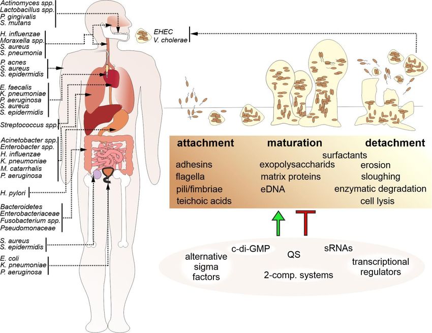
in a wide variety of microbial infections in the body (Fig. 1). tions in bacteria within the biofilm, decreased penetration
The National Institutes of Health (NIH) revealed that of antibiotics due to the extracellular matrix, inactivation of
among all microbial infections, 60-80% are linked to biofilm the antibiotic by compounds within the extracellular ma-
formation [2]. Biofilm formation not only occurs on medi- trix, inoculum effects related to the very large number of
cal devices such as contact lenses, catheters, prostheses, bacteria in the biofilm relative to the available antibiotic
heart valves and pacemakers, but also on a variety of body molecules and increased exchange of bacterial resistance
surfaces, including the skin or mucosal surfaces of the res- mechanisms as bacteria reside in close proximity to each
piratory and digestive tract. Moreover, biofilms formed in other. Bacterial biofilms also facilitate immune evasion, for
the environment are not only a likely survival and persis- example by preventing phagocytosis or immune cell modu-
tence stage for facultative pathogens outside the host, but lation and dysfunction via release of bacterial byproducts
also a relevant reservoir for the initiation of new infections. or toxins. Not surprisingly, hospitals have to deal with di-
FIGURE 1: Biofilm formation is a common feature among bacterial human pathogens. Bacterial biofilms by human pathogens are found on
various tissues of the human body, on medical devices, e.g. catheters or prostheses, and in the environment, representing a reservoir for
new infections. A schematic overview indicating representative bacterial species associated with biofilm-related diseases and their occur-
rence in the body (arrows) is presented on the left. Biofilm formation (upper right) is a multistep process organized in an attachment, matu-
ration and detachment phase. Biofilm formation is controlled and modulated by several factors including bacterial surface molecules, se-
creted matrix effectors, as well as environmental components and stressors. Thus, it is not surprising that bacterial biofilm regulation (lower
right) involves the interplay of several positive and negative regulatory cascades including quorum sensing systems (QS), regulatory small
RNAs (sRNAs), alternative sigma factors, two-component systems and second messengers, such as c-di-GMP.
OPEN ACCESS | www.microbialcell.com 29 Microbial Cell | FEBRUARY 2021 | Vol. 8 No. 2A. Schulze et al. (2021) Biofilms by bacterial human pathogens
verse nosocomial infections caused by biofilm-forming senting a health risk especially for immunocompromised
bacterial pathogens that may severely affect patients suf- patients. Especially in shower hoses Legionella spp. com-
fering from predispositions like immune suppression or monly produces biofilms, which are thought to promote
pre-existing diseases. the persistence and chlorine-resistance of the respiratory
pathogen [8].
ENVIRONMENTAL BIOFILMS AND THEIR IMPACT ON
TRANSMISSION CHRONIC AND ACUTE DISEASES CAUSED BY BIOFILM
Between outbreaks facultative human pathogens may FORMING BACTERIA
form biofilms outside of the host as a persistence mode. In contrast to biofilms formed outside of the human, bac-
Importantly, biofilm formation can facilitate environmental terial biofilms can also be key factors for the fitness of
survival and thereby allows to maintain a high infectious pathogenic strains during host colonization. These biofilms
dose even for prolonged inter-epidemic periods. Thus, can be either associated with medical devices or formed
biofilm communities can represent a reservoir for future independently from foreign body material via colonization
infections. Upon infection bacterial cells associated in bio- of host tissue, which is mainly observed along chronic in-
films are generally better protected against host defense fections.
mechanisms than their planktonic counterparts. Thus, bio-
films could be a likely form in which opportunistic bacterial Medical device-related bacterial biofilms
pathogens initiate the infection of a human host. In clinics, bacterial biofilm formation on foreign body im-
A representative example is Vibrio cholerae, the causa- plants, such as catheters (intravascular and urinary), or-
tive agent of the water borne diarrheal disease cholera. thopedic inserts as well as dental and breast implants, can
V. cholerae transits between the aquatic reservoir, where it result in severe infections. Most infections acquired in a
forms biofilms on chitinous surfaces, and the human host, hospital environment (nosocomial diseases) are implant-
where it efficiently colonizes the intestinal tract. Im- associated infections and comprise 50–70% of all nosocom-
portantly, not only intact biofilms, but also V. cholerae cells ial infections [9]. Biofilms on medical devices pose a huge
dispersed from a biofilm are more infectious than free- danger due to the high resistance to antibiotics, providing a
living, planktonic cells in the infant mouse model [3, 4]. reservoir of bacteria that can cause constant re-infections
These results suggest the existence of factors specifically and chronic inflammation that can also lead to tissue dam-
induced during biofilm formation that facilitate infection by age, clogging of devices and general resistance to treat-
V. cholerae even beyond the general idea of being better ment. Important microorganisms involved in health care
protected against host-derived antimicrobial factors within associated infections comprise Gram-positive bacteria, e. g.
a biofilm. The impact of biofilms on transmission of Staphylococcus aureus, Staphylococcus epidermidis, and
V. cholerae is highlighted by the fact that a simple sari cloth Enterococcus faecalis as well as Gram-negative bacteria,
filtration of drinking water, effectively removing biofilm- such as E. coli, Klebsiella pneumoniae, Proteus mirabilis,
associated bacteria, reduced the number of cholera cases and Pseudomonas. aeruginosa [10].
by approximately 50% in an Indian household study [5]. Roughly 80% of the microorganisms engaged in materi-
Thus, bacterial clumps or aggregates derived from mature al-related contaminations are S. epidermidis and S. aureus,
biofilms are a likely form in which clinically relevant the latter especially in connection with surgical site infec-
V. cholerae are taken up by humans, reinforcing the eco- tions, causing chronic wounds and other issues [11]. Nota-
logical and epidemiological role of biofilms. bly, the majority of these isolates exhibit multidrug re-
Another example is the enterohemorrhagic Escherichia sistance, posing an immense challenge for therapeutical
coli (EHEC) O104:H4 isolate, which showed increased bio- intervention in clinical practice [11].
film formation on fenugreek seeds and caused a severe Regarding vascular catheters, it has been documented
outbreak in Germany in 2011 with a higher rate of hemo- that within the initial seven days after catheterization, ex-
lytic-uremic syndrome than any recorded before [6]. Ge- traluminal biofilm by S. epidermidis, S. aureus, E. faecalis,
nome-wide sequence analyses revealed that the outbreak K. pneumoniae, and P. aeruginosa as well as the fungal
EHEC strain had acquired the novel diguanylate cyclase, pathogen Candida albicans considered a significant reason
DgcX, synthesizing the biofilm-promoting second messen- for catheter-related circulation system contaminations. In
ger c-di-GMP. Expression levels of DgcX are higher than any fact, vascular catheters that had been in situ for more than
other known E. coli diguanylate cyclase and it consequently 30 days showed proof of heavy luminal colonization and
fuels enhanced biofilm formation [7]. One explanation for biofilm development [12].
the unprecedented severity of this EHEC outbreak might be Along catheter-associated urinary tract infections, also
explained by the increased biofilm formation capacity of known as CAUTIs, P. aeruginosa is one of the main causes
O104:H4 providing a concentrative infective dose of the in device related bacterial infections. Another dangerous
pathogen organized in biofilm aggregates. biofilm producer linked to urinary tract infections, also
Environmental biofilms in drinking water systems serve known as UTIs, is K. pneumoniae. 63% of K. pneumoniae
as a reservoir for the respiratory tract pathogen Legionella isolates from urine samples of catheterized patients suffer-
pneumophila, causative agent of Legionnaires disease, and ing from UTIs were positive for in vitro biofilm production
opportunistic pathogens like Mycobacterium avium, repre- [13]. Chronic issues induced by device related infections
OPEN ACCESS | www.microbialcell.com 30 Microbial Cell | FEBRUARY 2021 | Vol. 8 No. 2A. Schulze et al. (2021) Biofilms by bacterial human pathogens
are often due to the biofilm production enabling a tena- that overproduce alginate. The conversion to mucoid
cious and persistent colonization. As such, urinary cathe- strains seems to be driven be thy lung microenvironment
ters have to be exchanged at least every three months. and is not observed outside of the human body.
Biofilms also play a huge role in ventilator-associated Notably, the extracellular polymers of P. aeruginosa
pneumonia that occurs in patients requiring mechanical biofilms are different for lung and UTIs described above as
ventilation breathing machines in hospitals after surgery or they contain higher amounts of the exopolysaccharide
various diseases, such as COVID-19. Due to the patients in alginate and extracellular DNA (eDNA) [21, 22]. Alginate
need of ventilator assisted breathing often suffering from protects P. aeruginosa against phagocytosis, opsonization,
underlying immune or lung problems, ventilator-associated antimicrobial compounds and clearance from the lungs
pneumonia can be a life-threatening condition. Ventilator- [23]. On the other hand, alginate fuels an immune com-
associated pneumonia has been recorded as pervasive plex-mediated inflammation via a pronounced antibody
after 48–72 h in patients who have been intubated and are response, which is characteristic for a Th2 polarized im-
on mechanical ventilation. The increased danger of trigger- mune response [24]. Overall, this results in severe lung
ing ventilator-associated pneumonia following intubation tissue damage. Notably, P. aeruginosa can reside asymp-
with mechanical ventilation is six to 20-fold. Especially en- tomatically within the human body until biofilm formation
dotracheal tubes are often associated with the develop- has reached a threshold and overwhelms the immune sys-
ment of biofilms and the methicillin-resistant S. aureus, tem. Mucoid strains are able to effectively colonize the
also known as MRSA, and Gram-negative bacilli, such as, K. lungs, stay persistent in the lungs of CF patients and are
pneumoniae, E. coli, P. aeruginosa, and Acinetobacter very difficult to treat. Consequently, biofilm production
baumanii [14 ]. Not surprisingly, mortality rates of ventila- seems to be the most important virulence factor for P.
tor-associated pneumonia are fundamentally higher than aeruginosa associated with high mortality and morbidity in
for UTIs and skin diseases [15]. CF patients.
The exact mechanisms how P. aeruginosa biofilms are
The respiratory tract effectively protected against antibiotics is still a question of
The large mucosal surface makes the respiratory tract a ongoing research. In the case of positively charged amino-
preferred niche for biofilm growth, which can result in glycosides, the negatively charged matrix components, e.
chronic inflammation of the mucosal tissue and reduced g. alginate or eDNA, allow only slow diffusion into the bio-
pulmonary function. For example, the widespread inflam- film and extend the adaption time for bacteria to mount a
matory disease chronic rhinosinusitis can be linked to stress response [25]. Other antibiotics don’t seem to be
presence of bacterial biofilms of the upper respiratory hindered by the barrier function of the biofilm matrix, but
tract. S. aureus biofilms have been found on the nasal mu- are yet still less effective against P. aeruginosa biofilms
cosal surface of 50% of patients [16], but additional causa- compared to planktonic bacteria. It is hypothesized that
tive agents include Streptococcus pneumoniae, Haemophi- the biofilm provides a privileged environment for drug-
lus influenzae and Moraxella catarrhalis [17]. The latter tolerant persister cells to survive, which can tolerate anti-
two tend to form inter-species biofilms, making treatment microbials for prolonged periods [23].
even more complicated. Exposure of P. aeruginosa to hydrogen peroxide or ac-
Chronic phenotypes of pharyngitis and laryngitis are tivated polymorphonuclear neutrophils induces a mutation
frequently associated with biofilm formation. A recent in the mucA gene, changing it to the characteristic mucoid
study identified biofilms in 62% patients with chronic lar- phenotype. A Brazilian study revealed that this mutation
yngitis [18], consisting of pathogens like S. aureus, H. influ- can be found in 93% of mucoid P. aeruginosa isolated from
enzae, C. albicans, Moraxella nonliquefaciens, Propionibac- CF patients [26]. In general, P. aeruginosa biofilm growth in
terium acnes, Neisseria meningitidis, and S. pneumoniae CF lungs is associated with an increased frequency of mu-
[18]. Substantial biofilm formation might explain the re- tations, slow growth and adaptation of the bacteria to the
quirement for extended and multiple deployment of anti- conditions in the lungs, and to antibiotic therapy. Thus,
biotics to treat certain cases of chronic laryngitis. P. aeruginosa biofilms in CF patients can only be prevented
Bacterial biofilms are also frequently associated with by early aggressive antibiotic prophylaxis or therapy, be-
chronic infections of the lower respiratory tract, mainly fore the biofilm is fully developed, or they can be treated
observed in predisposed patients suffering from abnormal by chronic suppressive maintenance therapy once the bio-
mucociliary clearance and other impaired host defenses, film is already fully developed to extend lung function for
such as cystic fibrosis (CF). Chronic infections of the lung several years [27].
can exacerbate the primary disease and result in destruc- Patients with chronic obstructive pulmonary disease
tive inflammation. The altered viscosity, lower sheer and (COPD) have a high risk of an acute excerbation triggered
nutrient richness of patient’s mucosa seems to promote by bacterial infections caused by Pseudomonas, Klebsiella,
biofilm formation [19]. While the lower respiratory tract of Acinetobacter, Enterobacter, Moraxella catarrhalis and
young patients with CF is prone to infections of H. influen- mixed infections such as Pseudomonas and Klebsiella or
zae and S. aureus, the main cause for infection in the lungs Pseudomonas and Acinetobacter [28].
of adult CF patients is P. aeruginosa [20]. If initial coloniza- Along these species enhanced biofilm production is of-
tion is not prevented, P. aeruginosa establishes perma- ten associated with clinical isolates. For example, around
nently in the lungs and often mucoid mutants are selected 85% of clinical isolates of K. pneumoniae exhibit robust
OPEN ACCESS | www.microbialcell.com 31 Microbial Cell | FEBRUARY 2021 | Vol. 8 No. 2A. Schulze et al. (2021) Biofilms by bacterial human pathogens
biofilm production, which is also associated with multiple tissues. However, if left untreated it may develop into in-
drug resistance [29]. Although biofilm production is often flammatory infections, such as pulpitis and apical perio-
described in the context of infections of COPD affected dontitis. While especially Streptococcus mutans, Actinomy-
lungs, direct demonstration of biofilm formation in lungs is ces, and Lactobacillus spp. were previously regarded as
mostly lacking and verification still remains mostly by indi- responsible for caries, the list of caries-associated bacteria
rect means. now includes species of the genera Actinomyces, Lactoba-
cillus, Dialister, Eubacterium, Olsenella, Bifidobacterium,
The urogenital tract Atopobium, Propionibacterium, Scardovia, Abiotrophia,
A healthy urinary tract is occupied by a diverse natural Selenomonas, and Veillonella in addition to carbohydrate-
bacterial flora resulting in relative high acidity by bacterial fermenting oral streptococci. Many of them are still not
metabolism and thereby fairly protected from bacterial cultivatable in the laboratory. Usually when S. mutans col-
infections. Thus, main causes of biofilm-associated bacteri- onizes tooth cavities caries follows after six to 24 months
al infections in the urogenital tract are device-related (see [37] The cariogenicity of S. mutans is due to the adherence
above). However, device-unrelated UTI through smear properties of its secreted extracellular polymeric substanc-
infection can occur. Notably, biofilm formation capacity of es (EPSs), production of which is fueled in part by fructose
uropathogenic E. coli (UPEC) and S. aureus isolates was [38].
correlated with genitourinary tract infections in several Periodontal diseases, such as gingivitis and periodonti-
studies [30, 31]. Biofilm producing bacteria can exacerbate tis are chronic inflammatory diseases of tissue around the
infections due to their relatively high antibiotic resistance, teeth. Gingivitis is an inflammation of the gums, frequently
which may turn acute infections into chronic or reoccurring observed as a response of the surrounding tissue to bacte-
infections. For example, about 20% of women with acute rial biofilm formation on the teeth. While under healthy
cystitis (inflammation of the bladder) suffer from reoccur- conditions the gingival sulcus is colonized with predomi-
ring UTI mostly caused by bacterial strains with strong bio- nantly Gram-positive streptococci at relative low level [39]
film production. Consistently, UPEC strains involved in re- the microflora can change within a couple of weeks in a
occurring UTIs are better biofilm producers than UPEC complex mixture of mainly anaerobic Gram-positive and -
strains causing only single episodes [32]. A recent study negative bacteria if biofilm formation is not prevented.
focusing on UTIs caused by S. aureus revealed that 69% of Prolonged colonization of the oral cavity facilitates further
patients’ isolates exhibit strong biofilm production, which invasion into the mucosal tissue and distribution of bacte-
resulted in increased resistance to nitrofurantoin, tetracy- rial toxins. As a consequence, gingivitis can exacerbate into
cline, erythromycin and ciprofloxacin compared to non- periodontitis, if no action in intervention of supragingival
biofilm producing strains [30]. Concordantly, a study focus- biofilm formation is taken. The growing biofilm can then
ing on chronic bacterial prostatitis demonstrated that ap- extend into the periodontal pocket and manifests as a sub-
prox. 85% of 150 different bacterial isolates from chronic gingival biofilm. Biofilms and the ongoing inflammation will
bacterial prostatitis patients were strong or moderate bio- gradually result in an opening of the periodontal pockets,
film producers, including strains like E. faecalis, Staphylo- disintegration of periodontal fibers and destruction of
coccus spp., E. coli, and 20 other Gram-negative rods [33]. bones, which will loosen the teeth and finally results in
their loss [40]. In contrast to gingivitis, the tissue destruc-
Digestive tract tion in periodontitis is irreversible. The subgingival biofilms
The digestive tract of the human body is colonized with a are dominated by diverse Gram-negative rods like Prevotel-
vast quantity and diversity of microbes, with the highest la spp., Porphyromonas gingivalis, and Fusobacterium nu-
concentration in the colon. Already more than 700 differ- cleatum, but also include motile bacteria and spirochetes
ent bacterial species reside in the oral cavity of humans in deeper layers close to the epithelial surface [35].
[34], which can initiate formation of dental biofilms, also Notably, the biofilm plaque serves as a constant reser-
known as dental plaque. The exact composition of the den- voir of microbes as well as their inflammatory effectors,
tal biofilm varies not only between different sites in the both of which can spread systematically in the body. Thus,
oral cavity, but also between individuals. Despite this, a dental biofilm bacteria are also directly and indirectly asso-
core microbiome has been proposed, and includes species ciated with several other systemic diseases such as cardio-
of the following genera: Streptococcus, Veillonella, Granu- vascular diseases, atherosclerosis, infective endocarditis,
licatella, Neisseria, Haemophilus, Corynebacterium, Rothia, aspiration pneumonia, diabetes mellitus, preterm birth,
Actinomyces, Prevotella, Capnocytophaga, Porphyromonas, and low birth weight babies [41].
and Fusobacterium [35] The gastric mucosa of approximately 50% of the human
The dental biofilm can cause diseases in the teeth and population is colonized by Helicobacter pylori [42]. Coloni-
their supporting tissues, i.e. dental caries and periodontal zation with H. pylori is linked to the initiation of peptic ul-
diseases. Regular removal of dental plaque is essential, as cer disease, corpus-predominant gastritis, and possibly also
with increasing biofilm thickness bacteria are better pro- esophageal, adenocarcinomas [42]. Organization of H. py-
tected against bactericidal activities of the saliva, which lori in biofilms has been visualized within the gastric muco-
can no longer penetrate or reach the whole tooth [36]. sa [43]. One of the best studied virulence factors of H. py-
Dental caries is characterized by a demineralization of lori is urease, neutralizing the acidic conditions in the im-
the teeth without concurrent inflammation of surrounding mediate gastric environment cells [44]. Notably, in patients
OPEN ACCESS | www.microbialcell.com 32 Microbial Cell | FEBRUARY 2021 | Vol. 8 No. 2A. Schulze et al. (2021) Biofilms by bacterial human pathogens
suffering from peptic ulcer disease more than 95% of the infections as well as improper wound healing due to chron-
mucosal gastric surface was covered by bacterial biofilms ic inflammation [54].
in urease-positive biopsies, while less than 2% of the sur- Many studies have confirmed that dermal tissues of
face was covered in urease-negative biopsies [45]. The chronic wounds contain several biofilm-forming bacteria,
importance of in vivo biofilm formation by H. pylori is also such as S. aureus, S. epidermidis, P. aeruginosa, E. coli, En-
highlighted by a recent study demonstrating that combina- terobacter spp., E. faecalis, and K. pneumoniae. Almost
tory treatment with antibiotics coupled with the biofilm 88–98% of wound infections have been found to be S. au-
disrupting compound N-acetylcysteine eradicated H. pylori reus positive [55]. S. aureus has fibrin receptors and thus
in 2/3 of the patients, while a sole antibiotic therapy only can bind to fibrinogen, which can start biofilm formation.
cleared the infection in 1/5 of the patients [46]. This affinity of S. aureus to bind to fibronectin, collagen
The residual intestinal mucosa is colonized with an and laminin makes it easy for the pathogen to colonize
enormous quantity and diversity of bacterial microbiota various host surfaces such as the skin. Patients having
generally growing as healthy biofilm communities [47]. S. aureus biofilm infections require extended healing times
While defined pathogens cause distinct acute diarrheal due to delay in re-epithelialization of the infected tissue
diseases, the etiology and link to defined bacterial species [56]. This can often exacerbate in patients that suffer from
for inflammatory bowel disease (IBD), irritable bowel syn- other diseases such as diabetes mellitus, which already
drome and colorectal cancer is less clear. However, it is damages the patients’ tissue. S. aureus biofilms are hard to
widely accepted that the intestinal microbiota can have deal with due to their incredible resistance to antibiotic
beneficial as well as adverse effects on these disease states therapy and host immune response, with biofilm produc-
[48, 49]. For example, in case of ulcerative colitis, a chronic tion even being promoted by the presence of ß-lactam
relapsing form of IBD, a variety of biofilm-producing spe- antibiotics and cytokines [57]. Generally, antibiotic re-
cies including Fusobacterium spp., Shigella spp. and adhe- sistant S. aureus strains, such as MRSA, pose a worldwide
sive E. coli have been implicated to promote initiation and problem in clinical medicine. S. aureus and P. aeruginosa
maintenance of disease [50]. Similarly, Crohn’s disease has are the two most common causes of chronic wound infec-
been associated with an overall increase of Enterobacteri- tions and are frequently co-isolated from the same wound.
aceae, Pseudomonas spp and Bacteroidetes, bacterial Chronic wounds don’t always only contain chronic infec-
groups known to have members with good biofilm forming tion of a single bacterial strain, but can co-occur with sev-
capabilities [51]. eral different biofilm producing strains such as S. aureus
It seems reasonable, that bacterial biofilms can pro- and P. aeruginosa. Analysis of 22 patient samples by using
mote chronic colonization of these bacterial groups in gut. specific peptide nucleic acid and fluorescence in situ hy-
Moreover, the relatively high antimicrobial resistance of bridization revealed that P. aeruginosa colonizes the deep-
biofilms would explain the observed intractability of IBD to er layers in the wound bed, while S. aureus was rather
antibiotic therapy. Finally, biofilm matrix components may found on the wound surface [58]. Recent data indicates
potentiate the proinflammatory response, which is a hall- that both bacteria benefit from each other in coinfected
mark of IBD. Importance of bacterial biofilms in the patho- wounds and synergistically increase antibiotic tolerance
genesis of ulcerative colitis and Crohn’s disease is indeed [59]. Wounds infected with P. aeruginosa are larger in size
suggested by several reports, but we are just at the begin- and require longer healing periods [58].
ning to understand their impact on IBD and a comprehen- Emerging data also suggests that biofilm formation is a
sive mechanistic understanding is currently lacking [52]. key colonization factor of the opportunistic pathogen
P. acnes associated with the inflammatory disease acne
Skin and wounds vulgaris as well as invasive infections of skin, the cardiovas-
More than 60% of the microbial load on the human skin is cular system, soft and deep organ tissue and implant asso-
composed of diverse biofilm producing bacteria. The pre- ciated infections [60]. Most likely biofilm formation in se-
dominant floras include Staphylococcus spp., Corynebacte- baceous follicles results in elevated resistance of P. acnes
rium spp., and Propionibacterium spp. [53]. Biofilm produc- against [61]. Biofilm-like aggregates of P. acnes are more
ing skin bacteria cause a number of skin diseases, such as frequently observed in skin biopsies of acne vulgaris pa-
acne vulgaris caused by P. acnes, cellulitis, erysipelas and tients compared to healthy control groups. Moreover, re-
erythema nodosum caused by Streptococcus pyogenes, cent data suggests that biofilm formation by P. acnes is
impetigo caused by S. pyogenes and S. aureus, necrotizing phylotype-dependent and isolates derived from invasive
fasciitis caused by S. pyogenes, Klebsiella and Clostridium infections are associated with better biofilm production
amongst others, staphylococcal scaled skin syndrome compared to healthy skin isolates [62, 63].
caused by S. aureus, chronic ulcers caused by Bacteroides,
Clostridium and Streptococcus, and finally otitis externa Biofilms associated with invasive disease
and chronic wounds caused by P. aeruginosa. In general, Invasive microbial infections occur at parts of the body that
biofilms increase the bacterial fitness against host immune are generally considered germ free, e. g. the blood or other
defenses, bacteria, antibiotic therapy and general hygiene internal fluids and internal body sites such as the brain or
treatment. Bacterial biofilms also impact the risk of infec- the heart. Even though the infection routes can vary, some
tion and progression of chronic wounds, as they have been invasive microbial infections correlate with the ability of
associated with increased wound development and skin the responsible microbes to form biofilms. Well known
OPEN ACCESS | www.microbialcell.com 33 Microbial Cell | FEBRUARY 2021 | Vol. 8 No. 2A. Schulze et al. (2021) Biofilms by bacterial human pathogens
biofilm associated invasive microbial diseases include en- of metabolic waste products. Thus, some bacteria will de-
docarditis caused by Streptococcus, osteomyelitis mainly tach from the mature biofilm to resume a planktonic life-
caused by S. aureus, otitis media caused by S. pneumonia style.
and H. influenzae and meningitis caused by A. baumannii Formation and maintenance of biofilms require extra-
and H. influenzae. cellular matrix components, which are responsible for sur-
Although bacterial endocarditis is mostly linked with face adhesion, cell binding and preserving the biofilm ar-
heart implants, it can also occur through microbes reaching chitecture (Fig. 1, Table 1). Not only is there a vast diversity
the heart either through wounds or in some cases through of the microbial community, but also the extracellular ma-
the bloodstream during the course of an invasive infection trix shows species-specific variability. The EPS secreted by
[64]. Microbes like Streptococcus spp. have fibronectin the constituent population of the biofilm is the major com-
receptors facilitating biofilm formation on different tissues ponent of bacterial biofilms. The EPS mainly consist of pol-
at various sites of injury, which can cause tissue damage of ysaccharides, but may also contain other biomolecules like
the valves and is especially detrimental in the case of en- proteins, nucleic acids, glycopeptides, lipids, lipopolysac-
docarditis [65]. Open fractures, beside posing immediate charides as well as sequestered metals.
danger to health, can also lead to chronic infections such Many bacterial species are forming biofilms helping
as the bone disease osteomyelitis. S. aureus is predomi- them to persist within the environment, protecting them
nantly present as a causative agent in cases of invasive against the host’s immune system and therefore promot-
osteomyelitis [66]. S. aureus has fibrin receptors and thus ing infection and the development of disease symptoms.
can bind to fibrinogen present in the bone matrix and can Here, the focus is laid on V. cholerae, P. aeruginosa, S. au-
start biofilm formation. This affinity of S. aureus to bind to reus and S. epidermidis due to their overlapping coverage
fibronectin, collagen and laminin makes it easy for the of the mentioned biofilm functions (Table 1). V. cholerae
pathogen to colonize the bone by forming a biofilm [67]. biofilms formed in the aquatic ecosystem not only facilitate
One of the more predominant invasive diseases is otitis environmental persistence, but also impact transmission of
media, an infection of the inner ear. S. pneumonia and the disease [72]. P. aeruginosa biofilms are found on medi-
H. influenzae both cause otitis media, with more and more cal devices as well as in the respiratory tract, i.e. in the
biofilm forming serotypes emerging as antibiotic treatment lungs of CF patients [73]. Finally, biofilms of S. aureus and
increases pointing to an important role of biofilms as pro- S. epidermidis are frequently associated with infections
tective factors in those cases [68]. derived from indwelling medical devices and chronic
Clinical isolates of invasive non-typeable H. influenzae wounds [74, 75]. The selected candidates are well charac-
and A. baumanii from bacterial meningitis patients, terized biofilm producers as well as genetically modifiable,
demonstrate higher biofilm production compared to iso- allowing deeper phenotypical analyses by the implementa-
lates of these species, derived from carriers, chronic dis- tion of loss-/gain-of-function constructions.
ease or respiratory tract infections [69, 70], which empha-
sizes the impact of biofilm formation for these pathogens Attachment
to cause invasive diseases. Although biofilm formation is Bacterial adhesion on surfaces consists of reversible and
not directly linked to bacterial meningitis caused by Neis- irreversible stages and involves numerous factors, ranging
seria meningitidis, biofilm production is an important mu- from flagella, pili, fimbriae, lipopolysaccharides, lipopro-
cosal survival and persistence factor for the bacterium. teins, membrane proteins, adhesins, and eDNA.
Approximately 30% of carriage isolates are strong biofilm The importance of flagella-mediated motility for initial
producers, a far greater percentage as observed for acute attachment has been reported for several pathogens, in-
disease isolates. This suggests that biofilms might be im- cluding V. cholerae and P. aeruginosa [76, 77]. V. cholerae
portant for the chronic carriage of the bacterium, which uses its single, polar, Na+-driven flagellum to swim near the
provides a reservoir for invasive meningococcal disease surface [78]. In close proximity to the surface hydrodynam-
[71]. ic forces acting on the flagellum and cell body re-direct
flagellar rotation into a clockwise direction resulting in
BACTERIAL BIOFILM FORMATION AND COMPOSITION circular swimming behavior [79]. Movement of V. cholerae
Based on the contribution of bacterial biofilms to bacterial becomes more restricted upon tethering to the surface by
infections, bacterial biofilm development and composition their flagella [79]. An elegant microscopical study by Utada
became a focal point of interest within the scientific com- and coworkers identified two motility modes named
munity. Bacterial biofilm formation is a multistep process “roaming“ and “orbiting” [80]. Besides flagellar motility
(Fig. 1): In general, initial surface attachment of planktonic these motion types require the mannose sensitive hemag-
bacteria is reinforced via adhesive surface appendages or glutinin type IV pili (MSHA) of V. cholerae promoting
proteins. Upon irreversible attachment and microcolony mechano-chemical attachment to surfaces. Weak interac-
formation bacteria induce factors for production and se- tions between the surface and MSHA enable bacteria to
cretion of extracellular matrix components, which results pass over the surface by long directional movements with
in the formation of a three-dimensional biofilm architec- only small curvatures, which define the “roaming mode”.
ture. Finally, a mature biofilm requires dispersal to avoid In contrast, the “orbiting mode” results from stronger in-
harmful overgrowth, nutrient limitation and accumulation teractions between the surface and MSHA visualized by
tight, repetitive movements with near-circular orbits with
OPEN ACCESS | www.microbialcell.com 34 Microbial Cell | FEBRUARY 2021 | Vol. 8 No. 2A. Schulze et al. (2021) Biofilms by bacterial human pathogens
TABLE 1. Overview of factors involved in the different stages of biofilm formation for the bacterial pathogens V. cholerae, P. aerugino-
sa, S. aureus and S. epidermidis discussed in this article. For details we kindly refer to the text (see chapter “Bacterial biofilm formation
and composition”).
stage in biofilm bacterial pathogen
formation
V. cholerae P. aeruginosa S. aureus/ epidermidis
flagella motility,
flagella/ twiching motility, hydrophobic surface,
type IV pili,
attachment type IV pili, teichoic acids,
adhesins and
Cup fimbrial adhesins and adhesins (e.g. Atl, Bap,
chitin-binding factors (e.g.
lectins MSCRAMMs, SERAMs)
GbpA, ChiRP, FrhA, CraA)
exopolysaccharide (VPS), exopolysaccharide (alginate, exopolysaccharide (PIA),
eDNA, Psl, Pel), eDNA,
proteinaceous factors (RbmA, eDNA, proteinaceous factors [e.g. SasG,
maturation proteinaceous factors (e.g. Aap, and other adhesins (see
RbmC, Bap1),
lipids CdrA, LecA/B), above)],
rhamnolipids teichoic acids
nucleases (Dns and Xds), Alginate lyase, exoproteases (e.g. SspA/ Esp,
detachment proteases, rhamnolipids, SspN/ SepA, SplA-F, ScpA)
predicted sugar lyase (RbmD) cell lysis
high curvatures. More and more MSHA-surface interac- environment than what is currently suggested by laborato-
tions may tether orbiting cells tighter to the surface. Even- ry studies mainly focusing on plastic material.
tually, bacteria attach irreversibly to the surface and initi- Similar to V. cholerae, P. aeruginosa is thought to get
ate production and secretion of the Vibrio exopolysaccha- into close proximity to the surface via flagella-mediated
ride (VPS) and biofilm matrix proteins resulting in micro- motility. Non-flagellated mutants show reduced attach-
colony formation followed by biofilm maturation (see be- ment especially under glucose- or amino acid-rich condi-
low “Three-dimensional biofilm formation and matura- tions [76, 85]. However, in contrast to V. cholerae, P. aeru-
tion”). Notably, non-motile mutants lacking the major fla- ginosa reversibly attaches to surfaces in an upright (verti-
gellin subunit FlaA are still capable of forming biofilms, but cal) position and moves along random trajectories in
aggregate first in liquid culture before the clumps immobi- “walking” mode using twitching motility mediated by type
lize on surfaces resulting in altered biofilm architecture IV pili [86]. Mutants with a defective type IV pilus form
[79]. Moreover, flaA mutants show increased VPS produc- aberrant biofilms [76]. Upon horizontal orientation to the
tion, which suggests that loss of the flagellum could induce surface, attachment transits into an irreversible state, but
biofilm formation [79]. Mutations in the flagellar motor bacterial cells are still active for two-dimensional move-
complex negate the VPS overproduction of flaA mutants, ment via twitching motility resulting in the organization of
indicating that the flagellar motor could act as a mechano- microcolonies. Comprehensive studies by the Tolker-
sensor involved in the transition to the irreversible attach- Nielsen group suggest that P. aeruginosa also uses twitch-
ment state and initiation of matrix production [81]. ing motility for climbing up microcolonies formed by a sub-
It should be emphasized that environmental biofilm population of non-motile cells to form the typical mush-
formation of V. cholerae in aquatic reservoirs occurs on room-like architecture of a mature biofilm [87]. P. aeru-
chitinous surfaces, consisting of β-1→4 linked N-acetyl-D- ginosa recognizes surface attachment via the WspA protein,
glucosamine (GlcNAc) [82]. Several factors promoting at- the membrane-bound receptor protein of the Wsp
tachment to chitin have been reported. For example, the chemosensory signal transduction system that activates c-
GlcNAc-binding protein GbpA, which seems quite specific di-GMP synthesis upon surface contact [88]. As it will be-
for GlcNAc-oligosaccharides, the chitin-regulated type IV come evident below (see chapter “Regulation”) the second
pili ChiRP promoting competitive attachment to chitinous messenger c-di-GMP is a central signal involved in biofilm
surfaces, and the MSHA pili, which generally facilitates regulation. In P. aeruginosa activation of the Wsp system
adhesion to abiotic and chitinous surfaces, e.g. borosilicate, and high c-di-GMP levels act positively on the production
zooplankton and crab shells [83]. Moreover, the flagellum- of CdrA (cyclic diguanylate-regulated two-partner secretion
regulated hemagglutinin FrhA and c-di-GMP-regulated partner A) adhesin and Cup fimbrial adhesins, which pro-
adhesin A (CraA) promote attachment and initial biofilm mote surface adherence, as well as the exopolysaccharides
formation on chitin [84]. Thus, it is likely that these factors Psl, Pel, and alginate, which are structural parts of the bio-
play more crucial roles for biofilm formation in the natural film matrix [89-91].
OPEN ACCESS | www.microbialcell.com 35 Microbial Cell | FEBRUARY 2021 | Vol. 8 No. 2A. Schulze et al. (2021) Biofilms by bacterial human pathogens
Regarding biofilm formation of non-motile bacteria, the Notably, S. epidermidis encodes for the membrane-
best studied representatives are probably S. aureus and spanning giant 1.1 mDa fibronectin-binding protein Embp,
S. epidermidis. In absence of a flagellum, adherence to while Ebh represents the homologue in S. aureus [99, 100].
hydrophobic surfaces is facilitated by the overall hydro- The current knowledge on staphylococcal adhesins was
phobic character of the bacterial envelope [74]. Further- recently reviewed in detail by Heilmann et al., which we
more, attachment to abiotic surfaces via hydrophilic and suggest for further reading [92].
ionic interactions is promoted by defined surface factors,
including wall teichoic acids, the major autolysin AtlE of Three-dimensional biofilm formation and maturation
S. epidermidis, its S. aureus homologue Atl, and the surface Upon surface attachment bacteria alter their expression
protein Bap of S. aureus, respectively [92, 93]. For example, profile from a planktonic to a sessile lifestyle highlighted by
atlE mutants in S. epidermidis exhibit a less hydrophilic the upregulation of components required for the biofilm
surface and reduced biofilm formation capacity on polysty- matrix formation. The exact biofilm matrix composition
rene [94]. S. aureus dtlA mutants lack an amino acid substi- differs between species, but generally includes a blend of
tution in the wall teichoic acids, which increases their neg- various secreted biomolecules, such as polysaccharides,
ative charge and thereby reduces initial attachment to eDNA, proteins, lipids, and teichoic acids.
hydrophobic glass or plastic surfaces [92].
Indwelling devices are rapidly surrounded by host tis- Exopolysaccharides
sue and coated with a host-derived matrix. To initiate bio- In many bacteria the development of a mature biofilm is
film formation, various staphylococcal surface factors not associated with the production of exopolysaccharides,
only adhere to host cell surfaces, but also bind extracellular which are frequently the major component of the biofilm
host matrix components, e.g. fibronectin, fibrinogen, vit- matrix.
ronectin, thrombospondin, bone sialoprotein, elastin, and For example, the VPS constitutes up to 50% of the ma-
collagen [92]. Aside of the above-mentioned wall teichoic ture Vibrio biofilm matrix and is required for the develop-
acids, autolysins and Bap, these largely comprise the cova- ment of a three-dimensional biofilm [101, 102]. It is a pol-
lently-linked microbial surface components recognizing ymer with a major repeating unit of 1→4 linked
adhesive matrix molecules (MSCRAMMs) and the non- α-L-GulpNAcAGly3OAc, β-D-glucose, α-D-glucose and
covalently surface-associated secretable expanded reper- α-D-galactose, with α-l-GulpNAcAGly being an amide be-
toire adhesive molecules (SERAMs). MSCRAMMs contain a tween C-6 of 2-acetamido-2-deoxy-α-l-gulopyranosyluronic
conserved domain organization including an N-terminal acid and an amino group of glycine, OAc being an
signal peptide, an outwardly exposed ligand-binding do- O-acetylation and NAc being a N-acetylation [103]. Re-
main with directly repeated sequences, a hydrophobic placement of α-D-Glc by an α-D-GlcNAc in approximately
membrane-spanning region, a C-terminal LPXTG motif re- 20% of the repeating units increases diversity [103]. The
quired for cell wall anchorage, and a positively charged tail two nearby chromosomal loci vps-I and vps-II encode pro-
[95]. Cell wall anchorage is predominantly mediated by the teins for VPS biosynthesis and export, which are activated
SrtA sortase, a membrane-bound transpeptidase covalent- shortly after surface attachment [104, 105]. Notably, the
ly linking the protein via the carboxyl group of threonine in vps-I and vps-II gene clusters are separated by the rbmA-E
the LPXTG motif to the amino group of the peptidoglycan operon [106, 107], encoding for the two matrix proteins
[96]. Due to its conserved role in anchoring virulence fac- RbmA and RbmC (see below).
tors to the cell wall, SrtA is suggested as a target for anti- P. aeruginosa produces three different types of exopol-
virulence drug development against staphylococci, entero- ysaccharides, i.e. alginate, Psl (polysaccharide synthesis
cocci and streptococci [97]. While S. aureus isolates encode locus) and Pel (pellicle) [108]. Alginate is an acetylated pol-
for more than 20 MSCRAMMs, there are currently only ymer of β-1,4-linked D-mannuronate and L-guluronate,
twelve identified in S. epidermidis. Representative exam- which is synthesized by enzymes encoded by the algACD
ples include the fibronectin-binding proteins FnbPA and gene cluster [109]. It is not only the most important struc-
FnBPB as well as the fibrinogen-binding proteins ClfA and tural component of P. aeruginosa biofilms, but also acts as
ClfB of S. aureus or the accumulation-associated protein a barrier for antimicrobial compounds and facilitates im-
Aap and Bhp of S. epidermidis, which are highly homolo- mune evasion, thereby contributing to in vivo persistence
gous to SasG and Bap in S. aureus. Attachment flexibility during lung colonization [110, 111]. Biofilm formation of
and diversity is ensured as one MSCRAMM can bind sever- P. aeruginosa independent of alginate production high-
al host factors and MSCRAMMs exhibit overlapping binding lights the existence of other polysaccharide matrix compo-
capacities. Not surprisingly, identification of the individual nents, e.g. Psl and Pel [112, 113]. While Pel is present in
binding spectra of MSCRAMMs is still ongoing. SERAMs are most P. aeruginosa strains, Psl is not wide-spread and is
a loosely defined group of secreted proteins, which bind only produced by few P. aeruginosa strains, most notably
back to bacterial surface by so far uncharacterized mecha- by strain PAO1, but not PA14. Moreover, while Psl is found
nism(s) and have relaxed binding specificity to host matrix mainly at the outer surface of microcolonies, Pel is mainly
factors [98]. Representative examples include the extracel- located at the stem of the mushroom structure [114, 115].
lular adherence protein Eap (also known as Map or P70) The polysaccharide synthesis locus (psl) harbors 15 genes
and the extracellular matrix and plasma binding protein involved in biosynthesis of the extracellular sugar polymer
Emb of S. aureus, which are absent in S. epidermidis [98]. Psl, containing D-mannose, D-glucose and L-rhamnose
OPEN ACCESS | www.microbialcell.com 36 Microbial Cell | FEBRUARY 2021 | Vol. 8 No. 2A. Schulze et al. (2021) Biofilms by bacterial human pathogens
[116]. Psl can be found in a larger cell-associated form and nent and enables bacteria to adapt to environmental
in a smaller soluble form. Especially, the smaller variant is changes via eDNA modulation.
thought to facilitate intercellular interactions and cell- For example, in V. cholerae biofilms eDNA levels are
surface attachment, but the exact mechanism yielding in controlled by the extracellular endonuclease Dns and the
the smaller variant is currently unclear [117]. Psl not only exonuclease Xds, which is important for the development
supports adherence during initial biofilm stages, but also of a typical sponge-like biofilm architecture and detach-
contributes to the structural stability of mature biofilms. In ment from mature biofilms [4]. Similar observations have
doing so Psl interacts with other abundant biofilm matrix been reported for P. aeruginosa with its secreted EndA
components including the matrix protein CdrA and eDNA nuclease, and for S. aureus releasing two thermostable
[89, 118]. Moreover, P. aeruginosa deposits a chemtrail of nucleases Nuc1 and Nuc2 [129-131]. Due to extracellular
Psl as it moves on a surface, which guides subsequent cells nucleases and respective nucleotide uptake systems, eDNA
to microcolony formation [119]. By exploiting the released can also serve as a carbon, nitrogen and phosphate source
DNA from the host’s neutrophil extracellular traps, origi- [132-134]. Not surprisingly, phosphate starvation activates
nally a defense system against pathogens, the eDNA-Psl nucleases in V. cholerae resulting in eDNA degradation and
interaction acts as biofilm scaffold and facilitates survival biofilm dispersion [132]. Indeed, this perception is con-
of P. aeruginosa during lung colonization [118]. Similarly, firmed by results showing that phosphate limitation nega-
the Pel polysaccharide can also bind to eDNA due to its tively impacts biofilm formation in V. cholerae [135, 136].
cationic amino sugars, which might explain why Pel can Besides its contribution to the biofilm architecture, eDNA is
partially compensate a lack of Psl in P. aeruginosa biofilms also a major proinflammatory factor of P. aeruginosa bio-
[120]. Mutant strains lacking Pel are more susceptible to films, limits penetration of antimicrobial compounds and
aminoglycoside antibiotics either because Pel binds amino- allows horizontal gene transfer [25, 133, 137].
glycosides to reduce their activity or blocks their penetra-
tion into the biofilm [121]. The pel locus comprises a seven Matrix proteins
gene operon encoding for proteins with predicted func- Another important component of the bacterial biofilm ma-
tions for biosynthesis of the glucose-rich polysaccharide, trix are proteins. Most proteins studied in the context of
but the exact chemical composition of Pel remains to be biofilm matrix contribute to the adhesive properties, stabil-
elucidated [120]. ity and morphology of the biofilm. However, it should be
The major biofilm exopolysaccharide class in staphylo- noted that some proteins associated with biofilms exhibit
cocci is the polysaccharide intercellular adhesin (PIA) or, enzymatic properties, e.g. sugar hydrolases, proteases and
according to its chemical composition, a polymer of 1→6 the above-mentioned nucleases, which actively degrade
linked N-acetylglucosamines (PNAG), respectively [122]. It and modulate other matrix components resulting in biofilm
is considered to be the most important intercellular adhe- reorganization and dispersal [4, 126, 129-131, 138-142].
sin of staphylococci and is crucial for biofilm formation and In V. cholerae three major biofilm matrix proteins with
virulence in S. epidermidis [123, 124]. PIA is synthesized by predicted carbohydrate-binding domains have been identi-
the proteins expressed from the icaADBC (intercellular fied, i.e. Bap1 (Biofilm-associated protein 1) as well as
adhesion) operon [124]. The N-acetylglucosamine transfer- RbmA and RbmC (rugosity and biofilm structure modulator
ase IcaA, together with IcaD, synthesizes an N-acetyl- A and C) [106]. Importantly, they exhibit individual spatio-
glucosamine oligomer [125]. Chain growth is dependent on temporal expression profiles and consequently fulfill dif-
IcaC, which is suggested to act as PIA exporter. PIA is par- ferent roles in biofilm formation, which was comprehen-
tially deacetylated on the bacterial surface by the PIA sively characterized by an elegant microscopical study by
deacetylase IcaB [126]. This step is crucial for PIA retention Berk and coworkers [104]. The 26 kDa RbmA appears first
and thus for the various functions PIA fulfills, not only bio- on the cell surface after cells have attached to the surface
film formation, but resistance to antimicrobial peptides and VPS production was initiated [104]. At later stages
and neutrophil phagocytosis [126]. Moreover, partial loss RbmA can be found on cell surfaces throughout the entire
of the N-acetyl groups after secretion results in a cationic mature biofilm [104]. RbmA exhibits binding specificity to
character facilitating electrostatic interactions with other sugars including sialic acid derivates, which can be found in
extracellular molecules and adhesive properties of the lipopolysaccharides as well as to galactose, which is a com-
biofilm matrix [126]. ponent of VPS [105, 143]. This suggests that surface-
located RbmA can act mainly as a scaffold protein mediat-
eDNA ing intercellular and cell-matrix interactions. Along initial
It is becoming increasingly evident that eDNA is a polymer- biofilm formation, RbmA secretion is followed by the
ic matrix component of many bacterial biofilms and most 75 kDa Bap1, predominantly at sites were cells have con-
likely originates from cell lysis [127]. The highly polymeric tact with the surface or other bacteria. Even in mature
and anionic features of DNA allow cell-to-cell interactions biofilms, Bap1 is mainly found at the bottom of the biofilm
via surface molecules in the matrix network [128]. Im- with highest concentrations close to the founder cells, sug-
portantly, several bacteria secrete nucleases to degrade gesting that it is predominantly secreted by these early
eDNA, which makes it a rather flexible structural compo- biofilm members. Thus, anchoring the biofilm to the sur-
face seems an important and unique feature of Bap1.
Moreover, Bap1 was shown to bind outer membrane vesi-
OPEN ACCESS | www.microbialcell.com 37 Microbial Cell | FEBRUARY 2021 | Vol. 8 No. 2You can also read