CytoBrowser: a browser-based collaborative annotation platform for whole slide images version 1; peer review: awaiting peer review
←
→
Page content transcription
If your browser does not render page correctly, please read the page content below
F1000Research 2021, 10:226 Last updated: 22 MAR 2021

SOFTWARE TOOL ARTICLE

CytoBrowser: a browser-based collaborative annotation
platform for whole slide images [version 1; peer review:
awaiting peer review]
Christopher Rydell             , Joakim Lindblad
MIDA Group, Department of Information Technology, Uppsala University, Uppsala, 751 05, Sweden

v1   First published: 22 Mar 2021, 10:226                                    Open Peer Review
     https://doi.org/10.12688/f1000research.51916.1
     Latest published: 22 Mar 2021, 10:226
     https://doi.org/10.12688/f1000research.51916.1                          Reviewer Status AWAITING PEER REVIEW

                                                                             Any reports and responses or comments on the

Abstract                                                                     article can be found at the end of the article.
We present CytoBrowser, an open-source (GPLv3) JavaScript and
Node.js driven environment for fast and accessible collaborative
online visualization, assessment, and annotation of very large
microscopy images, including, but not limited to, z-stacks (focus
stacks) of cytology or histology whole slide images. CytoBrowser
provides a web-based viewer for high-resolution zoomable images
and facilitates easy remote collaboration, with options for joint-view
visualization and simultaneous collaborative annotation of very large
datasets. It delivers a unique combination of functionalities not found
in other software solutions, making it a preferred tool for large scale
annotation of whole slide image data. The web browser interface is
directly accessible on any modern computer or even on a mobile
phone, without need for additional software. By sharing a "session",
several remote users can interactively explore and jointly annotate
whole slide image data, thereby reaching improved data
understanding and annotation quality, effortless project scaling and
distribution of resources to/from remote locations, efficient creation
of "ground truth" annotations for methods' evaluation and training of
machine learning-based approaches, a user-friendly learning
environment for medical students, to just name a few. Rectangle and
polygon region annotations complement point-based annotations,
each with a selectable annotation-class as well as free-form text fields.
The default setting of CytoBrowser presents an interface for the
Bethesda cancer grading system, while other annotation schemes can
easily be incorporated. Automatic server side storage of annotations
is complemented by JSON-based import/export options facilitating
easy interoperability with other tools. CytoBrowser is available here:
https://mida-group.github.io/CytoBrowser/.

Keywords
virtual microscopy, digital cytology, whole slide image, annotation,
assessment, visualization, bioimage informatics, JavaScript

                                                                                                                        Page 1 of 9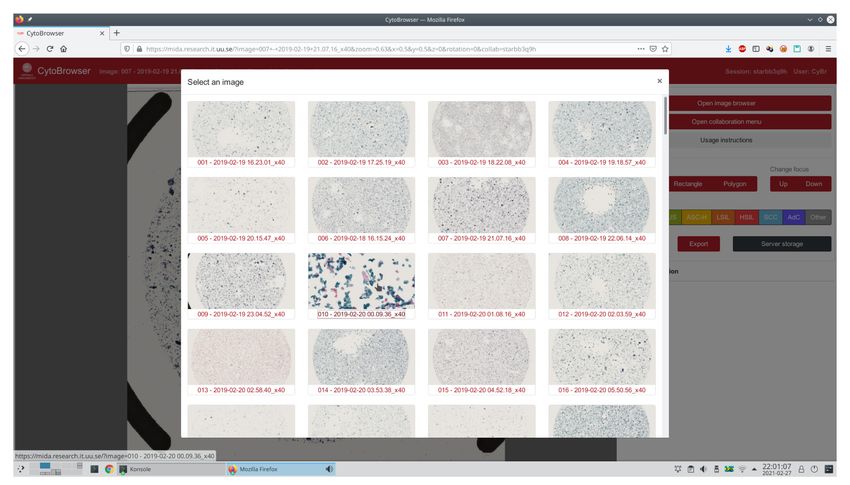
F1000Research 2021, 10:226 Last updated: 22 MAR 2021

               This article is included in the NEUBIAS - the
               Bioimage Analysts Network gateway.

Corresponding author: Joakim Lindblad (joakim@cb.uu.se)
Author roles: Rydell C: Conceptualization, Software, Validation, Visualization, Writing – Original Draft Preparation, Writing – Review &
Editing; Lindblad J: Conceptualization, Data Curation, Funding Acquisition, Project Administration, Resources, Software, Supervision,
Validation, Visualization, Writing – Original Draft Preparation, Writing – Review & Editing
Competing interests: No competing interests were disclosed.
Grant information: The project is financially supported by Sweden’s Innovation Agency, VINNOVA, through grants 2017-02447 and
2020-03611.
The funders had no role in study design, data collection and analysis, decision to publish, or preparation of the manuscript.
Copyright: © 2021 Rydell C and Lindblad J. This is an open access article distributed under the terms of the Creative Commons
Attribution License, which permits unrestricted use, distribution, and reproduction in any medium, provided the original work is properly
cited.
How to cite this article: Rydell C and Lindblad J. CytoBrowser: a browser-based collaborative annotation platform for whole slide
images [version 1; peer review: awaiting peer review] F1000Research 2021, 10:226 https://doi.org/10.12688/f1000research.51916.1
First published: 22 Mar 2021, 10:226 https://doi.org/10.12688/f1000research.51916.1

                                                                                                                                   Page 2 of 9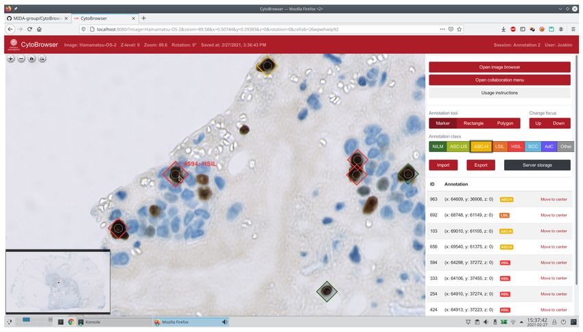
F1000Research 2021, 10:226 Last updated: 22 MAR 2021

Introduction                                                          of z-stacks, where changing focus is as easy as in a light
The acquisition and use of whole-slide images (WSIs) is rap-          microscope.
idly growing in research and medical practice1,2. The use of
                                                                      A great benefit of virtual microscopy over optical micros-
WSIs is often referred to as virtual microscopy, due to the
                                                                      copy is the possibilities for sharing and collaboration. Digitized
appealing ability to digitally recreate the microscopy experi-
                                                                      WSIs can be made accessible via the Internet, thereby ena-
ence. WSIs are produced by high-resolution scanning of entire
                                                                      bling efficient usage of distributed resources and reaching
microscopy glass slides. The resulting images are often larger
                                                                      remote parts of the world. Tools for interactive visualization and
than 100,000 × 100,000 pixels and many gigabytes in size;
                                                                      collaborative assessment of WSIs facilitate improved under-
one single image of a cytological or histological specimen
                                                                      standing as well as increased quality of analysis, where several
may contain more than 100,000 cells. The ability to inspect and
                                                                      experts may share experiences. Collaborative tools for WSI
annotate WSIs and therefrom derive results has become a cru-
                                                                      annotations are also gaining popularity in medical educa-
cial component of many bioimage analysis workflows and is
                                                                      tion; in a mixed methods trial13 it was found that collabora-
also making headway into modern clinical practice. The sheer
                                                                      tive WSI annotation provided superior understanding of key
size of WSI data, where a single image may be larger than
                                                                      microscopic features by senior medical students as compared to
the available RAM memory of a standard workstation, is prob-
                                                                      individual WSI annotation.
lematic for many standard visualization tools, imposing a need
for specialized software solutions.
                                                                      Despite the growing number of tools for WSI visualization and
                                                                      annotation, we find that there is a lack of light-weight, easy to
There is lately a strong desire within the scientific community       use OSS tools which are well suited for collaborative annotation
towards increased use of open and collaborative solutions and         of cytological data. The majority of existing tools are directed
the development of open-source software (OSS)3. A strong              towards histopathology, with different needs and requirements.
argument for the open philosophy is that it efficiently reduces       Aiming to gather the experience and knowledge of experts
the risk for vendor lock-in situations, where a customer using        from all over the globe, an easy to use, browser-based solu-
a proprietary product or service cannot easily transition to          tion, which may be directly utilized on any computer or tablet
another technology due to incompatibilities, inefficient processes,   without need for software installation, is highly desired. The
or contract constraints.                                              ability for several users to simultaneously view and comment
                                                                      the same sample improves understanding and analysis qual-
A variety of OSS project that support WSIs exist, examples            ity. The CytoBrowser software, presented here, aims to meet
include caMicroscope4, Digital Slide Archive5, Sedeen viewer6,        all of these needs and desires. It delivers easy and flexible
TissUUmaps7, Cytomine8, OMERO9, and QuPath10. The vast                access to WSIs through a browser-based interface, see Figure 1
majority of existing WSI tools are developed with histology           for an example view. CytoBrowser aims to well replicate the
in mind, whereas cytology is only recently gaining increased          optical microscope feeling, with good support for z-stacks.
attention.                                                            A growing set of annotation tools are well suited for cyto-
                                                                      logical data. Efficient collaboration is achieved through shared
One specificity of cytopathological analysis, as compared to tis-     views, with option to follow the view of another user. Annota-
sue based histopathological analysis, is the need for several         tions may be added and commented on concurrently by sev-
focus levels. Where a tissue sample provides a reasonably flat        eral users. The software, completely in JavaScript and Node.js,
surface, cells of a cytological sample are distributed in a layer     is light-weight and very easy to install and run. CytoBrowser
of a certain thickness, from a few micrometers for a liquid-          is typically hosted as a web service, but may also be used
based sample, up to several tens of micrometers for a smear.          locally. We share the source code of CytoBrowser openly
The depth of field of a high resolution light microscope is often     and freely under the GNU General Public License v3.0, with
below 1 µm, and microscopic analysis of cytological specimen          the hope that a growing community of users will jointly
typically involves adjusting focus individually for each cell.        contribute to its further improvement.
A skilled cytopathologist utilizes the limited depth of focus
as a way to separate overlapping cells and also to get a 3D           Methods
feeling of the sample. To accommodate this property in virtual        CytoBrowser is a fork and extension of TissUUmaps7 (GPLv3
microscopy, it is common to acquire several focal layers of a         license), which in turn builds on the OpenSeadragon14 (BSD-
specimen and create a so called z-stack. Several studies have         3-Clause license) web-based viewer for high-resolution zoom-
examined the importance of z-stacking for whole slide imag-           able images. Although TissUUmaps is a very capable tool
ing; Hanna et al.,11 conclude that z-stacks offer a superior          for visualization of cytology WSIs, it is developed with tis-
mechanism for overcoming focusing problems encountered                sue analysis in primary focus and the user interface and tools
with digital cytology slides, while Lu et al.,12 demonstrated         are not always ideal for cytological data. CytoBrowser instead
that even such a small deviation from the in-focus plane as           puts cytological analysis in the main focus. An important fea-
0.8 µm may have a clear negative impact on deep learn-                ture for cytological data is efficient handling of z-stacks (cur-
ing-based classification performance. Visual assessment of            rently not supported in TissUUmaps); CytoBrowser delivers
scanned samples requires easy interactive inspection at mul-          fast browsing through focus levels by use of transparent lay-
tiple resolutions and focus levels, but only very few of the          ers. CytoBrowser also allows z-level differentiated annotations,
above mentioned OSS solutions offer smooth and fast handling          useful, e.g., for annotating overlapping cells.

                                                                                                                              Page 3 of 9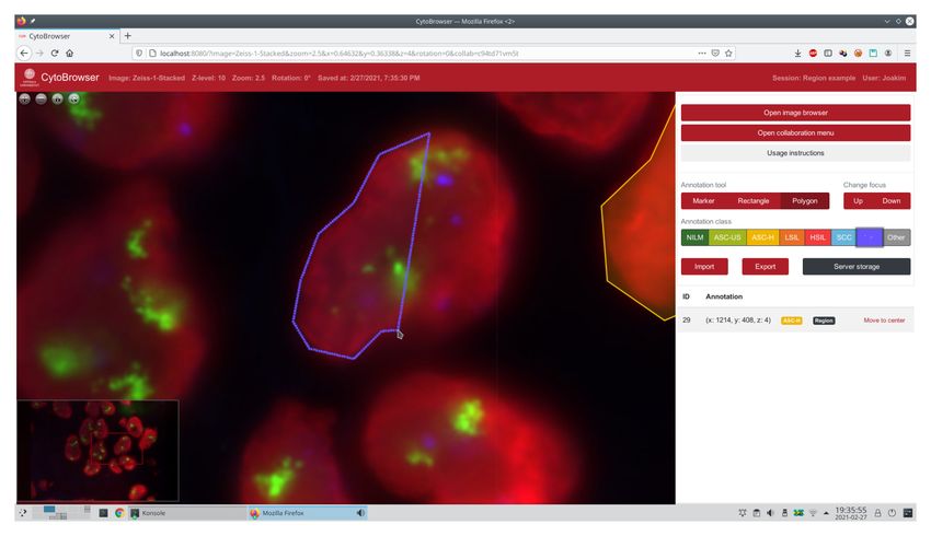
F1000Research 2021, 10:226 Last updated: 22 MAR 2021
Figure 1. Example of typical usage – annotation of a brightfield WSI in a standard web browser. The user has just placed a marker
on a cell nucleus, indicating the Bethesda grade HSIL (High-grade squamous intraepithelial lesion). An overview of the WSI is presented in
the lower left part of the screen, with the current zoomed-in viewport indicated as a small red rectangle.
The development of CytoBrowser is aimed towards ease of use an image belong to one of several annotation classes. By
by cytotechnologists and cytopathologists on a global scale. default these classes follow the Bethesda grading system16.
Implemented tools for shared multi-user sessions facilitate Classes are defined in a separate file and can easily be modified
efficient remote collaboration, including options for joint-view to suit the specific task.
visualization and collaborative annotation and assessment of
very large datasets. CytoBrowser delivers this through a web- CytoBrowser utilizes the Deep Zoom Image (DZI) format for
browser interface which is functional on any modern computer fast rendering of very large WSIs. Currently, each z-layer is
or tablet, facilitating efficient utilization of distributed resources. provided as a separate DZI file, where a filename of the form
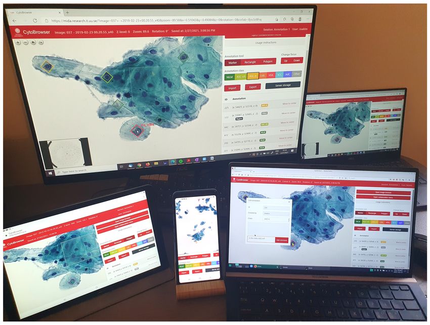
A demonstration setup of CytoBrowser running on several imagename_zNNN.dzi indicates an image with name
devices (Microsoft Edge browser under Windows, Firefox ’imagename’ at z-level ’NNN’, and where ’NNN’ is any
browser under Linux, Chrome browser on Android tablet and number, typically an offset in nanometer.
phone) is shown in Figure 2. The multi-user design of Cyto-
Browser offers concurrent access to samples, where different The backend of CytoBrowser is run in Node.js17, with a web
experts may assess, comment on, or edit each others annota- server implemented with the Express18 module. The server pro-
tions, or where an educator may, e.g., show students different vides HTTP endpoints for getting information about the avail-
medically relevant examples. able images as well as for writing and reading annotation
data in server-side storage. While these endpoints are mainly
Implementation intended to be used by the CytoBrowser client, they could
The frontend of CytoBrowser is implemented in JavaScript. also be used to easily access this information from other
The implementation makes use of the Revealing Module15 applications. The server also provides a WebSocket endpoint
design pattern to clearly separate different parts of functionality for full-duplex communication between the client and server
and to make it both easily maintainable and expandable. Open- to facilitate the collaboration functionality. This communi-
Seadragon14 is used to display a viewport in the user interface cation is done using JSON messages. The only mandatory
and to dynamically load different parts of the image from the field in a such a message is the type field, which informs the
server as needed. The annotations that the user can place on recipient how to process any other fields that may be present.
Page 4 of 9F1000Research 2021, 10:226 Last updated: 22 MAR 2021
Figure 2. Demonstration of several users collaboratively annotating a cytology WSI using different devices. The tablet and the
laptop in the foreground follow the view displayed on the larger monitor in the background, while the phone is showing a somewhat larger
view. The user of the laptop in the foreground is adding a text comment to the upper left marker, while the (followed) user has just placed
a HSIL marker on the nucleus in the lower part of the screen. CytoBrowser is compatible with all modern browsers.
When a user opens an image in CytoBrowser, they either select image. When data is altered by a connected client, only the
an existing set of annotations to work on or start a new set. alteration is broadcast to other collaborators.
Different sets of annotations for the same image may, e.g.,
originate from different experts or be imported from a machine For additional details regarding the implementation, we refer
learning-based classification. The annotation set also defines the reader to the implementation.md file of the software.
a collaboration session, consisting of all users currently
working on the same annotation set, much like users collabo- Operation
rate on the same document in web-based applications such Opening images stored on the server is easy; available images
as Google Docs19. By encoding the current state of the inter- are presented to the user as a gallery of thumbnails, see
face, including the active image and annotation set, but also Figure 3 for an example. By default these thumbnails dis-
viewport location and zoom level, in the dynamically updated play a lower-resolution version of the full image, but by hov-
URL, it becomes very easy to invite colleagues to discuss a ering over a specific entry the user is presented a zoomed-in
sample by simply sharing the active URL. In the same man- view from the middle part of the image. After selecting an
ner, it is easy to return to a previous image location through a image, the user is prompted to either start a new annotation
stored browser bookmark. session or to continue on a previously created.
The protocol implemented for collaborations has been cre- CytoBrowser enables placing several different kinds of anno-
ated with robustness in mind. The backend stores a canoni- tations on an image. Markers can be placed by clicking a sin-
cal copy of the shared data, including information about the gle point, rectangular regions can be placed by clicking two
participating users, the active image and the placed annota- opposite corners, and polygonal regions can be placed by
tions, that the clients request when entering a collaboration. The clicking each vertex, see Figure 4 for an example of the latter.
backend also verifies that clients are displaying the correct While these three annotation tools are currently present in
Page 5 of 9F1000Research 2021, 10:226 Last updated: 22 MAR 2021
Figure 3. Example of a user selecting which WSI to open, the active image (3rd row, 2nd column) is shown zoomed-in.
Figure 4. Example of a user in the process of outlining a polygonal region in a fluorescence microscopy z-stack.
Page 6 of 9F1000Research 2021, 10:226 Last updated: 22 MAR 2021
CytoBrowser, the system is designed to allow easy addition Example Bash scripts, demonstrating how to download
of new tools. and convert publicly available images in Zeiss (.zvi) and
Hamamatsu (.ndpi) format into the Deep Zoom Image (.dzi)
Once annotations are placed, there are different ways of inter- format, suitable for CytoBrowser, are provided together with
acting with them. They can be moved or deleted using the the software in the examples directory. Software packages
mouse. By right-clicking an annotation, the user gets access which may be useful for converting WSIs include libvips,
to more actions in a context menu. Here they can change the Bio-Formats, and NDPITools.
class assigned to the annotation or leave text comments on it
to provide information that is not captured by the class label Conclusions
alone. In Figure 2, the user of the laptop in the foreground is We present CytoBrowser, an OSS developed in response to
adding such a comment. the request for light-weight, open, flexible, and easy to use
tools for collaborative visualization, annotation and assessment
Three different options for saving and loading annotation of whole slide image data. CytoBrowser is developed particu-
data are provided. The easiest option is for the user to rely larly with cytological analysis on mind, where efficient han-
on the autosave functionality of CytoBrowser, where the cur- dling of z-stacks is a central requirement. The combination of
rent annotation set is continuously stored on the hosting server. a JavaScript based browser component and a Node.js driven
The second option is to manually store the data on the server. server-side component enables fast access to large image data
For this option, we also provide a simple versioning system. together with easy and flexible storage and sharing of anno-
If a file is saved to a directory that contains a file with the same tations. Being a tool particularly designed for collaborative
name, the user gets the option to either rewrite the latest ver- interactive multi-user access, it facilitates knowledge exchange
sion of the file or to create a new version of it. The user is then and alleviates interdisciplinary research.
able to choose which version of a file to load when brows-
ing the server files. The third option is to store the annotation In addition to being a much desired resource for the creation
set on the local machine of the user, using the “import” and of large annotated datasets, crucial for training and evaluation
“export” buttons in the interface. For all three options, the of machine and deep learning-based approaches to bioimage
annotations are stored as JSON files, making the data eas- analysis, as well as the visualization and qualitative evaluation
ily transferable between different tools. This enables the anno- of the results of such methods, we also envision that Cyto-
tations to be used for data selection or training of machine Browser may be a useful tool for distributed education, quality
learning methods. Inversely, CytoBrowser may be used to improvement, competency assessment, proficiency testing, and
visualize the classification or segmentation results generated by performance evaluation20.
other tools.
By delivering a powerful virtual microscope environment which
We believe that easy collaboration between experts from dif- is easily accessed through a web-browser interface and func-
ferent fields is important for an efficient workflow. For Cyto- tional on any modern computer or tablet, we hope that Cyto-
Browser, we have implemented functionality that enables such Browser, facilitating efficient utilization of distributed resources
collaboration to be performed remotely, where two or more and expert knowledge, may contribute towards fair and
users can browse and annotate the same image together in a universally accessible health-care.
shared session. Any changes made by one user will be seen by
the other users in real-time. Users can see each other’s mouse Software availability
cursors in the viewport, and they have the option to lock their • Software available from: https://mida-group.github.io/
view to follow that of another user. This may be useful, e.g., CytoBrowser/
in cases where an expert from one domain needs to visually • Source code available from: https://github.com/MIDA-
explain something about the data to an expert in another group/CytoBrowser
domain. This capability may thus enhance the transfer of knowl-
edge between different fields and simplify multidisciplinary • Archived source code at time of publication: https://doi.
research. org/10.5281/zenodo.458276021
• License: GNU General Public License v3.0
Use cases
To download and install the software, follow the instruc-
tions at https://mida-group.github.io/CytoBrowser/. It is very Acknowledgements
easy to set up a local CytoBrowser server to be accessed from We thank Leslie Solorzano and Prof. Carolina Wählby for their
the same machine (localhost), or, e.g., through an SSH tun- support in forking TissUUmaps.
nel. For larger and more persistent use cases, we recommend
to rely on one of popular open-source web servers, such as This publication was supported by COST Action NEUBIAS
Apache and Nginx, as a secure loadbalancing frontend for the (CA15124), funded by COST (European Cooperation in Science
server. and Technology).
Page 7 of 9F1000Research 2021, 10:226 Last updated: 22 MAR 2021
References
1. Aeffner F, Zarella MD, Buchbinder N, et al.: Introduction to digital image digital-slide modalities for cytopathology screening and interpretation.
analysis in whole-slide imaging: a white paper from the digital pathology Cancer Cytopathol. 2017; 125(9): 701–709.
association. J Pathol Inform. 2019; 10: 9. PubMed Abstract | Publisher Full Text
PubMed Abstract | Publisher Full Text | Free Full Text 12. Lu J, Sladoje N, Stark CR, et al.: A deep learning based pipeline for efficient
2. Kumar N, Gupta R, Gupta S: Whole slide imaging (WSI) in pathology: current oral cancer screening on whole slide images. In International Conference on
perspectives and future directions. J Digit Imaging. 2020; 33(4): 1034–1040. Image Analysis and Recognition. 2020; 249–261. Springer.
PubMed Abstract | Publisher Full Text | Free Full Text Publisher Full Text
3. Marée R: Open practices and resources for collaborative digital pathology. 13. Sahota M, Leung B, Dowdell S, et al.: Learning pathology using collaborative
Front Med (Lausanne). 2019; 6: 255. vs. individual annotation of whole slide images: a mixed methods trial.
PubMed Abstract | Publisher Full Text | Free Full Text BMC Med Educ. 2016; 16(1): 311.
4. Saltz J, Sharma A, Iyer G, et al.: A containerized software system for PubMed Abstract | Publisher Full Text | Free Full Text
generation, management, and exploration of features from whole slide 14. OpenSeadragon: An open-source, web-based viewer for high-resolution
tissue images. Cancer Res. 2017; 77(21): e79–e82. zoomable images, implemented in pure JavaScript, for desktop and
PubMed Abstract | Publisher Full Text | Free Full Text mobile. Accessed on: Feb. 2021.
5. Gutman DA, Khalilia M, Lee S, et al.: The digital slide archive: A software Reference Source
platform for management, integration, and analysis of histology for 15. The revealing module pattern in javascript. Accessed on: Feb. 2021.
cancer research. Cancer Res. 2017; 77(21): e75–e78. Reference Source
PubMed Abstract | Publisher Full Text | Free Full Text 16. Nayar R, Wilbur DC: The Bethesda system for reporting cervical cytology:
6. Martel AL, Hosseinzadeh D, Senaras C, et al.: An image analysis resource definitions, criteria, and explanatory notes. Springer, 2015.
for cancer research: Piip—pathology image informatics platform for Reference Source
visualization, analysis, and management. Cancer Res. 2017; 77(21): 17. Node.js: A javascript runtime built on Chrome’s V8 JavaScript engine.
e83–e86. Accessed on: Feb. 2021.
PubMed Abstract | Publisher Full Text | Free Full Text Reference Source
7. Solorzano L, Partel G, Wählby C: TissUUmaps: Interactive visualization 18. Express: Fast, unopinionated, minimalist web framework for Node.js.
of large-scale spatial gene expression and tissue morphology data. Accessed on: Feb. 2021.
Bioinformatics. 2020; 36(15): 4363–4365. Reference Source
PubMed Abstract | Publisher Full Text | Free Full Text
19. Announcing Google Workspace, everything you need to get it done, in one
8. Marée R, Rollus L, Stévens B, et al.: Collaborative analysis of multi-gigapixel location. Google Cloud Blog. Accessed on: Feb. 2021.
imaging data using cytomine. Bioinformatics. 2016; 32(9): 1395–1401. Reference Source
PubMed Abstract | Publisher Full Text | Free Full Text
20. Quinn AM, Minhajuddin AT, Hynan LS, et al.: Agreement between
9. Allan C, Burel JM, Moore J, et al.: OMERO: flexible, model-driven data cytotechnologists and cytopathologists as a new measure of
management for experimental biology. Nat Methods. 2012; 9(3): 245–253. cytopathologist performance in gynecologic cytology. Cancer Cytopathol.
PubMed Abstract | Publisher Full Text | Free Full Text 2017; 125(7): 576–580.
10. Bankhead P, Loughrey MB, Fernández JA, et al.: QuPath: open source software PubMed Abstract | Publisher Full Text
for digital pathology image analysis. Sci Rep. 2017; 7(1): 16878. 21. Rydell C, Lindblad J: MIDA-group/CytoBrowser: Bugfix release v1.0.1, URL
PubMed Abstract | Publisher Full Text | Free Full Text location encoding (Version v1.0.1). Zenodo.
11. Hanna MG, Monaco SE, Cuda J, et al.: Comparison of glass slides and various http://www.doi.org/10.5281/zenodo.4582760
Page 8 of 9F1000Research 2021, 10:226 Last updated: 22 MAR 2021
The benefits of publishing with F1000Research:
• Your article is published within days, with no editorial bias
• You can publish traditional articles, null/negative results, case reports, data notes and more
• The peer review process is transparent and collaborative
• Your article is indexed in PubMed after passing peer review
• Dedicated customer support at every stage
For pre-submission enquiries, contact research@f1000.com
Page 9 of 9You can also read