Cytomorphological Parameters of Hashimoto Thyroiditis and Its Clinical Correlation - A Prospective Six Years Study in a Tertiary Care Hospital of ...
←
→
Page content transcription
If your browser does not render page correctly, please read the page content below
IOSR Journal of Dental and Medical Sciences (IOSR-JDMS)
e-ISSN: 2279-0853, p-ISSN: 2279-0861.Volume 18, Issue 6 Ser. 2 (June. 2019), PP 32-37
www.iosrjournals.org

 Cytomorphological Parameters of Hashimoto Thyroiditis and Its
    Clinical Correlation – A Prospective Six Years Study in a
     Tertiary Care Hospital of Western Uttar Pradesh, India
1
    Dr Rajnish Kumar, Associate Professor, 2Dr Alok Mohan, Professor, 3Dr Purva
      Sharma, Assistant professor, 4Dr Pradeep Kr Sharma, Assistant Professor,
    5
      Dr Roopanshi Mittal, Junior resident, 6Dr Vaseem Ansari, Junior resident
     1, 2, 4, 5,6
             (Department of Pathology, Muzaffarnagar Medical College, Muzaffarnagar Uttar Pradesh,India)
         3
          (Department of Pathology, Mulayam Singh Yadav Medical College, Meerut, Uttar Pradesh, India)
                                     Corresponding Author: Dr Alok Mohan

Abstract: Background: Among non-neoplastic lesions of thyroid, Hashimoto thyroiditis is the most common
thyroid lesion after goiter. It is an autoimmune disorder characterized by destruction of thyroid follicles by
cellular as well as humoral immunity.It has an incidence of 3-6/10000 population per year and prevalence rate
of 1-4%. It is more common in 3rd to 4th decade of life with female sex predilection. There is rising trend of
Hashimoto thyroiditis either due to dietary iodine supplementation or due to improved diagnostic modalities or
both.
Methods: All patients presented with anterior neck swelling moving with deglutition were selected in the study.
Total 287 patients diagnosed as Hashimoto thyroiditis on cytology in six years from May 2013 to April 2019
were prospectively included. Patients with Hashimoto thyroiditis were categorized into three grades
cytologically using Bharti et al grading system.
Results: Majority of the patients were females in 3r to 5 th decade of life. Majority of patients showed diffuse
neck swelling and hypothyroidism. Grade II thyroiditis was seen in maximum number of cases. Plasma cells
were very helpful in diagnosing Hashimoto thyroiditis where lymphocytic infiltration was insignificant.
Conclusions: Clinical presentation as well as cytomorphological features varies depending on the stage of the
disease. Lymphocyte impingement of thyroid follicular cells is must for diagnosis of Hashimoto thyroiditis but
careful and diligent evaluation of cytomorphological features along with clinical presentation and thyroid
hormonal assay are essential for diagnosis of this entity.
Keywords: Hashimoto thyroiditis, chronic lymphocytic thyroiditis, Fine needle aspiration cytology, thyroid
function test
----------------------------------------------------------------------------------------------------------------------------- ----------
Date of Submission: 21-05-2019                                                                            Date of acceptance: 06-06-2019
---------------------------------------------------------------------------------------------------------------------------------------------------

                                                            I. Introduction
          Hashimoto thyroiditis (HT) is an autoimmune disease described by Hakaru Hashimoto in 1912 and
hence so named.[1] Hashimoto thyroiditis has an incidence of 3-6/10000 population per year and prevalence
rate of 1-4%.[2] Fine needle aspiration cytology (FNAC) is highly sensitive in diagnosing Hashimoto thyroiditis
with 92% of diagnostic accuracy.[3,4] Among non neoplastic conditions of thyroid, HT is the most common
autoimmune thyroiditis after goitre. It is characterised by increased number of mature and transformed
lymphocytes impinging on follicular cells and Hurthle cell change.[5] Number of patients with Hashimoto’s
thyroiditis seems to be increasing and this rising trend has been linked to excess iodine intake.[6]

                                                     II. Material And Methods
Study Design: Prospective hospital based study
Study Location: This study was done in Department of Pathology in a tertiary care teaching hospital in western
Uttar Pradesh, India.
Study Duration: Six years (May 2013 to April 2019)
Sample size: 287 patients
Inclusion criteria: All the cases of thyroid FNAC diagnosed as Hashimoto thyroiditis in between the specified
period of time interval.
Exclusion criteria: Smears diluted with excessive blood, known case of Hashimoto thyroiditis and subjects
taking thyroxine or any other drug interfering with thyroid function.
DOI: 10.9790/0853-1806023237                                          www.iosrjournals.org                                            32 | Page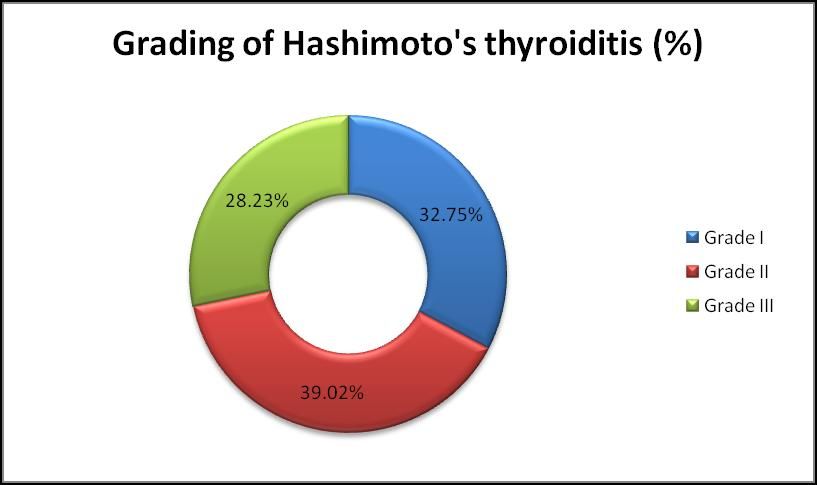
Cytomorphological parameters of Hashimoto thyroiditis and its clinical correlation – A prospective

Procedure methodology:
          Ethical committee clearance from the college was taken for the study. FNAC procedure was performed
on an outpatient basis after informed consent. For FNAC, 23 gauze needle was used for thyroid swelling. The
material thus obtained was spread over a clean glass slides. Smears were prepared and fixed by two methods –
one air dried fixed in methanol and stained with May Grunwald Giemsa (MGG) stain and the other one alcohol
fixed and stained with Papanicolaou(Pap)
          Smears with were categorized into three grades using Bhatia et al grading system as follows[7]. Grade I
[Mild]: Few lymphoid cells infiltrating the follicles/increased number of lymphocytes in the background. Grade
II [Moderate]: Moderate lymphocytic infiltration/ mild lymphocytic infiltration with Hurthle cell change/giant
cells/anisonucleosis. Grade III [Severe]: Florid lymphocytic inflammation with germinal centre formation, very
few follicular cells left.

                                                         III. Result
        Out of 1046 cases of thyroid FNAC performed in the last six years, 287 cases (27.43%) were
diagnosed as Hashimoto thyroiditis on cytology. There were increasing cases of cytologically proven cases of
Hashimoto thyroiditis from 2013 (13.79%) to 2019 (41.21%). (Chart 1)

              Chart 1. Line diagram showing rising trend of Hashimoto thyroiditis (n=287 cases)

         The patients studied were categorized into 2nd to 7th decade of life with predilection of this disease
from 3rd to 5th deacade of life (74.22% cases). Maximum number of cases were noted in 4th decade (n=84,
29.27%) followed by 3rd decade (n=73, 25.44%). No case was seen in 1 st decade and after 7th decade of life. In
our study female patients outumbered male patients with a ratio of female to male was 6.17:1. (Table 01)
         On local examination, patient presented with anterior neck swelling moving with deglutition. Swelling
was diffuse in 191 patients (66.55%), multinodular in 62 patients (21.60%) and solitary nodule in 34 patients
(11.85%). (Table 01)

       TABLE 01 Age, Sex distribution and Clinical presentation of 287 cases of Hashimoto Thyroiditis
    Age in years   Total cases of     Sex                                        Local examination
                   Hashimoto          Female                  Male                                    Nodularity
                   thyroiditis                                                   Diffuse
                   No. of cases (%)   No. of cases (%)        No. of cases (%)             Multinodular      Solitary
    10-19          24                 22                      02                 16        05                03
                   (8.36%)            (7.67%)                 (0.69%)
    20-29          73                 60                      13                 52        20                01
                   (25.44%)           (20.91%)                (4.53%)
    30-39          84                 68                      16                 56        19                09
                   (29.27%)           (23.69%)                (5.58%)
    40-49          56                 53                      03                 37        12                07
                   (19.51%)           (18.46%)                (1.05%)
    50-59          31                 27                      04                 18        05                08
                   (10.80%)           (9.40%)                 (1.40%)
    60-69          19                 17                      02                 12        01                06
                   (6.62%)            (5.92%)                 (0.70%)
    Total          287                247                     40                 191       62                34
                   (100%)             (86.06%)                (13.94%)           (66.55)   (21.60%)          (11.85%)

DOI: 10.9790/0853-1806023237                                www.iosrjournals.org                                   33 | Page
Cytomorphological parameters of Hashimoto thyroiditis and its clinical correlation – A prospective
Out of 287 cases, 208 patients (72.47%) got their thyroid function tests done. (Chart 2)
Chart 2. Thyroid Hormone Evaluation of Hashimoto thyroiditis (n=208 cases)
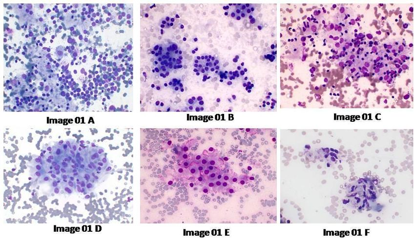
Smears showed thyroid follicular cells in form of clusters, sheets and microfollicular pattern as well as
scattered singly. Lymphoid cells comprising of either mature lymphocytes or reactive lymphoid cells were seen
in all cases of Hashimoto thyroiditis. Lymphoid cells were seen impinging on thyroid follicular cells in fair
number of smears and in the background in some.[Image 01A, 01B & 01C]
Out of total 287 cases, smears showed plasma cells in 224 cases (78%), Hurthle cell change in 100
cases(34.84%), and eosinophils in 37 cases (12.89%).[Image 01 B]
Image 01A: Reactive lymphoid cells and scattered follicular cells.(MGG x 400) Image 01B: Follicular cells in
clusters and forming microfollicular pattern, scattered plasma cells and lymphocytes.(MGG x400) Image 01C:
Poorly cohesive Hurthle cells, lymphocytes adherent to them and epithelioid cells. (MGGx400) Image 01D:
Epithelioid cells forming giant cells.(MGGx400) Image 01E: Hurthle cells with fire flares.(MGGx400) Image
1F: Fibroblasts in advanced stage of Hashimoto thyroiditis (MGGx400)
DOI: 10.9790/0853-1806023237 www.iosrjournals.org 34 | PageCytomorphological parameters of Hashimoto thyroiditis and its clinical correlation – A prospective
Maximum number of cases (n=198, 68.98%) did not show any colloid and revealed colloid in 89 cases
(31.01%) in the present study. Epithelioid cells and giant cells were seen in 132cases (46%) and 46 cases
(16.03%) of cytologically diagnosed Hashimoto’s thyroiditis.[Image01C & 01D]
In the present study, fire flares (marginal vacuoles) were seen in 63 cases (21.95%). [Image 01E]
In our study, smears were moderately cellular in 170 cases (59.24%) and richly cellular in 72 cases (25.08%).
Smears were sparsely cellular in 45 cases (15.68%). Fibroblasts were seen in majority of smears having sparse
cellularity. [Image 01F]
Table 02 Cytomorphological features of Hashimoto’s thyroiditis (n=287)
Cytomorphological
Number of cases
features
Richly cellular 72 (25.08%)
Moderately cellular 170 (59.24%)
Sparsely cellular 45 (15.68%)
Plasma cells 224 (78%)
Hurthle cells 100 (34.84%)
Eosinophils 37 (12.89%)
Epithelioid cells 132 (46%)
Giant cells 46 (16.03%)
Fire flares 63 (21.95%)
89 (
Colloid
31.01%)
Chart 3 Grading of Hashimoto’s thyroiditis (n=287 cases)
Cytomorphological features of all snears were studied and graded accordingly. One hundred and
twelve patients (39.02%) were graded as Grade II thyroiditis. Ninety four patients (32.75%) had grade I
thyroiditis while grade III thyroiditis were seen in 81 patients (28.23%). ( Chart 3)
IV. Discussion
Hashimotos thyroiditis or chronic lymphocytic thyroiditis is an autoimmune non-neoplastic lesion
characterised by destruction of thyroid follicles by cell mediated as well as antibody mediated immune
processes. Cytomorphological features include varying number of follicular cells, lymphocytic infiltration,
Hurthle cells, fibroblasts and colloid depending on the stage of the disease. Three main morphologic types
corresponding to different stages of the disease namely juvenile, hypertrophic and fibrous lymphocytic
thyroiditis were identified.[8]
There was a rising trend being 13.79% (2013) to 41.21% (2019). Benvenge et al showed in their study
that Hashimoto thyroiditis increased to 10 times than it was until early 1990.[9] The cause of this rising trend
remains uncertain. Whether it is because of high iodine intake mainly in coastal areas.[4,6]
DOI: 10.9790/0853-1806023237 www.iosrjournals.org 35 | PageCytomorphological parameters of Hashimoto thyroiditis and its clinical correlation – A prospective
Majority of the patients (74.22%) were seen in 3 rd decade to 5th decade. Bhatia et al and Sood et al also
showed same results.[7,10] In my study female to male ratio was 6.17:1. In the previous study conducted by
Rathi M et al and Pachrupe A et al female preponderance was observed.[11,12]
On local examination, thyroid swelling was diffuse in 66.55% in the present study. Rathi M et al also
showed similar result in their study (68%).[4,11] In a study conducted by Friedman M et al, anterior neck
swelling was nodular in 80% cases but nodular presentation (solitary as well as multinodular) was only in
33.45% cases in our study.[14]
In the present study hypothyroidism was seen in 71.63% of patients. Similar results (62.78%) were
also seen in a study conducted by Uma P et al.[13]
In our study, smears were moderately cellular in 170 cases (59.24%) and richly cellular in 72 cases
(25.08%). Abundant cellularity obtained may correspond to an early stage as opposed to a later fibrotic
lymphocytic thyroiditis. Smears were sparsely cellular in 45 cases (15.68%). In late stage of Hashimoto
thyroiditis also known as fibrous lymphocytic thyroiditis, cytological smears are sparsely cellular comprising of
fibroblasts, lymphocytes, follicular cells and Hurthle cells on cytological examination.[8,14]
Lymphoid cells infiltrate is an essential but not the sole cytological feature for diagnosis of this entity
and lymphoid cells were seen in all cases of Hashimoto thyroiditis in the present study. Lymphoid infiltrate can
also be seen in Graves disease, papillary thyroid carcinoma and lymphoma. Presence of other associated
cytological features like Hurthle cells, epithelioid cells and giant cells etc along with clinical correlation play a
significant role for definitive diagnosis. Absence of monotonous lymphoid cell population and polyclonal nature
of the infiltrate can distinguish this entity from Non-Hodgkin lymphoma.[7,12,15]
In the present study grade II thyroiditis were seen in majority of cases (39.02%) . Jayaram et al 1987
and Bhatia et al also showed mainly grade II thyroiditis in 62.16% and 44% cases respectively while Kumar et
al showed grade I thyroiditis in majority of cases (61.09%).[2,5,7]
Plasma cells were seen in 78% of our study. Thomas et al also observed similar results (75%) in their
study. In diagnosing early stage of Hashimotos thyroiditis plasma cells are useful where lymphocytic infiltration
of follicles are insignificant.[16]
Present study showed eosinophils in smears in 12.89% of cases. But peripheral eosinophilia should be
ruled out before making decision that increased number of eosinophils are associated with Hashimoto
thyroiditis. Ekambaram et al and Rathi M et al also showed higher association of eosinophils in cases of
Hashimoto’s thyroiditis.[6,11]
Mohan A et al observed that in their study of 154 cases of Hurthle cell containing lesions of thyroid, 94
cases (61.04%) were cytologically diagnosed as Hashimoto thyroiditis.[17] In the present study, Hashimoto
thyroiditis having Hurthle cells was noted in 100 cases (34.84%). While Rathi et al showed Hurthle cell
change in 74% cases of Hashimoto’s thyroiditis.[11] Hurthle cells in Hurthle cell neoplasm in contrast to
Hashimoto’s thyroiditis show more uniform appearance, more dyshesive pattern, more prominent nucleoli and
usually lack of lymphocytic inflammatory component.[15]
Absent or scant colloid is an usual feature of Hashimoto thyroiditis as seen in our study but presence of
colloid paradoxically is not unusual as there may be a combined autoimmunity and iodine supplementation.
[2,16]
Marginal vacuoles were seen in 21.95% cases in our study. These are cytoplasmic signs of
hyperstimulation which were seen in non-neoplastic conditions. Mohan etal showed fire flares in 19.15% of
cases.[17]
In the present study, epithelioid cells and giant cells were seen in 46% and 16.03% cases.
Multinucleated giant cells and epithelioid cells can be seen in 40% of cases.[5] These cases need to be
differentiated from granulomatous thyroiditis. The inflammatory infiltrate in granulomatous thyroiditis is mixed
not uniformly lymphocytic and multinucleated giant cells are seen surrounding leaked colloid and epithelioid
cells[18]. In overlapping cases, thyroid hormone evaluation and antibodies are must to diagnosis.[19]
V. Conclusion
FNAC continues to be of significance in diagnosing Hashimoto thyroiditis especially in developing
countries. Correct diagnosis can be achieved in majority of cases by combination of clinical examination,
thyroid function test and cytological features obviating need for unnecessary surgical intervention.
References
[1]. Takami HE, Miyabe R, Kameyama K. Hashimoto’s thyroiditis. World J Surg 2008;32:688-692.
[2]. Kumar N, ray C, Jain S. aspiration cytology of Hashimoto thyroiditis in an endemic area. Cytopathol 2002;13:31-39
[3]. Kocjan G.Lymphoid infiltrate. In:Schroder G, editor.Fine needle aspiration cytology : diagnostic principles and dilemmas. 1 st ed
Germany: Springer;2006.p 99-101.
[4]. Chandanwale SS, Gore CR, Bamanikar SA, Gupta N, Gupta K. Cytomorphologic spectrum of Hashimoto’s thyroiditis and its
clinical correlation: A retrospective study of 52 patients. CytoJournal 2014 dec 9;11(9).
DOI: 10.9790/0853-1806023237 www.iosrjournals.org 36 | PageCytomorphological parameters of Hashimoto thyroiditis and its clinical correlation – A prospective
[5]. Jayaram G, Marwaha RK, Gupta RK, Sharma SK. Cytomorphologic aspects of thyroiditis. Acta Cytol 1987;31: 687-93
[6]. Ekambaram M , Kumar B , Chowdhary N , Siddaraju N , Kumar S.Significance of eosinophils in diagnosing Hashimoto’s
thyroiditis on fine needle aspiration cytology.Indian J Pathol Microbiol 2010 ; 53:476-9.
[7]. Bhatia A, Rajwanshi A, dash RJ, Mittal BR, Saxena A. Lymphocytic thyroiditis –is cytological grading significant? A correlation of
grades with clinical, biochemical, ultrasonographic and radionuclide parameters. CytoJournal 2007 april 30;4:10.
[8]. Doniach I.The thyroid gland. In : Symmers WStC (ed) Systemic Pathology. 2 nd edition vol 4. Edinburgh: churchill livingstone;
1978: 1975-2037.
[9]. Gayathri BN, Kumar MLH, Kalyani R, Prasad KK.Fine needle aspiration cytology of Hashimoto’s thyroiditis- A diagnostic pitfall
with review of literature. Journal of cytology 2011;28(4):210-3.
[10]. Sood N, Nigam JS. Correalation of fine needle aspiration cytology fidings with thyroid function test in cases of lymphocytic
thyroiditis.Journal of Thyroid Research;2014:article ID430510, 5 pages.
[11]. Rathi M, Ahmad F, Budania SK, Awasthi S, Kumar A, Dutta S. Cytomorphological aspects of Hashimoto’s thyroiditis: our
experience at a tertiary centre. Clin Med Insights Pathol.2014;7:1-5.
[12]. Pachrupe AP, Dhume VM, Kavishvar V, Varthakavi P. Cytomophological study of chronic lymphocytic thyroiditis. Indian Journal
of Pathology: Research and Practice 2018 May 5; 7(5)579-84.
[13]. Uma P, Kartheek B.V.S., Himaja S, Lekha JC, Babu AK, Lakshmi B. Lymphocytic thyroiditis: a correlation of cytological grades
with clinical, biochemical and ultrasound findings. Int J Res Med Sci 2013 nov;1(4):523-31
[14]. Friedman M, Shimaoka K, Rao U, Tsukada K, Gavigan M, Tamura K. Diagnosis of lymphocytic thyroiditis (nodular presentation)
by needle aspiration. Acta Cytol 1981;25:5:13-22.
[15]. Bibbo M, Wilbur DC. Comprehensive Cytopathology. 4 th ed.Elsevier Saunders;518-20.
[16]. Thomas T, Sreedharan S, Khadilkar UN, Deviprasad D, Kamath MP, Bhojwani KM, Alva A. Clinical, biochemical &
cytomorphologic study on Hashimoto’s thyoiditis. Indian J Med Res. 2014 Dec; 140(6):729-35.
[17]. Mohan A, Kumar R, Sharma P, Mishra P, Sharma PK.Oncocytic lesions in thyroid: A prospective cytomorphological study in uttar
Pradesh India. Indian Journal of Pathology: Research and Practice 2018 May 5; 7(5)595-601.
[18]. Persson PS. Cytodiagnosis of thyroiditis. A comparative study of cytological, histological, immunological and clinical findings in
thyroiditis. Acta Med Scand 1968;483(Suppl):7-100
[19]. Bhalotra R, Jayaram G. Overlapping morphology in thyroiditis (Hashimoto’s and subacute) and Grave’s disease. Cytopathology
1990;1:371-2.
Dr Rajnish Kumar. “Cytomorphological parameters of Hashimoto thyroiditis and its clinical
correlation – A prospective six years study in a tertiary care hospital of western Uttar
Pradesh, India.” IOSR Journal of Dental and Medical Sciences (IOSR-JDMS), vol. 18, no. 6,
2019, pp 32-37.
DOI: 10.9790/0853-1806023237 www.iosrjournals.org 37 | PageYou can also read