De Novo' Brain AVMs-Hypotheses for Development and a Systematic Review of Reported Cases - MDPI
←
→
Page content transcription
If your browser does not render page correctly, please read the page content below
Review

‘De Novo’ Brain AVMs—Hypotheses for Development and a
Systematic Review of Reported Cases
Ioan Alexandru Florian 1,2,*,†, Lehel Beni 1,†, Vlad Moisoiu 1,†, Teodora Larisa Timis 3,†, Ioan Stefan Florian 1,2,
Adrian Balașa 4,5 and Ioana Berindan-Neagoe 6

                                           1 Clinic of Neurosurgery, Cluj County Emergency Clinical Hospital, 400012 Cluj-Napoca, Romania;
                                             lehelbeni@yahoo.com (L.B.); vlad.moisoiu@gmail.com (V.M.); stefanfloriannch@gmail.com (I.S.F.)
                                           2 Department of Neurosurgery, Iuliu Hatieganu University of Medicine and Pharmacy, 400012 Cluj-Napoca,

                                             Romania
                                           3 Department of Physiology, Iuliu Hatieganu University of Medicine and Pharmacy, 400006 Cluj-Napoca,

                                             Romania; doratimis@gmail.com
                                           4 Clinic of Neurosurgery, Tîrgu Mureș County Clinical Emergency Hospital, 540136 Tîrgu Mureș, Romania;

                                             adrian.balasa@yahoo.fr
                                           5 Department of Neurosurgery, Tîrgu Mureș University of Medicine, Pharmacy, Science and Technology,

                                             540139 Tîrgu Mureș, Romania
                                           6 Research Center for Functional Genomics, Biomedicine, and Translational Medicine, Institute of Doctoral

                                             Studies, “Iuliu Hatieganu” University of Medicine and Pharmacy, 400012 Cluj-Napoca, Romania;
                                             ioananeagoe29@gmail.com
                                           * Correspondence: florian.ioan.alexandru@gmail.com
                                           † Equal contributions as first author.

                                           Abstract: Background and Objectives: Brain arteriovenous malformations AVMs have been consist-
                                           ently regarded as congenital malformations of the cerebral vasculature. However, recent case re-
                                           ports describing “de novo AVMs” have sparked a growing debate on the nature of these lesions.
Citation: Florian, I.A.; Beni, L.;
                                           Materials and Methods: We have performed a systematic review of the literature concerning de novo
Moisoiu, V.; Timis, T.L.; Florian, I.S.;
Balasa, A.; Berindan-Neagoe, I. ‘De
                                           AVMs utilizing the PubMed and Google Academic databases. Termes used in the search were
Novo’ Brain AVMs—Hypotheses for            “AVM,” “arteriovenous,” “de novo,” and “acquired,” in all possible combinations. Results: 53 arti-
Development and a Systematic               cles including a total of 58 patients harboring allegedly acquired AVMs were identified by research-
Review of Reported Cases. Medicina         ing the literature. Of these, 32 were male (55.17%), and 25 were female (43.10%). Mean age at de
2021, 57, 201.                             novo AVM diagnosis was 27.833 years (standard deviation (SD) of 21.215 years and a 95% confi-
https://doi.org/10.3390/medic-             dence interval (CI) of 22.3 to 33.3). Most de novo AVMs were managed via microsurgical resection
ina57030201                                (20 out of 58, 34.48%), followed by radiosurgery and conservative treatment for 11 patients (18.97%)
                                           each, endovascular embolization combined with resection for five patients (8.62%), and emboliza-
Academic Editor: Stavros J. Balo-
                                           tion alone for three (5.17%), the remaining eight cases (13.79%) having an unspecified therapy. Con-
yannis
                                           clusions: Increasing evidence suggests that some of the AVMs discovered develop some time after
                                           birth. We are still a long way from finally elucidating their true nature, though there is reason to
Received: 16 January 2021
                                           believe that they can also appear after birth. Thus, we reason that the de novo AVMs are the result
Accepted: 22 February 2021
Published: 26 February 2021
                                           of a ‘second hit’ of a variable type, such as a previous intracranial hemorrhage or vascular pathol-
                                           ogy. The congenital or acquired characteristic of AVMs may have a tremendous impact on progno-
Publisher’s Note: MDPI stays neu-          sis, risk of hemorrhage, and short and long-term management.
tral with regard to jurisdictional
claims in published maps and insti-        Keywords: arteriovenous malformation; de novo; acquired; seizure; hemorrhage
tutional affiliations.

                                           1. Introduction
Copyright: © 2021 by the authors. Li-
                                                Arteriovenous malformations (AVMs) of the brain consist of feeder arteries that
censee MDPI, Basel, Switzerland.
                                           amalgamate and form a nidus, shunting the oxygenated blood directly into the venous
This article is an open access article
                                           system without an interposing capillary network [1–4]. The most common and undenia-
distributed under the terms and con-
                                           bly the most severe clinical presentation is intracranial hemorrhage, AVMs representing

Medicina 2021, 57, 201. https://doi.org/10.3390/medicina57030201                                                    www.mdpi.com/journal/medicina
Medicina 2021, 57, 201 2 of 23
ditions of the Creative Commons At- an important source of mortality and morbidity especially in the younger population. Ep-
tribution (CC BY) license (http://crea- ilepsy, progressive neurological deficit, and chronic headache are less frequently encoun-
tivecommons.org/licenses/by/4.0/). tered in patients harboring this pathology. Asymptomatic patients may benefit from sim-
ple clinical and imagistic observation, whereas those with minimal symptoms can be man-
aged conservatively. Currently the most effective and permanent treatment method or
ruptured or symptomatic AVMs consists of microsurgical resection of the nidus and rig-
orous hemostasis, but radiosurgery and endovascular embolization are also viable man-
agement options.
AVMs are traditionally considered congenital, an estimated 95% of these lesions be-
ing sporadic in nature, whereas the remaining 5% are familial and most often associated
with hereditary hemorrhagic telangiectasia (HHT) [4]. The congenital aspect derives from
the observation that these lesions may occur at any age, from infants and children to adults
and elderly alike, even though there is a scarcity of conclusive data to demonstrate this
fact [5–7].
Our current grasp of cerebral AVMs has dramatically improved over the last few
decades, by delving into the genetic and epigenetic intricacies of their pathogenesis [8–
10]. Data concerning the molecular underlying of AVMs come from AVM syndromes as
well as from the analysis of sporadic cases of AVMs. For instance, genetic events involving
members of the transforming growth factor-β (TGF-β) signaling, namely Endoglin (ENG)
and Activin A receptor type I (ACVR1 or ALK1) are causative in HHT type 1 and type 2,
respectively [11,12]. Mutations in SMAD4, which is a downstream effector of TGF-β,
causes combined syndrome of HHT with juvenile polyposis [13]. In agreement with such
an interpretation, conditional knock out of either ENG or ALK1 resulted in arteriovenous
malformations of the skin in mice only after a secondary injury, for example wounding
[14,15]. Moreover, vascular endothelial growth factor (VEGF) has the ability to mimic this
wounding effect on skin AVM development, at least in animal models [16]. In the case of
the more common sporadically occurring AVMs, somatic KRAS mutations were discov-
ered in approximately 55% of brain AVM specimens, also linked to a lower patient age,
and a larger nidus mean size [17–19], whereas the non-coding polymorphism in ALK1
(ALK1 IVS3-35A>G) was established as a genetic risk factor in two independent studies
[20,21]. Other polymorphisms linked to sporadic AVMs include polymorphisms in ENG
[20], interleukin-1β (IL1B) [22] and VEGF [23]. Even so, we are currently unable to effec-
tively validate whether these malformations actually develop during embryogenesis and
evolve during one’s lifetime, or if they appear at a later point in time when certain micro-
environmental conditions are met.
There is a mounting number of pertinent case reports detailing AVMs that occurred
sporadically after credible evidence of their initial absence. Either adequate imaging stud-
ies or intraoperative examinations preceded the certain appearance of these vascular mal-
formations, which were then discovered at a variable interval. A diversity of preexisting
circumstances or pathologies either influenced or initiated the development of AVMs in
already susceptible patients. Ischemic stroke and transient ischemic attacks (TIA) were
among the most common ailments preceding de novo AVMs [24], as were seizures [25].
Certain individuals previously harbored another type of intracranial vascular pathology,
or even a differently located AVM, highlighting the irrefutable predisposition for such an
occurrence [26]. Intracranial tumors with a highly vascular component have also been de-
scribed as potential triggers for previously absent AVMs [27]. An earlier hemorrhagic ep-
isode [28] and even head trauma of variable severity have also been reported as generators
for de novo AVMs [29].
Notwithstanding the fact that cerebral AVMs are a rare pathology, they are cited as
the most common cause of intracranial hemorrhage in children and young adults, leading
to a lifelong threat of death or disability [2–4,30,31]. As such, elucidating their true natural
history is crucial in order to establish the best possible course of diagnosis and manage-
ment, and help lessen their socio-economic encumberment. In this systematic review weMedicina 2021, 57, 201 3 of 23
have included, to the best of our knowledge, all such incidences reported in the available
and relevant literature.
2. Material and Methods
We performed a rigorous examination of the PubMed and Google Academic medical
databases utilizing the terms “AVM”, “arteriovenous malformation”, “brain”, “cerebral”,
“intracranial”, “acquired”, and “de novo”, and all of their possible combinations. Our
search only identified articles in English, and we excluded animal studies from the sys-
tematic review. Other systematic reviews were also discarded. Afterwards, we methodi-
cally assessed the references of the manuscripts gathered for any additional articles sig-
nificant to our research. Among acquired AVMs that appeared after a previous pathology
or injury, we have also included AVMs that recurred at a different site after a previous
angiographically-demonstrated occlusion. We paid special attention not to include data
from overlapping series that were published at a different time or in another journal. Nev-
ertheless, certain redundancies cannot be completely dismissed.
Microsoft® Excel for Mac, version 16.37, was used to create a spreadsheet wherein all
relevant cases were gathered in order to create our database. The same program was uti-
lized to perform descriptive statistics, as well as to create the appropriate graphs.
3. Results
3.1. Literature Data
As of June 2020, our search yielded 218 results on PubMed. After careful considera-
tion of titles and abstracts, we have eliminated 163 articles which were not relevant to the
subject at hand, followed by the exclusion of an additional 16 that were either in vitro or
animal studies, other systematic reviews, case reports detailing other vascular pathologies
such as arteriovenous fistulas, aneurysms, cavernomas, or AVMs outside of the cranial
cavity. Among these, two articles which represented correspondence regarding the same
article, as well as a letter to the editor concerning another article, were eliminated. More-
over, 19 further articles were identified subsequent to the scrupulous reference review of
the previously retrieved studies. As such, we have obtained a total of 58 cases of presumed
de novo AVMs from 53 articles matching our inclusion criteria. We then divided the pa-
tients according to pathological subgroup of the presumed predisposing disease, and in
alphabetical order of the first author, as seen in Table 1. The flow-chart for our literature
gathering is summarized in Figure 1.
Figure 1. Analytical flow-chart of the literature search process used.Medicina 2021, 57, 201 4 of 23
3.2. Demographic Data and Descriptive Statistics
We identified 58 patients harboring de novo AVMs (AVMs that appeared on subse-
quent imaging studies after a definitive negative one), out of which 32 were male (55.17%),
and 25 were female (43.10%). Mean age at de novo AVM diagnosis was 27.833 years
(standard deviation (SD) of 21.215 years and a 95% confidence interval (CI) of 22.3 to 33.3),
with the extremes being 2.5 years [32] and 80 years [26], respectively (Figure 2). The gen-
der and age of one patient (1.72%) was unspecified in the respective case report [33] and
was therefore not included when calculating mean age and standard deviation.
Whereas original investigations varied between CT (either contrast-enhanced or oth-
erwise), MRI (including magnetic resonance venography), tran-sfontanellar ultrasonogra-
phy (US) in one case, or DSA, or any combination of these, all purportedly de novo AVMs
were subsequently confirmed via angiography, with the exception of two (3.44%) that
only had MRI scans [34,35] or CT scans [1,36]. The time from initial imaging study, which
did not reveal any AVMs, and the diagnostic study varied from three months [37] to 25
years [36], the average being 7.67 years and with a SD of 6.008 years (95% CI of 6.11 to
9.23).
We grouped the patients according to the major underlying pathology allegedly as-
sociated with the development of de novo AVMs. We considered ten major categories, in
descending order of frequency: preexisting cerebrovascular malformations (including
other AVMs, cavernomas and aneurysms)—13 patients (22.41%); hemorrhagic stroke—11
cases (20.69%); epileptic seizures—nine individuals (15.52%); brain tumors—six patients
(10.34%); ischemic stroke—five patients (8.62%); Moyamoya disease—four cases (6.9%);
traumatic brain injury—three patients (5.17%); and genetic syndromes, inflammatory dis-
eases, and liver cirrhosis, each with two individuals (3.45%). This is also illustrated in Fig-
ure 3.
Once diagnosed, the majority of de novo AVMs were treated via microsurgical resec-
tion (20 out of 58, or 34.48%), followed by radiosurgery and conservative management for
11 patients (18.97%) each (Figure 4). Endovascular embolization combined with subse-
quent resection was the choice method of therapy for five patients (8.62%), whereas only
three (5.17%) underwent embolization alone. For the other eight cases (13.79%), therapy
was not mentioned within their respective articles.
Half of the patients had a favorable outcome after treatment, either being neurologi-
cally intact, or recovering partially or completely. The natural history of five (8.62%) cul-
minated with death, the shortest period being only six days after surgery due to massive
pulmonary embolism [38], and the longest at 3.5 years after diagnosis and endovascular
embolization of the AVM [32]. The outcome was not specified for the remaining 24 cases
(41.38%).Medicina 2021, 57, 201 5 of 23
Figure 2. Distribution of patients with de novo arteriovenous malformations according to gender and decade of age at the
time of diagnosis. Abscissa: age group (decade of life) for male (dark grey) and female (light grey) patients. Ordinate:
number of patients included.
Figure 3. Pathologies allegedly associated with de novo arteriovenous malformations, distributed in ascending order of
frequency. Abscissa: total number of patients. Ordinate: associated pathology.Medicina 2021, 57, 201 6 of 23
Figure 4. Treatment methods utilized for de novo arteriovenous malformations.Medicina 2021, 57, 201 7 of 23
Table 1. illustrates each individual case reported, categorized according to the underlying pathology.
Diagno-
Age (yrs.)—De
Authors Sex Initial Symptoms Imaging (−) Associated Pathology Symptoms—De Novo Imaging (+) sis Inter- AVM location Treatment Outcome
Novo AVM
val (yrs.)
CTA Cingulate gyrus
Intraventricular hemor- CTA Splenium AVM & L Occipital
Akimoto et al., 2003 F 27 Headache, R sided numbness DSA 17 & corpus callo- MS Favorable
rhage (10 yrs. old) DSA AVM − ruptured + resected
MRI sum
CT MRI
Alvarez et al., 2012 M 8 Seizures Giant R T-O cavernoma ASX 2 Pineal C Favorable
MRI DSA
Dural AVF, embolization;
Generalized tonic-clinic MRI MRI L medial Occipi-
Bai et al., 2012 M 7 multiple dAVFs simultane- Seizure relapse 4 E Favorable
seizure DSA DSA tal
ous with AVM
MRI Ruptured R occipital cav- MRI
de Oliveira et al., 2020 F 16 Headache Headache 10 R Temporal E+MS Favorable
DSA ernoma, operated DSA
CT CT
R tentorial DAVF, treated via ASX; 2 years later (age 70)-
Friedman et al., 2000 M 68 Vertigo and vomiting DSA DSA 7 Sup. Vermis SRS Favorable
SRS and embolization vertigo and nausea
MRI MRI
CT CT
Cerebellar hemorrhage, R cerebellar AVM, ruptured, headache, vomiting, and R cerebellar, ec-
Kawashima et al., 2019 F 29 DSA DSA 20 E+MS Favorable
ataxia treated with SRS worsened ataxia topic
MRI MRI
MRI Midbrain cavernoma, rup- MRI
Lopez-Rivera et al., 2020 M 6 Midbrain hemorrhage Headaches 2 R Thalamus C N/S
DSA tured DSA
Severe headache, sei- R frontal AVM, E+MS; ec- MRI
Nagm et al., 2015 F 23 DSA ASX 18 R Frontal, ectopic C N/S
zures topic recurrence, reoperated DSA
Headache, vertigo, R cerebellar AVM, C treat- Recurrence of headache and R cerebellar, ec-
Nussbaum et al., 1998 M 34 DSA DSA 10 E Favorable
blurred vision ment vertigo topic
CT
R parietal AVM, treated via MRI R Parietal (me-
Rodriguez-Arias, 2000 F 11 Seizures DSA ASX 2 SRS Favorable
SRS DSA dial)
MRI
CTA L Frontal AVM (ruptured), CTA R Temporooccipi-
Shi et al., 2018 M 72 Gait instability L Hemianopsia 11 MS Favorable
DSA resected DSA tal
Unruptured R MCA aneu- MRI R Parietal (post-
Shidoh et al., 2016 F 73 ASX DSA ASX 5 SRS Favorable
rysm, clipped DSA central)
R superior cerebellar aneu-
CT
Torres-Quinones et al., rysm, hypertension, vermian MRI
M 80 Vertigo DSA Gait instability 10 L Thalamus N/S N/S
2019 AVM treated via radiosur- DSA
MRI
gery
CT R frontal cerebral hemor- MRI
Fuse et al., 2001 M 23 L hemiparesis ASX 4 R Frontal MS Favorable
DSA rhage DSAMedicina 2021, 57, 201 8 of 23
CT L temporal subcortical hem- Second L temporal hemor-
Isayama et al., 1991 M 20 Subcortical hemorrhage DSA 2 L Temporal MS N/S
DSA orrhage, evacuated surgically rhage
Headache, facial droop, CT
Facial droop, aphasia, vomit- CT
Jeffree et al., 2009 M 18 speech deficit, vomit- DSA L Parietal hematoma 10 R Parietooccipital MS Favorable
ing, LOC DSA
ing, LOC MRI
R Temporoparie-
Jeffree et al., 2009 M 15 N/S DSA R temporo-parietal IPH ASX DSA 5 MS Favorable
tal
Irritability, poor feed- TUS Unfavorable,
R temporo-parietal IPH at 17 CT R Sylvian and ba-
Jeffree et al., 2009 M 5 ing, vomiting and jaun- CT Vomiting, episodes of LOC 5 SRS died 3 months
days after birth DSA sal ganglia
dice MRI later
MRI
Lv et al., 2018 F 22 IPH DSA Hemorrhage from N/S cause Seizures and headaches 4 R Parietal N/S N/S
DSA
Mendelow et al., 1987 N/S N/S SAH Angio. SAH of N/S origin SAH Angio. 14 R Occipital C N/S
CT
Headache, vomiting, L CT R Frontal, R Pari-
Miyasaka et al., 2003 F 58 R parietal IPH Severe headache, vomiting DSA 8 SRS N/S
hemiparesis Angio. etal, L Occipital
MRI
Generalized seizures, pos- CT
Morioka et al., 1988 M 23 SAH, LOC Angio. SAH of N/S origin 7 L Frontal MS Favorable
tictal R hemiparesis Angio.
IPH, evacuated; local implan-
MRI Aphasia, focal seizures, new CTA
Nakamura et al., 2016 M 53 L frontal IPH tation of MSCs producing 3 L Frontal MS Favorable
DSA L frontal cerebral hemorrhage DSA
GLP-1
Peeters, 1982 M 26 SAH Angio. SAH of N/S origin Seizures Angio. 23 R Frontal N/S N/S
Porter and Bull, 1969 M 24 SAH Angio. SAH of N/S origin SAH Angio. 2 R Occipital N/S N/S
R parietal parafalcine arach- MRI
Dogan et al., 2019 M 25 Seizures, recurrent MRI Recurrent seizures 14 L Frontal C N/S
noid cyst, seizures DSA
CTA
CT HCP, absence seizures, au- Headache, vomiting followed
Kilbourn et al., 2014 M 18 N/S DSA 17 Pons C N/S
MRI tism by LOC
MRI
MRI
Markham et al., 2015 M 4 Seizures MRI Seizures Severe headache 3 L Temporal MS N/S
DSA
MRI
Neil et al., 2014 M 24 Seizures MRI Epilepsy and head trauma Seizures 9 L Parietal E+MS Favorable
DSA
Generalized tonic-clonic sei- AH (Pink Floyd’s “Another L Temporoparie-
Ozsarac et al., 2012 M 50 Epilepsy Angio. CTA 25 C N/S
zures Brick in the Wall”) tal
CT
Seizures, developmen- Seizures, behav. change,
Stevens et al., 2009 F 9 MRI SBH (L temporo-occipital) DSA 3 L Parietooccipital SRS N/S
tal delay ataxia, and aphasia
MRI
MRI
Wu et al., 2014 M 7 Fever, convulsions MRI Febrile seizure Seizures, L visual scotoma 4 R Occipital MS Favorable
DSAMedicina 2021, 57, 201 9 of 23
Single episode of sei-
Yeo et al., 2014 F 16 MRI Epileptic seizure of N/S cause Intermittent headaches MRI 9 L Temporal MS N/S
zures
MRI
Yeo et al., 2014 M 7 Seizures MRI Epilepsy Recurrent seizures 6 L cerebellar SRS N/S
DSA
CTA
headache, vertigo, nau- CT L cerebellar (pos-
Bennet et al., 2016 F 45 Vermian hemangioblastoma headaches, dizziness DSA 1 MS Favorable
sea, vomiting MRI terior)
MRI
headaches, confusion, L CT Unfavorable,
R carotid stenosis, malignant Nausea, vomiting, weight MRI
Harris et al., 2000 M 57 sided weakness and carotid Doppler 0,25 R Thalamus Biopsy, SRS died 4 months
R thalamic astrocytoma loss Angio.
numbness Angio. later
4th ventricle ependymoma,
developmental regres- MRI
Koch et al., 2016 F 24 MRI HCP, treatment with surgery Partial complex seizure 23 L Choroidal SRS N/S
sion DSA
+ radiotherapy
Headaches, L frontal Recurrent meningioma, oper- MRI
Lo Presti et al., 2018 F 67 CT L pulsatile tinnitus 7 L Frontal MS Favorable
lump ated, acrylic cranioplasty DSA
4th ventricle MB, operated, MRI
Mathon et al., 2013 M 9 N/S MRI ASX 4 R Sylvian E+MS N/S
chemo+radio DSA
L thalamic hemorrhage; ana- Unfavorable,
Transient L-sided L sided weakness, dysarthria, MRI
McKinney et al., 2008 F 58 MRI plastic ODG in the same re- 3 R Frontoparietal MS died on
weakness L visual field defect DSA
gion postop day 6
Morales-Valero et al.,
M 56 TIA DSA TIA, cause N/S Transient neurological event DSA 14 L Frontal N/S N/S
2014
CT
Headache, vomiting, MRI
Ozawa et al., 1998 M 39 DSA Thrombosis of SSS and R TS ASX 2 R Parietal E+MS Favorable
weakness in L arm DSA
MRI
R hemiplegia and se- CTA Acute L frontal ischemic Generalized tonic-clonic sei- CTA
Pabaney et al., 2016 F 52 8 L Frontal MS N/S
vere dysarthria MRI stroke zure, L frontal IPH DSA
MRI R PComA aneurysm, R hemi-
Santos et al., 2018 M 7 Mild L hemiparesis ASX DSA 6 R Thalamus C N/S
DSA spheric stroke;
MRI L Temporooccipi-
Shi et al., 2018 F 33 Headache MRV L TS thrombosis ASX 2 C Favorable
DSA tal
Weakness in upper MMD, bilateral revasculariza- MRI
Fujimura et al., 2014 F 14 MRI ASX 4 R Occipital C Favorable
limbs tion DSA
MRI R Frontal interhe-
Noh et al., 2014 F 15 TIA on R side MMD ASX DSA 8 C Favorable
DSA mispheric
Infarction, dysphasia, R SS-disease, MMD, L frontal Mild dysphasia, mild R hemi- MRI
O’Shaughnessy, 2005 M 6 MRI 3 R Sylvian MS Favorable
hemiparesis cerebral infarction paresis DSA
MRI
R arm and leg weak- CT
Schmidt et al., 1996 M 11 MMD, infarction ASX DSA 8 L Parietal N/S N/S
ness Angio.
SPECTMedicina 2021, 57, 201 10 of 23
Traffic accident, L frontal MRI
Gonzalez et al., 2005 F 7 Head trauma MRI Unremitting seizures 4 R Temporal SRS Favorable
hemorrhage DSA
Headache, vomiting, SAH of N/S origin, from head Headache, LOC, meningeal
Krayenbuhl, 1977 M 12 Angio. Angio. 9 R Frontal MS Favorable
stiffness of the neck trauma irritation
Severe TBI, L SDH; cranio- MRI
Miller et al., 2014 F 12 Head trauma CT Seizures, increased frequency 11 L Parietal MS Favorable
plasty DSA
ASX—screening for Recurrent epistaxis, thunder- MRI
Shimoda et al., 2015 M 5 MRI HHT 4 R Parietal E+MS Favorable
HHT clap headache DSA
Initially fa-
vorable; died
Congestive heart fail- Cardiomegaly and CHF; he- Intermittent downward gaze, MRI at the age of 6
Song et al., 2007 F 2,5 MRI 2,5 L CPA E
ure patic hemangiomas gait instability, DD DSA due to cere-
bral hemor-
rhage
Intermittent monocular
vision loss, ataxia, di- DSA Inflammation/demyelinating CTA
Severe headache, nausea,
Bulsara et al., 2002 F 32 plopia, ptosis, blurred MRI lesion in brainstem and dien- DSA 6 R Temporal MS Favorable
vomiting, ruptured AVM
vision, aphasia, gait PET cephalon MRI
disturbance
CT
Migraines, R-sided facial
Mahajan et al., 2009 F 30 Facial nerve palsy MRI Bell’s palsy DSA 14 L Frontoparietal N/S N/S
weakness, aphasia
MRI
Unfavorable,
CT
Cirrhosis, R pre-central DVA, Gait imbalance dysarthria, L R Frontal precen- died 2 weeks
Gondar et al., 2019 F 57 Mild head trauma MRI DSA 2 SRS
TBI after alcohol poisoning hemiparesis tral later due to
MRI
PE
Morales-Valero et al., MRI
F 35 Confusion MRI Liver cirrhosis, PSE IPH 4 L Parietooccipital N/S N/S
2014 DSA
Reported cases of de novo AVMs in pediatric and adult patients, arranged according to probable correlated pathology. All patients included had an initial negative imaging study for
the vascular malformation to have been labeled as ‘de novo.’ Imaging (−) reflects the initial negative study that did not show the presence of the AVM, whereas Imaging (+) signifies the
study that revealed the later-diagnosed AVM. An ectopic label for the location of the de novo AVM denotes a lesion in the same general area as a previous lesion, but outside of the
boundaries of that lesion. Abbreviations: AH, auditory hallucinations; Angio., angiography; AVM, arteriovenous malformation; ASX, asymptomatic; behav. change, behavioral change;
C, conservative; chemo+radio, chemotherapy and radiotherapy; CHF, congestive heart failure; CPA, cerebellopontine angle; CT, computed tomography; CTA, computed tomography
angiography; dAVF, dural arteriovenous fistula; DD, developmental delay; DSA, digital subtraction angiography; E, embolization; E+MS, embolization followed by surgical resection;
GLP-1, glucagon-like peptide 1; HCP, hydrocephalus; HHT, hereditary hemorrhagic telangiectasia; IPH, intraparenchymal hemorrhage; L, Left; LOC, loss of consciousness; MB, me-
dulloblastoma; MCA, middle cerebral artery; MMD, Moyamoya disease; MRI, magnetic resonance imaging; MRV, magnetic resonance venography; MS, microsurgical resection; MSC,
mesenchymal stem cells; N/S, not specified; ODG, oligodendroglioma; PComA, posterior communicant artery; PE, pulmonary embolism; PSE, portosystemic encephalopathy; R, right;
SAH, subarachnoid hemorrhage; SBH, subcortical band heterotopia; SDH, subdural hematoma; SRS, stereotactic radiosurgery; SS-disease, sickle-cell disease; SSS, superior sagittal sinus;
TBI, traumatic brain injury; TFUS, transfontanellar ultrasonography; TIA, transient ischemic attack; TS, transverse sinus; yrs., years.Medicina 2021, 57, 201 11 of 23
4. Discussion
4.1. Data Review
As evidenced by the demographic data, the majority of patients reported with de
novo AVMs (64.91%) were under the age of 30 at the time of diagnosis, highlighting that
even under the circumstances of acquired vascular malformations, this remains a pathol-
ogy of the young. Male-to-female ratio was 1.28, with a slight yet statistically insignificant
predominance in the male gender, which corresponds to the patient populations de-
scribed in the majority of reported series [2–4,30,31]. The interval between the initial in-
vestigation and definitive identification of acquired AVMs varied widely, the shortest be-
ing only three months in a patient who harbored an anaplastic astrocytoma immediately
adjacent to the AVM itself [37], and the lengthiest reaching 25 years in a patient who had
been treated with phenytoin for epileptic seizures during this time [36]. However, we
point out that in the report of Ozsarac et al., the initial angiographic studies were not
available, and it was the patient himself who stated upon recollection that those investi-
gations had been normal. As such, it is difficult to properly ascertain the moment when
the AVM appeared.
It may be worthwhile mentioning that some critics believe de novo AVMs are in fact
occult vascular lesions, only later visible as the result of low sensitivity and specificity of
certain imaging techniques, specifically DSA [29,39]. Even so, the majority of case reports
illustrating de novo AVMs have been published within the last two decades, during an
ever-increasing accessibility to high-resolution and, serial cross-sectional imaging tech-
niques, as well as an upsurge in detection of once-occult intracranial lesions.
In the following section, we will discuss the cases according to the underlying pa-
thologies and emit a hypothesis on their probably pathogenesis. Due to the rare nature of
acquired AVMs and, hence of their pathogenesis, the current theories regarding congeni-
tal AVM development may not apply here.
4.2. Finding the Culprit—Hypothesized Causes for De Novo AVMs
4.2.1. Intracranial Vascular Malformations and Aneurysms
The majority of the reported patients included in this systematic review had a previ-
ous cerebral vascular malformation or degenerative lesion before the occurrence of a de
novo AVM (13, or 22.41%). These ranged from dural arteriovenous fistulas (dAVFs),
preexisting AVMs or cavernous malformations, or even aneurysms. Patients with Mo-
yamoya disease are covered in a separate section, due to its frequency and implications.
Among these patients, seven (12.07%) had a previous AVM that was treated either inva-
sively or conservatively, the acquired lesion appearing either in the same location, or sep-
arately [12,26,40–42]. Nagm et al. suspected that the occurrence of de novo AVMs was the
result of certain environmental stimuli that triggered angiogenesis via the upregulation of
TGF-β and VEGF [43]. They also entertained the concept of increased immunoactivity
processes, since the analysis of the malformation samples acquired from the second inter-
vention in their patient revealed heightened levels of ENG in the perivascular area, as well
as accentuated immunoreactivity for phosphorylated extracellular signal-regulated ki-
nase (pERK).
In three of the patients, it was hypothesized that entrance radiation originating from
stereotactic radiosurgery (SRS)-treated vascular lesions either facilitated or triggered the
development of de novo AVMs [26,41,43]. After the removal of the recurrent nidus, Ka-
washima et al. discovered a slightly increased Ki-67 expression, in conjunction with neg-
ative expression of both ENG and VEGF, whereas raised ENG expression was noticed in
the area surrounding the nidus and exhibiting radiation-induced inflammation [41]. An-
other prospect is that, in the scenario of a recurring AVM, the recanalization of throm-
bosed vessels can ensue at some point in time after radiosurgery [43]. Correspondingly,
the blood flow redistribution ensuing gradual obliteration of vascular malformations mayMedicina 2021, 57, 201 12 of 23
expedite immature vessels around the original lesion, generating a secondary AVM. Ther-
apeutic exposure to radiation in various pathologies has been demonstrated to generate
endothelial cell injury, thus prompting vascular remodeling and VEGF and TGF-β pro-
duction [24,26,44–48]. In addition, radiation itself may produce mutations in the genes
involved in AVM development. It is therefore likely that radiation indeed plays as a veri-
table ‘second hit’ in the generation of de novo AVMs.
Prior to the discovery of acquired AVMs, three of the cases presented suffered from
cavernomas, all of which were situated in different parts of the brain than the secondary
malformation [27,49,50]. While cavernomas and AVMs possess distinct genetic back-
grounds in their pathogenesis [51,52], these cases may suggest that there might be a com-
mon origin within the signaling pathway of these vascular malformations, supplementing
the already enthralling complexity of intracranial AVMs [50]. Perhaps a less alluring but
nonetheless credible theory is that behind these de novo lesions lies an angiogenically
dynamic microenvironment stemming from a mixture of insults brought on by surgical
trauma and cerebral hemorrhage [28].
Disturbances within the cerebral venous drainage may also play a part in the genesis
of de novo AVMs, as it does for dAVFs [42]. A child and an adult, both male, harbored
dAVFs and were treated via embolization and radiosurgery combined with embolization,
respectively [53,54]. While it could be reasoned that both AVMs were simply missed on
the initial scans, an intriguing possibility is that the local venous hypertension brought
about by the occlusion of the dAVFs led to the growth of de novo AVMs.
A single case of surgically clipped unruptured intracranial aneurysm, without any
other concomitant vascular malformations, was linked to an acquired AVM in the terri-
tory of that respective artery [55]. The authors argued that certain physiological stimuli
such as vascular shear stress and prolonged cerebral hypoxia after aneurysmal clipping
might reactivate angiogenesis. As this is an isolated incidence, other possibilities have yet
to be expounded.
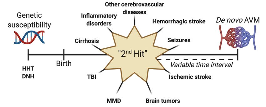
Figure 5. Schematization of the “second hit” theory for the development of de novo arteriovenous malformations (AVM).
Genetic susceptibility can be represented by syndromes such as hereditary hemorrhagic telangiectasia (HHT) or diffuse
neonatal hemangiomatosis (DNT). After birth, a second may occur, leading to the formation of an acquired AVM after a
variable point in time. This second hit can be a preexisting vascular lesion or malformation, hemorrhagic stroke, seizures,
ischemic stroke, brain tumors, Moyamoya disease (MMD), traumatic brain injury (TBI), cirrhosis, or inflammatory lesions.
4.2.2. Hemorrhagic Stroke
According to the information presented, hemorrhagic stroke from various sources is
the single most common primary brain injury leading to the formation of de novo AVMs
within the same site. Although angiographically occult fistulas masked by hematomas,
thrombosis, arterial feeder vasospasm, or posthemorrhagic brain edema cannot be dis-
missed, a possible pathophysiological mechanism for AVM generation in a hemorrhagicMedicina 2021, 57, 201 13 of 23
cerebral environment should be postulated. It is likely that hemorrhagic stroke in conjunc-
tion with surgical trauma fosters appropriate angiogenetic conditions [28,56,57]. It may
also seem likely that at least some of these AVMs stemmed from a small and previously
missed angioma [58]. After surgical resection, the available empty space and probable re-
peated silent bleeds resulting in a progressive vascular enlargement in the hematoma cav-
ity may also explain these occurrences [59]. Jeffree et al. conjectured that AVMs might
develop not though the means of dilatation of preexisting vessels, but via the generation
of brand-new vessels [60]. They proposed that the levels of certain growth factors increase
upon endothelial cell exposure to shear stress, later resulting in the development of a vas-
cular network and recruitment of new vessels within a previously occult and asympto-
matic malformation. The authors also speculated an alternative, namely that the hema-
toma itself triggers de novo AVM formation concurrently with its own resolution.
Nevertheless, it appears that acquired AVMs may develop even remotely from the
location of the initial hemorrhage. One of the patients in the series by Jeffree et al. suffered
from a left parietal hematoma, followed by a right parieto-occipital de novo AVM found
ten years afterward [60]. The case described by Miyasaka et al. had a right parietal hema-
toma eight years before three AVMs were revealed [56]. Only one grew near the hematic
cavity, whereas one was situated in the frontal lobe of the same hemisphere, and the re-
maining one in the left occipital lobe. Four more individuals who experienced SAH of
unknown origin developed de novo AVMs in various areas of the brain, any time between
2 and 23 years after the original injury [33,57,58,61]. Nakamura et al. reported a more
unique instance, wherein a left frontal intraparenchymal hemorrhage was surgically evac-
uated, followed by the therapeutic local implantation of mesenchymal stem cells secreting
the neuroprotective glucagon-like peptide-1 (GLP-1) [62]. Despite some criticism [62], the
authors clearly infirmed the presence of any vascular malformations at the site of surgery
[62,63]. At the three-year mark after the intervention, a ruptured AVM was diagnosed in
the same region of the brain. They hypothesized that aside from VEGF, GLP-1 might also
play a role in promoting angiogenesis and vessel proliferation. Validating the angioge-
netic role of intracranial hematomas may be possible in the near future.
4.2.3. Seizures
Several instances of epileptic convulsions with no discernible underlying causes have
been associated with acquired AVMs after a variable period of time since the onset of the
seizures [25,35,64–68]. According to numerous authors, as well as our own experience,
seizures represent the second most common form of manifestation of cerebral AVMs
[1,31,69]. It has been previously demonstrated that VEGF is upregulated within neural
and glial cells following epileptic seizures, most likely acting as a mechanism to combat
postictal neurodegeneration [25,35,67,70–74]. Moreover, preclinical studies have revealed
a variety of beneficial effects of VEGF elevation in epilepsy and status epilepticus [75–77].
On the other hand, pathological upsurge of VEGF can result in a disrupted blood-brain-
barrier integrity, and may also further the progression of certain diseases such as demye-
linating injuries and vascular malformations [71,73,75].
One particular case was preceded by a single episode of seizures nine years prior to
the discovery of an AVM, which was absent on the initial MRI scan [35]. Similarly, a seven-
year-old boy diagnosed with a de novo AVM experienced febrile seizures four years prior
to definitive imaging study [68]. These two cases may suggest that even a solitary episode
of epileptic convulsions creates the appropriate angiogenetic conditions for AVM growth.
Withal, subcortical band heterotopia (SBH), a neural migration disorder, is also associated
with pharmacoresistant epilepsy [78–80]. A nine-year-old girl with SBH also developed
an acquired AVM after three years of seizures and developmental delay [67]. Further-
more, it seems that there is no requirement for the seizures to have a tonic-clonic charac-
teristic, as exemplified in a case by Kilbourn et al. [64]. This patient was diagnosed with
hydrocephalus at an infant age, and later developed autism and absence seizures, a de
novo AVM being discovered at the age of 18 years. As such, it is possible that epilepsy, inMedicina 2021, 57, 201 14 of 23
any of its forms, may play a decisive role in the appearance of acquired AVMs through an
exaggerated and detrimental protective mechanism against neurodegeneration.
4.2.4. Brain Tumors
Although rare instances, intracranial neoplasms with a high vascularity coexisting
with AVMs have been described in the recent past [81–84]. AVMs occurring after the treat-
ment of a previous brain tumor are also remarkably rare [27,37,44,85,86]. In the case re-
ported by Bennet et al. of a de novo cerebellar AVM occurring after the surgical resection
of a vermis hemangioblastoma, it is likely that the altered hemodynamic conditions,
which stemmed from the removal of the tumor, played a crucial role in the evolution of
the vascular lesion [85]. It may also seem plausible that the AVM coexisted with the he-
mangioblastoma but was occult beforehand. Medvedev et al. described a case in which a
cerebellar AVM and a hemangioblastoma coincided, also theorizing a common ancestry
linking these two pathologies [87]. A similar mechanism might also be involved in the
appearance of an acquired brain AVM after recurrent meningioma surgery [27]. Likewise,
malignant glial tumors foster a highly proangiogenic environment, as quantified by the
enhanced production of VEGF, which could prove a crucial stimulus for a de novo AVM
occurrence [37,38]. A second possible reason is that an environmental exposure gave rise
to both the neoplasm and the malformation, although the incriminated factor is uncertain
[38,70]. Radiosurgery has been incriminated as a probable etiologic factor for malignant
brain tumors, even after therapy for AVMs, despite the risks being relatively low [88–90].
The exposure to radiation could also be considered a stimulus for these malformations,
since tumors of the fourth ventricle (an ependymoma and a medulloblastoma, respec-
tively) undergoing postoperative radiotherapy later developed de novo AVMs [44,86].
This might be supported by the vasculopathy and vessel remodeling following radiation
therapy. Regardless of etiological considerations, the simple association with such neo-
plasms warrants a poor prognosis and a low short-term survival from the start
[27,37,38,44,81–87].
4.2.5. Ischemic Stroke, Venous Sinus Thrombosis, and Transient Ischemic Attacks (TIA)
Only very few cases of de novo AVMs have been correlated with ischemic events of
the brain. Pabaney et al. argued that an inflammatory or ischemic lesion could have has-
tened the development of acquired AVMs in contrast with congenital AVMs [24]. They
reasoned that the stroke acted as a ‘second-hit’ that resulted in the formation a de novo
AVM. Previously, it has been demonstrated that ischemia has the potential to trigger vas-
cular proliferation via an amplified expression of hypoxia-inducible factor-1 (HIF-1), re-
sulting in an uninhibited vascular proliferation and the occurrence of arteriovenous
shunts within a patient with genetic susceptibility and alterations regarding angiogenesis
and inflammatory cascades [24,51,91]. The appearance of de novo vascular lesions could
also be a response to injury mechanism after stroke [92]. This might actually be the case,
as other vascular malformations (i.e., dural and pial arteriovenous fistulas) can originate
from infection, inflammation, or traumatic injury, advocating the concept of environmen-
tal factors promoting angiogenesis [52,66]. Regarding venous sinus thrombosis, it may
seem plausible that the increased venous pressure and subsequent hypoxia in the adjacent
tissues arising from such an event leads to an enhanced angiogenic activity [14,45,66,93].
During embryological growth, venous hypertension could be the result of occlusion, ste-
nosis or agenesis. Otherwise, cerebral ischemia itself may offer a physiological incentive
for angiographically occult AVMs to enlarge and become patent [94]. It is worthwhile
mentioning that among this group of de novo AVM patients, only one had an ischemic
event during infancy [92], the other individuals being adults at the time of initial injury.Medicina 2021, 57, 201 15 of 23
4.2.6. Moyamoya Disease (MMD)
MMD is defined as a rare chronic occlusive pathology of the cerebral vasculature,
having an unidentified etiology [95–98]. It is characterized by a series of bilateral steno-
occlusive alterations in the terminal portion of the internal carotid artery (ICA) and its
branches, as well as a coexisting abnormal network of collateral vessels at the base of the
brain. Its presentation differs according to age, children most often manifesting epileptic
seizures and ischemic events, while in adults it generally leads to hemorrhagic stroke.
There have been a few instances in which AVMs preceded the development of MMD,
hatching the hypothesis that the increased amount of blood traveling within the malfor-
mation itself creates a turbulent flow level of the common carotid artery bifurcation, in
turn generating the hyperplasia of the intima and the ensuing collateralization and pro-
gression of an acquired form of MMD [99–101]. The association between MMD and ulte-
rior de novo AVMs has been described in a small number of case reports, all of which
were pediatric, potentially turning the aforementioned hypothesis on its head [95–101]. It
is possible that the analogous biological backgrounds of these two pathologies regarding
the enhanced expression of proangiogenic molecules such as VEGF, or certain inflamma-
tory molecules including tumor necrosis factor α (TNFα), MMP and IL-6, led to the occur-
rence of AVMs after the progression of MMD [95]. This might be the case, as the patient
described by Fujimura et al. harbored an AVM supplied by the posterior circulation,
which by definition was not affected by MMD. Additionally, this patient concomitantly
suffered from sickle cell disease, yet no explanation could be offered whether this condi-
tion had any influence on either the appearance of MMD or the AVM. Another plausible
explanation is that MMD in these patients produced an angiogenic failure, which in turn
resulted in the formation of anomalous arteriovenous shunts [96]. As Schmit et al. pointed
out, it could also be stipulated that the hyperangiogenic microenvironment predominant
in MMD, in conjunction with a local proangiogenic stimulus (for instance VEGF or fibro-
blastic growth factor—FGF) as a consequence of cerebral infarction, would have been suf-
ficient for an AVM to develop on that precise spot [98]. The role of genetic factors, whether
congenital or acquired, in the presence of prolonged hypoperfusion should also be con-
sidered [97]. Nevertheless, the probability that the AVMs coexisted with MMD but were
angiographically occult at the time of initial angiography cannot be entirely ruled out.
As observed in these collection of case reports, brain infarction, and associated cere-
brovascular pathologies associated, may generate sufficient stimuli for an AVM to de-
velop postnatally, at least if the patient possesses a certain susceptibility. More research is
needed in this field to precisely determine the nature of these predispositions.
4.2.7. Traumatic Brain Injury (TBI)
Whether head trauma itself actually contributes to the formation of a proangiogenic
environment, or its association to de novo AVMs is purely coincidental, remains to be
proven. Traumatic injury has, however, been reported as an etiologic factor for arteriove-
nous fistulae [102–104]. The case described by Gonzalez et al., of a three-year-old girl who
suffered a mild TBI as a result of traffic accident, developed a delayed hemorrhagic lesion
that manifested through generalized epileptic seizures [29]. These seizures became in-
creasingly difficult to control, lasting for four years before the diagnosis of a de novo AVM
was established. As anteriorly discussed, both the hemorrhagic and the epileptic compo-
nents could be held accountable for the appearance of an acquired AVM, although the
hematoma had a left frontal placement, whereas the vascular lesion was situated in the
right posterior temporal lobe. No hemorrhagic collections were identified in the vicinity
of the AVM to mask this anomaly. A comparable case, in which an 11-month-old girl ex-
perienced a severe TBI with an acute subdural hematoma on the left convexity, was diag-
nosed with an ipsilateral de novo AVM almost 11 years later, after suffering from seizure
disorder for around six years [105]. In this instance, the patient was also diagnosed withMedicina 2021, 57, 201 16 of 23
posttraumatic encephalomalacia, which could in theory act as a suitable terrain for an ab-
normal vasculature in a developing brain. Similarly, a young boy suffered a blunt head
injury and subsequent subarachnoid hemorrhage from an unknown source, and devel-
oped an AVM in the right frontal lobe nine years after the initial incident [106]. We hy-
pothesize that the presence of TBI resulting in intracranial hemorrhage or seizure disorder
may act as a ‘second hit’ that generates a de novo AVM in patients with genetic suscepti-
bility via VEGF upregulation (Figure 5). This theory needs to be further studied.
4.2.8. Genetic Syndromes
HHT is a systemic autosomal dominant genetic syndrome with a marked predispo-
sition to develop intracranial AVMs [6,7,11,12,46,51]. Two genes that have been linked to
the development of HHT are the ENG gene, which is correlated with HHT1 as well as a
higher prevalence of AVMs, and the ACVRL1 or ALK1 gene, which is associated with the
HHT2 phenotype and a lower brain AVM incidence [11,12]. A single patient in the re-
ported literature was confirmed to have HHT and developed a de novo AVM [46]. At five
months of age, he underwent screening for HHT, as both his mother and older brother
suffered from this syndrome and harbored a symptomatic AVM of the brain and of the
spine respectively, despite being asymptomatic himself. At the age of five years, a de novo
AVM became manifest and the patient underwent preoperative embolization and micro-
surgical resection. This case underlines the potential of these vascular malformations to
appear and expand within highly vulnerable individuals, without necessarily requiring
the involvement of external factors. AVMs in HHT have been shown to have a bidirec-
tional evolution, possessing both the possibility of expansion and to regression [34]. Ko-
miyama entertained the hypothesis that congenital brain AVMs could develop until the
age of two years [93]. We, however, believe that there is no predefined interval within
which these lesions materialize.
Additionally, a newborn diagnosed with hepatic hemangiomatosis and congestive
heart failure suffered from a de novo AVM in the left cerebellopontine angle (CPA), which
was treated via endovascular embolization at three and four years of age respectively [32].
Before the vascular malformation was found, MRI revealed two enhancing lesions in the
left CPA and pineal region, which could have been brain hemangiomas. It was not men-
tioned whether or not she was tested for HHT or any other genetic syndrome, and she
died because of brain hemorrhage at six-years-old. Brain AVMs and hemangioblastomas
have been described in patients with diffuse neonatal hemangioma-tosis (DNH) in the
past [107,108]. The authors of the respective case report proposed two possibilities: either
the enlargement and progression of a coincidental AVM adjacent to the preexisting CPA
hemangioma, or the conversion of said hemangioma into an AVM [32]. As there are only
a finite number of individuals with diffuse neonatal hemangioma-tosis, it might prove
challenging to screen them for acquired cerebral AVMs.
4.2.9. Inflammatory Diseases
Only two cases of inflammatory diseases not related to the cerebral vasculature and
de novo AVMs were described in the literature. The first was of an unidentified inflam-
mation or demyelinating lesion in the brainstem [109], and the second was associated with
a previously diagnosed Bell’s palsy on the same side [39]. A probable mechanism is the
overexpression of VEGF as a result of the inflammatory process, similar to the mechanism
explained in ischemic stroke [39,52,66]. Another explanation could be increasing hemody-
namic stress due to the inflammation, leading to vascular remodeling.
4.2.10. Liver Cirrhosis
Cirrhosis defines a diffuse fibrosis of the liver and the alteration of the hepatic micro-
architecture into structurally anomalous nodules. Despite that, to the extent of ourMedicina 2021, 57, 201 17 of 23
knowledge, only two instances of de novo brain AVMs were described in patients suffer-
ing from cirrhosis, and that the correlation between these two pathologies was poorly
studied, we nevertheless consider it worthwhile mentioning [6,110]. Peripheral systemic
vascular malformations are already considered a mark of advanced chronic liver disease;
hence it is tantalizing to assume that these lesions and cerebral AVMs share a common set
of mechanisms and etiologic factors. Among these we point out an excessive production
alongside a clearance reduction of factors such as VEGF, TNF- α, IL-6, MMP-3 and MMP-
9, or nitric oxide synthetase (NOS), as well as a decreased catabolism of estrogens [5,110].
To further support this assumption, Shimoda et al. illustrated the case of spontaneous
AVM disappearance two years after a successful liver transplantation in a patient with
alcohol-induced cirrhosis [111]. It is therefore likely that the alterations instigated by the
state of venous hypertension and thrombosis, combined with a hepatic “second hit,” pro-
vides the necessary stimuli for an intracranial AVM to develop in cirrhosis. More such
patients should be investigated for cerebral vascular malformations further along the
course of their chronic liver disease before finally establishing this correlation.
4.3. Past, Present, Future, and Personal Opinions
Up until recently, AVMs were adamantly considered congenital disorders with the
capacity to evolve, rupture, regress, and even recur after treatment [52]. Contemporary
evidence, including the case reports incorporated in this study, suggests that we are still
a long way from fully elucidating these lesions, which could actually also be acquired. In
research on adult mice, Chen et al. demonstrated that the deletion of ACVRL1 within en-
dothelial cells resulted in elevated local endothelial cell proliferation throughout brain
angiogenesis, as well as the induction of de novo AVMs [112]. Similarly, Walker et al.
obtained lesions resembling human AVMs in adult mice after co-injecting adenoviral vec-
tor exhibiting Cre recombinase and adeno-associated viral vectors containing VEGF
within their basal ganglia, thereby producing ACVRL1 mutation [113]. In their study, Park
et al. demonstrated that either physiological or environmental stimuli such as injuries,
alongside the respective genetic deletion, are needed for ACVRL1-deficient blood vessels
to evolve into AVMs within adult mice [15]. Zhu et al. also managed to trigger the for-
mation of AVMs in adult mice, this time via CRISPR/Cas9-mediated ACVRL1 gene mu-
tations [114]. Although other experimental models exist, it is beyond the scope of this re-
view to present them exhaustively. It is, however, safe to assume that a number of genetic
mutations, in conjunction with the appropriate environmental factors, may lead to the
development of de novo AVMs.
Evidence from several of the reported case studies and series suggest that radiother-
apy-related injury triggers an increased VEGF production as a response-to-injury type of
adaptation, which can result in the spontaneous formation of brain vascular malfor-
mations [26,41,52,115–117]. While the majority of these lesions are cavernomas, it cannot
be fully disproved that radiation can also induce the formation of AVMs. We believe that
the adaptive changes in the cerebral vasculature occurring after stereotactic radiosurgery
can also induce the exaggerated angiogenesis necessary for de novo AVMs.
Individuals in which intraparenchymal hemorrhage (IPH) were surgically removed
serve as irrefutable evidence that de novo AVMs can develop in the hematic cavity, with-
out preexisting vascular lesions in the respective regions [56,59,60,118]. We tend to con-
sider these cases especially because the intraoperative aspect cannot be replicated entirely
by imaging studies, at least with current technology.
Seizures present a dual quality, on the one hand being the second most common
symptom for brain AVMs, and on the other as a possible generator for de novo AVMs
[25,35,36,64,66–68]. Although we cannot contradict the fact that epilepsy can cause the
expansion of these lesions, the possibility that a preexisting angiographically occult cere-
bral vascular malformation triggered the aforementioned seizures cannot be completely
negated.Medicina 2021, 57, 201 18 of 23
However, arguably the most important aspect is that all of the reported cases of de
novo AVMs had an initial symptom or a preexistent underlying pathology. It is therefore
tempting to believe that the actual number of such individuals is much higher, yet for
various reasons, many of these patients are not investigated prior to the occurrence of
AVMs. The fact that not all patients had an initial DSA might be a confounding factor, as
the diagnosis of brain AVM might have been missed in this case. As the majority of these
patients suffered from vascular conditions such as MMD, hemorrhagic stroke, aneurysms
or other allegedly congenital vascular anomalies, the predisposition for developing AVMs
was indubitably present, requiring a ‘second hit’ stimulus for AVMs to properly appear
(Figure 5) [66].
One of the major limitations regarding the alteration of the ‘congenital AVM doc-
trine’ arises from the limited number of de novo lesions, which are mostly found in sin-
gular case reports. Additionally, the exact pathophysiological mechanisms of AVM
growth are conjectured, although not fully understood. A recent consensus meeting of
international neurosurgical experts debated whether AVMs should be regarded as con-
genital or acquired [7]. The authors all agreed that the genetic predisposition is insufficient
to induce AVM formation by itself, and that a ‘second hit’ of variable nature is always
required. Despite the fact that the majority of patients with brain AVMs harbor mutations
of the KRAS gene [17,18], certain somatic mutations have very low frequencies within
these lesions. This in itself raises the concern whether these mutations actually possess
functionality in AVMs development, or whether they can be accurately quantified. An-
other limitation that the authors acknowledge is the hypothetical nature of this review.
This stems from the fact that de novo AVMs are extremely scarce, and the data surround-
ing their development is reduced. However, we hope that the mechanisms proposed in
this work will be more adequately studied in the near future.
Therefore, we propose that brain AVMs can be either present at birth and able to
evolve or regress, or acquired but in the presence of preexisting genetic susceptibility.
There needs to be an association between clinical, genetic, and animal studies in order to
verify this hypothesis.
5. Conclusions
Increasing evidence suggests that at least some of the AVMs discovered develop
some time after birth. We are still a long way from finally elucidating their true nature,
though there is reason to believe that they can also appear after a proposed ‘second hit’
during a patient’s lifetime (Figure 5). The congenital or acquired characteristic of AVMs
may have a tremendous impact on prognosis, risk of hemorrhage, and short and long-
term management.
This systematic review presents the reported cases of de novo brain AVMs in the
current literature. Despite being a rare pathology, AVMs represent the most frequent
cause of intracranial hemorrhage in young patients. Furthermore, it is possible that the
real number of ruptured de novo AVMs is underrepresented, since such lesions present-
ing with acute hemorrhage in patients that have never undergone a previous CT or MRI
scan are automatically labeled as congenital in nature.
It must be noted that all of the diagnoses of de novo AVMs were made in conjunction
with a previous associated pathology or injury. Had this not been the case, imaging stud-
ies prior to the symptomatic manifestation of the acquired AVMs would have been un-
necessary, and the initial absence of these vascular lesions could not have been docu-
mented. Patients who experience seizures with or without apparent underlying causes,
hemorrhagic or ischemic strokes, TBI of varying severity, other cerebrovascular malfor-
mations, or highly vascular tumors should receive regular imaging screening which in-
cludes DSA more often to highlight or exclude the formation of de novo AVMs. Further
research should be conducted to more adequately ascertain the congenital or acquired
characteristic of brain AVMs.You can also read