Detection of Oesophageal Fistula by Radionuclide Salivagram SPECT/CT
←
→
Page content transcription
If your browser does not render page correctly, please read the page content below
ORIGINAL RESEARCH
                                                                                                                                                 published: 24 August 2021
                                                                                                                                            doi: 10.3389/fonc.2021.612122

                                              Detection of Oesophageal Fistula by
                                              Radionuclide Salivagram SPECT/CT
                                              Yingwei Wang 1,2,3, Chao Wang 4, Lin Liu 1,2,3, Xinwen Huang 5, Zhaoyou Guo 5, Wei Zeng 5,
                                              Rui Sun 1,2,3 and Yue Chen 1,2,3*
                                              1 Department of Nuclear Medicine, Affiliated Hospital of Southwest Medical University, Luzhou, China, 2 Nuclear Medicine

                                              and Molecular Imaging Key Laboratory of Sichuan Province, Luzhou, China, 3 Academician (Expert) Workstation of Sichuan
                                              Province, Luzhou, China, 4 Department of Thoracic Surgery, Affiliated Hospital of Southwest Medical University, Luzhou, China,
                                              5 Department of Radiology, Affiliated Hospital of Southwest Medical Universitye, Luzhou, China

                                              Purpose: Videofluoroscopic swallowing study (VFSS) is currently the most widely used
                                              clinical examination method for diagnosis of oesophageal fistula, but it has many
                                              limitations. Therefore, we evaluated radionuclide salivagram single-photon emission
                                              computed tomography (SPECT/CT) as a new method of oesophageal fistula diagnosis.
                                              Methods: We retrospectively evaluated the data of 11 patients (10 men and 1 woman,
                        Edited by:
                                              aged 41 to 70 years, with an average age of 58.6 years) who had clinically suspected
                         Haibin Shi,
           Soochow University, China          oesophageal fistula from January 2019 to October 2020. They underwent radionuclide
                       Reviewed by:           salivagram SPECT/CT and VFSS examinations, and we analysed and compared the
           Prasanta Kumar Pradhan,            results of the two examinations.
Sanjay Gandhi Post Graduate Institute
   of Medical Sciences (SGPGI), India         Results: A total of 11 patients were included in this study. Ten underwent both salivagram
                          Jianguo Lin,
                                              and VFSS examinations. One patient was unable to swallow the contrast agent; therefore,
Jiangsu Institute of Nuclear Medicine,
                                China         only salivagram was performed, and we excluded this patient from the VFSS analysis. A
                 *Correspondence:             total of 11 patients underwent salivagram examinations, of which 6 were positive and 5
                         Yue Chen             were negative. A total of 10 patients were tested by VFSS, of which 6 results were positive
             chenyue5523@126.com
                                              and 4 were negative.
                   Specialty section:         Conclusion: Radionuclide salivagram SPECT/CT and VFSS are complementary, which
         This article was submitted to
                                              can greatly improve the clinical diagnosis and prognosis of oesophageal fistula. When the
                   Cancer Imaging and
        Image-directed Interventions,         patient cannot perform the VFSS, or the clinical symptoms are inconsistent with the VFSS
               a section of the journal       imaging findings, the salivagram is an ideal test method.
                 Frontiers in Oncology

      Received: 30 September 2020             Keywords: salivagram, oesophageal fistula, SPECT/CT, VFSS,            99mTc-DTPA

         Accepted: 05 August 2021
        Published: 24 August 2021

                            Citation:
                                              INTRODUCTION
                   Wang Y, Wang C,
              Liu L, Huang X, Guo Z,
                                              Oesophageal fistula is a severe complication of oesophageal or lung cancer treatment, especially after
  Zeng W, Sun R and Chen Y (2021)
 Detection of Oesophageal Fistula by
                                              radiation therapy (1–3). It has been reported that foreign bodies in the oesophagus can also cause
 Radionuclide Salivagram SPECT/CT.            oesophageal fistulas (4). There are many types of oesophageal fistulas, including pleural, aortic, and
            Front. Oncol. 11:612122.          atrial. Among them, oesophageal-mediastinal fistula and tracheo-oesophageal fistula are the most
     doi: 10.3389/fonc.2021.612122            common, with the latter more common than the former (5).

Frontiers in Oncology | www.frontiersin.org                                           1                                         August 2021 | Volume 11 | Article 612122
Wang et al.                                                                                                    Detection of Oesophageal Fistula by SPECT/CT

    At present, clinical diagnosis of oesophageal fistula is                       requirement for informed consent was waived due to the
mainly based on clinical symptoms and imaging, the latter                         study’s retrospective design.
including multidetector computed tomography (CT) and
videofluoroscopic swallowing study (VFSS) (6, 7). Many                             Salivagram
diseases can have the same clinical symptoms. It is difficult to                   We selected technetium-99m diethylenetriaminepentaacetic acid
establish the diagnosis or the location and size of the fistula by                 (99mTc-DTPA) as a radionuclide imaging agent. Before the
clinical symptoms alone. CT lacks sensitivity to detect a small                   examination, the patient needed no special preparation. 99mTc-
fistula, and VFSS is the most commonly used examination.                           DTPA with a dose of 1 mCi and a volume of about 1-2 mL was
However, clinical application of VFSS has limitations.                            dropped onto the patient’s tongue twice with a syringe. The time
For example, the procedure is difficult if 1) the patient cannot                   interval between the two applications was 30 min. After the
cooperate with bodily position changes during the examination;                    administration, the patient was instructed to move around and
2) the patient has a large tracheo-oesophageal fistula or more                     try to swallow as much as possible. A Siemens Symbia T16
severe complications; 3) a gastric tube is present; or 4) the patient             SPECT/CT (Siemens, Munich, Germany) was used for static and
has severe dysphagia. Patients in these circumstances cannot                      forward acquisition at 30 min, 60 min, and 120 min after the last
achieve the expected diagnostic results if tested by VFSS.                        dose. The peak energy was 140 Kev, the window width was 20%,
Therefore, we sought a diagnostic method without the                              the matrix was 128 × 128, the magnification was 1.0, the
above limitations.                                                                acquisition count was 500 K, and the patient was supine. In
    The advantages of radionuclide inspection are safety,                         advanced position, the visual field included the oral cavity,
convenience, high patient tolerance, and no special requirement                   oesophagus, bilateral lung fields, and gastric cavity.
for patient location during the procedure. Also, a combination with                  If a patient’s oesophageal fistula is suspected to be unilateral, the
SPECT/CT can more accurately determine the location, size, and                    patient can be ordered to lie on the affected side to increase the
tissue surrounding the lesion. In this study, the oesophageal fistula              positivity rate. During the acquisition process, if the imaging agent
was diagnosed using radionuclide salivagram and the results were                  is observed outside the digestive tract, local SPECT/CT
compared with those of VFSS.                                                      tomosynthesis imaging can be performed immediately, and the
                                                                                  scanning range is consistent with that of the static acquisition:
                                                                                  voltage 120 Kv, current 100 mAs, layer thickness 5 mm, pitch 0.8
                                                                                  mm, rotation time 1.0 s, reconstruction using FBP algorithm,
MATERIALS AND METHODS                                                             convolution kernel B40s, reconstruction of soft tissue window,
We retrospectively examined the data of 11 patients who                           layer thickness 3.0 mm, layer spacing 3.0 mm. Finally, Siemens
underwent radionuclide salivagram SPECT/CT and VFSS in                            post-processing software Syngo MI VA70A was used for image
our hospital from January 2019 to October 2020 (Table 1).                         fusion analysis.
Two associate chief physicians read the films independently to
minimise the impact of subjective factors. The final diagnostic                    Diagnosis
result was established by consensus between the two physicians.                   A positive diagnosis was determined when any images in the
The patients had one or more of the following medical                             acquisition showed imaging agent concentrated outside the
conditions: 1) oesophageal tumours; 2) history of radiation                       digestive tract. A negative diagnosis was determined when
therapy for oesophageal tumours; 3) history of incarcerated                       during the entire examination, only normal digestive tract
oesophagus with sharp foreign bodies; 4) prominent cough                          images were found.
and fever; and 5) CT examination for suspected oesophageal                           For patients with obvious clinical symptoms and history of
fistula. This study involved no identifying patient characteristics                oesophageal surgery or radiotherapy, but no abnormalities on
and was approved by the hospital ethics committee. The                            imaging, we can repeat the administration of 1 mCi 99mTc-DTPA

TABLE 1 | Basic patient information and results of salivagram and VFSS.

Patient             Age             Sex                  Cause of disease                     Salivagram          VFSS                Final diagnosis

1                    67              M         Surgery for Carcinoma oesophagus                    +                +            Oesophageal fistula
2                    55              M         Surgery for Carcinoma oesophagus                    +                +            Oesophageal fistula
3                    67              M         Surgery for Carcinoma oesophagus                    +                +            Oesophageal fistula
4                    41              M         Surgery for Carcinoma oesophagus                    –                –            Without oesophageal   fistula
5                    54              M         Oesophageal tumour                                  +                +            Oesophageal fistula
6                    65              M         Oesophageal tumour                                  +                +            Oesophageal fistula
7                    55              M         Oesophageal tumour                                  –                –            Without oesophageal   fistula
8                    70              M         Radiotherapy for oesophageal carcinoma              –                –            Without oesophageal   fistula
9                    49              M         Radiotherapy for oesophageal carcinoma              +                N            Oesophageal fistula
10                   66              F         Oesophageal foreign body                            –                –            Without oesophageal   fistula
11                   56              M         Surgery for Carcinoma oesophagus                    –                +            Without oesophageal   fistula

+, Positive; −, Negative; N, No examination.

Frontiers in Oncology | www.frontiersin.org                                   2                                   August 2021 | Volume 11 | Article 612122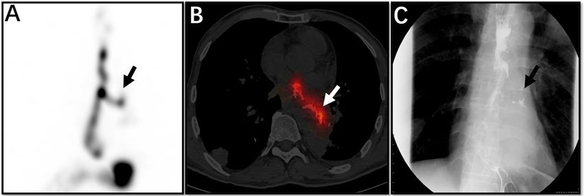
Wang et al.                                                                                                       Detection of Oesophageal Fistula by SPECT/CT

once and repeat imaging after 2-4 hours, and routinely perform                    positive (6 men; mean age, 60.6 years) and 4 were negative (3
tomographic fusion imaging. We analysed the positive cases and                    men and 1 woman; mean age, 58 years). In one patient with a
found that the highest positivity rate was between 60 and 120                     positive diagnosis, tomographic fusion images confirmed that it
min after the second dose of imaging agent.                                       was a false positive.
                                                                                     We compared the results of the two tests in the patient group,
Videofluoroscopic Swallowing Study                                                 each test having its own advantages and disadvantages. Among
Patients need no special preparations (although gastric tubes                     the 11 patients who underwent salivagram, there were 6 positive
must be removed). The procedure was performed according to                        diagnoses and 5 negative diagnoses. The demonstrated image
the routine VFSS protocol, and we observed whether there was                      was acquired after 2 hours of administration of the
leakage of contrast medium outside the gastrointestinal tract                     radiopharmaceutical. Comparing the images of all patients, we
under fluoroscopy.                                                                 found that images taken at 60-120 min after dosing had the
                                                                                  highest positivity rate (Figure 1). Among the patients, one
                                                                                  (patient 9) had severe clinical symptoms (wheezing) and was
RESULTS                                                                           unable to swallow the contrast agent for VFSS testing. Two
                                                                                  patients had indwelling gastric tubes, and salivagrams were
Salivagram: Among the 11 patients analysed (10 men and 1                          performed without removing the tubes (Figure 2). Therefore,
woman, aged 41 to 70 years, with mean age 58.6 years), 6 were                     only the salivagram was performed, which illustrated the
positive (6 men; mean age, 59.5 years), and 5 were negative (4                    advantage of salivagram (Table 1 and Figure 3). Patient 11
men and 1 woman; mean age, 57.6 years).                                           (Table 1) was examined in post-oesophageal tumour surgery.
   VFSS: Ten of the 11 patients analysed (9 men, 1 woman; aged                    VFSS examination suggested that an upper oesophageal fistula
41 to 70 years, with mean age 59.4 years) completed VFSS; 6 were                  had formed. However, in the tomographic fusion images, we

 FIGURE 1 | The patient was a 53-year-old man with oesophageal cancer and recurrent cough. Whole SPECT imaging (A), flued SPECT/CT imaging (B), and VFSS
 imaging (C) are demonstrated. These strip imaging agents are sites of oesophageal fistula (arrows). This patient underwent a barium swallow of the upper
 gastrointestinal tract before salivagram, so the high-density substance in the image is barium (B).

 FIGURE 2 | This is a 48-year-old male patient with an oesophageal tumour and requirement for a gastric tube due to inability to eat. The patient underwent
 salivagram without gastric tube removal. Whole SPECT imaging (A), flued SPECT/CT imaging (B), and VFSS imaging (C) are demonstrated. The gastric tube (A, B,
 straight arrow) is visible. An abnormal concentration of imaging agent is present in the left thoracic cavity (B, C, triangular arrow).

Frontiers in Oncology | www.frontiersin.org                                   3                                      August 2021 | Volume 11 | Article 612122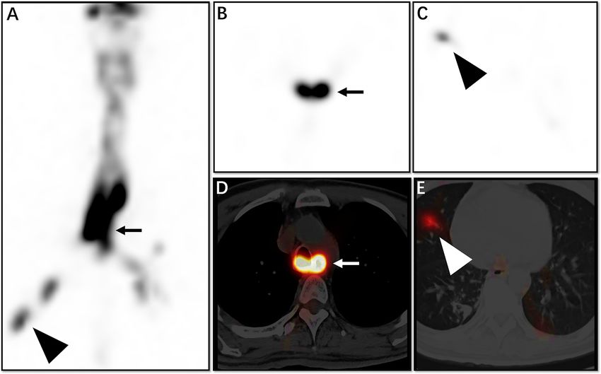
Wang et al. Detection of Oesophageal Fistula by SPECT/CT
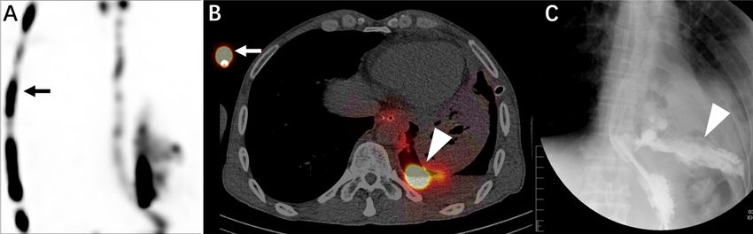
FIGURE 3 | This 43-year-old man had inability to swallow and recurrent coughing. Whole SPECT imaging (A), tomography SPECT imaging (B, C), and flued
SPECT/CT imaging (D, E) are demonstrated. We see from the image that the opening of the oesophageal fistula is located at the plane of the main bronchial
bifurcation (A, B, D, straight arrow), and imaging agent is also seen at the distal bronchus (A, C, E, triangular arrow).
recognised that the fistula suggested by VFSS was a diverticulum tumour and a partial oesophageal resection. The patient was
at the oesophageal anastomosis (Figure 4). Through examined by VFSS and found to have an apparent small fistula in
tomographic fusion imaging, we were able to diagnose the the upper oesophagus. To further evaluate the apparent fistula,
lesion more accurately. the patient was examined by salivagram. On the maximum
intensity projection (MIP) image, we saw the abnormal
concentration of the imaging agent outside the lumen of the
DISCUSSION upper oesophagus, but our tomographic fusion showed that this
was not a fistula. Rather, it was a small diverticulum of the
One of our patients was examined after surgery for oesophageal gastro-oesophageal anastomosis.
cancer. The physician sought to evaluate the patient’s In our study, we found that the optimal time for image
postoperative recovery. He was examined by salivagram in our acquisition after administration of imaging agent was 60-120 min.
department. Two hours after the administration of the imaging In some patients, although an oesophageal fistula is highly suspected
agent, we found a limited outward projection of the middle and clinically, imaging results can be negative. With salivagram, we can
lower oesophagus. The agent concentration was focal, but it was use a consistent method, one repeat dose of imaging agent, and
uncertain whether it represented an artefact. Therefore, we asked acquire images after 2-4 h. If necessary, we can also perform
the patient to undergo another examination after 2 hours. We tomographic fusion imaging to minimise misdiagnosis.
were surprised to find that the previously visualised lesion had Bivins et al. proved in their 1977 study that radionuclide
disappeared; therefore, we considered it to have been an artefact. imaging has potential value in the diagnosis of oesophageal
We found no oesophageal fistula in this patient. We followed up fistula (10). Radionuclide imaging has long been used
the patients and the final clinical diagnosis was consistent with clinically, and its effectiveness was proven in 1980 (11). Dunn
our conclusions. We believe that there are many causes for this et al. proved in a 1983 study that radionuclide imaging has
performance: 1) various factors, such as reduced oesophageal further research value in the diagnosis of an oesophageal fistula
peristalsis frequency; 2) decreased saliva production; 3) (12). Since then, many studies have shown that this method is
oesophageal diverticulum. When the presence of a lesion is highly sensitive for detecting aspiration of saliva (11, 13). Our
unclear in early imaging, delayed imaging can be performed to research is also based on the above report. However, so far, most
improve the accuracy of the diagnosis (8). salivagram examinations have been performed for children and
With extension of the imaging time, improvement in the rarely for adults (13–18).
accuracy of disease diagnosis has been confirmed in many Previous studies have shown that salivagram is often used in
reports (9). Another patient had a malignant oesophageal the diagnosis and treatment of congenital oesophageal atresia,
Frontiers in Oncology | www.frontiersin.org 4 August 2021 | Volume 11 | Article 612122Wang et al. Detection of Oesophageal Fistula by SPECT/CT
FIGURE 4 | This 53-year-old male patient had a partial oesophageal resection due to an oesophageal tumour. The patient had a VFSS examination, which
suggested an upper oesophageal fistula (A, arrow). The patient also underwent salivagram. From the MIP image, we see a small concentration of imaging agent
outside the lumen of the upper oesophagus (B, arrow). On tomographic fusion imaging, we see that the abnormality represents a diverticulum at the anastomosis of
the oesophagus (C, arrow) rather than an oesophageal fistula.
tracheo-oesophageal fistula, and aspiration pneumonia in with changes in posture during the examination; 2) the contrast
children (19–22). From these reports, we can also infer that agent swallowed by the patient does not achieve the diagnostic
salivagram is a safer method of inspection. Reports regarding the effect; 3) the clinical diagnosis is inconclusive with VFSS; 4) the
diagnosis of an oesophageal fistula using salivagram are scarce. patient has tracheo-oesophageal fistula or other serious
Judging from the current equipment in nuclear medicine complications; 5) the physician must evaluate the anatomy
departments, the popularity of SPECT is high, and almost all around the fistula; 6) the patient has a gastric tube; and 7) the
nuclear medicine departments perform it. 99mTc-DTPA is a patient has severe dysphagia.
commonly used radiopharmaceutical in nuclear medicine. This In this study, we found that salivagram is more tolerable for
combination provides good equipment and imaging preparation patients, and its rate of diagnosis of oesophageal fistula is also high.
to improve the popularity of salivagram. It effectively addresses the limitations we have found with VFSS.
As mentioned earlier, many diseases can lead to oesophageal For patients with a clear history of oesophageal tumours and
fistulas, which have a high mortality rate, especially those with oesophageal radiotherapy, but no obvious abnormalities on the
delayed diagnosis and treatment. Therefore, timely diagnosis and salivagram, we added SPECT/CT tomography, which can show the
treatment are needed for a better prognosis. Although VFSS is anatomical location of the lesion more clearly, minimise false
the most common method for diagnosis of oesophageal fistulas, negatives, and provide physicians with more disease information.
we found that it has many limitations on clinical application. To It can be seen from the previous cases that SPECT/CT
better solve clinical problems, we conducted this study and found tomographic fusion imaging can help us to maximise the
that salivagram can be preferable for diagnosis of oesophageal sensitivity for diagnosis and minimise false positives. 99mTc-
fistulas in the following situations: 1) the patient is unable to cope DTPA is a conventional clinical imaging agent, and its safety has
Frontiers in Oncology | www.frontiersin.org 5 August 2021 | Volume 11 | Article 612122Wang et al. Detection of Oesophageal Fistula by SPECT/CT
been widely recognised (23). We use oral administration, which is DATA AVAILABILITY STATEMENT
also the most natural means of consumption (11).
However, our study also has some limitations. First, because The original contributions presented in the study are included in
our study sample size is small, the positivity and negativity rates the article/supplementary material. Further inquiries can be
of the two methods are not comparable. This provides scope for directed to the corresponding author.
further study. Second, compared with VFSS, the examination
time for salivagram is longer, especially for patients with very
small fistulas, the waiting time for examination is longer, and it ETHICS STATEMENT
may require repeated imaging agent administration and imaging.
Our research work also demonstrates new use of a commonly The studies involving human participants were reviewed and
used radiopharmaceutical. We hope to explore the potential approved by Affiliated Hospital of Southwest Medical University,
value of salivagrams in overcoming the challenges encountered Luzhou, Sichuan, China. The patients/participants provided
in clinical practice and to therefore reduce patient suffering. their written informed consent to participate in this study.
In conclusion, salivagram is a highly tolerable, safe, and reliable
examination for patients. Radionuclide salivagram SPECT/CT and
VFSS complement each other, which can greatly improve the
clinical diagnosis and prognosis of the oesophageal fistula. When AUTHOR CONTRIBUTIONS
the patient cannot perform the VFSS or the clinical symptoms are
inconsistent with the imaging findings, radionuclide salivagram All authors listed have made a substantial, direct, and intellectual
SPECT/CT is an ideal testing method. contribution to the work, and approved it for publication.
REFERENCES for Evaluating Patients With Aspiration Pneumonia. Ann Nucl Med (2013) 27
(3):247–52. doi: 10.1007/s12149-012-0680-6
1. Kim TH, Lee IJ, Kim JH, Lee CG, Lee YC, Kim JW. High-Dose Versus 12. Dunn EK, Man AC, Lin K, Kaufman HD, Solomon NA. Scintigraphic
Standard-Dose Radiation Therapy for Cervical Esophageal Cancer: Demonstration of Tracheo-Esophageal Fistula. J Nucl Med (1983) 24
Retrospective Single-Institution Study. Head Neck (2019) 41(1):146–53. (12):1151–4.
doi: 10.1002/hed.25483 13. Baikie G, Reddihough DS, South M, Cook DJ. The Salivagram in Severe
2. Guan X, Liu C, Zhou T, Ma Z, Zhang C, Wang B, et al. Survival and Cerebral Palsy and Able-Bodied Adults. J Paediatr Child Health (2009) 45
Prognostic Factors of Patients With Esophageal Fistula in Advanced (6):342–5. doi: 10.1111/j.1440-1754.2009.01496.x
Esophageal Squamous Cell Carcinoma. Biosci Rep (2020) 40(1). 14. Lee DY, Kim KM, Kim JS. H-Type Tracheoesophageal Fistula Detected by
doi: 10.1042/BSR20193379 Radionuclide Salivagram. Nucl Med Mol Imaging (2012) 46(3):227–9.
3. Marone EM, Coppi G, Kahlberg A, Tshomba Y, Chiesa R. Combined doi: 10.1007/s13139-012-0148-6
Endovascular and Surgical Treatment of Primary Aortoesophageal Fistula. 15. Kaya M, Inan M, Bedel D. Detection of Tracheoesophageal Fistula Caused by
Tex Heart Inst J (2010) 37(6):722–4. Ingestion of a Caustic Substance by Esophageal Scintigraphy. Clin Nucl Med
4. Zhao S, Tinzin L, Deng W, Tong F, Shi Q, Zhou Y. Sudden Unexpected Death (2005) 30(5):365–6. doi: 10.1097/01.rlu.0000159912.41578.e6
Due to Left Subclavian Artery-Esophageal Fistula Caused by Fish Bone. 16. Vatansever U, Acunas B, Salman T, Altun G, Duran R. A Premature Infant With
J Forensic Sci (2019) 64(6):1926–8. doi: 10.1111/1556-4029.14092 H-Type Tracheoesophageal Fistula Demonstrated by Scintigraphic Technique.
5. Palmes D, Kebschull L, Bahde R, Senninger N, Pascher A, Laukotter MG, Clin Nucl Med (2006) 31(8):451–3. doi: 10.1097/01.rlu.0000226898.53461.90
et al. Management of Nonmalignant Tracheo- and Bronchoesophageal Fistula 17. Kapoor S, Ratan SK, Gathwala G, Mehndiratta S, Kashyap R.
After Esophagectomy. Thorac Cardiovasc Surg (2021) 69(3):216–22. Bronchoesophageal Fistula in a Neonate: Radionuclide Study as Diagnostic
doi: 10.1055/s-0039-1700970 Modality. Am J Perinatol (2003) 20(7):341–5. doi: 10.1055/s-2003-45281
6. Alharbi SR. Tuberculous Esophagomediastinal Fistula With Concomitant 18. Thomas EJ, Kumar R, Dasan JB, Chandrashekar N, Agarwala S, Tripathi M,
Mediastinal Bronchial Artery Aneurysm-Acute Upper Gastrointestinal et al. Radionuclide Scintigraphy in the Evaluation of Gastro-Oesophageal
Bleeding: A Case Report. World J Gastroenterol (2019) 25(17):2144–8. Reflux in Post-Operative Oesophageal Atresia and Tracheo-Oesophageal
doi: 10.3748/wjg.v25.i17.2144 Fistula Patients. Nucl Med Commun (2003) 24(3):317–20. doi: 10.1097/
7. von Rahden BH, Stigler B, Weiss W, Stein HJ. Thyroid Artery 00006231-200303000-00012
Erosion by Esophageal Cancer: Management With Interventional 19. Cuestas G, Rodriguez V, Millan C, Bellia Munzon P, Bellia Munzon G. H-
Radiology. J Gastrointest Surg (2007) 11(7):945–7. doi: 10.1007/s11605- Type Tracheoesophageal Fistula in the Neonatal Period: Difficulties in
007-0118-y Diagnosis and Different Treatment Approaches. A Case Series. Arch Argent
8. Levin K, Colon A, DiPalma J, Fitzpatrick S. Using the Radionuclide Pediatr (2020) 118(1):56–60. doi: 10.5546/aap.2020.eng.56
Salivagram to Detect Pulmonary Aspiration and Esophageal 20. Klouda TM, Lindholm E, Poletto E, Rani S, Varlotta L, Velasco J. Presentation
Dysmotility. Clin Nucl Med (1993) 18(2):110–4. doi: 10.1097/00003072-1993 of an H-Type Tracheoesophageal Fistula in an Adolescent Male With Cystic
02000-00003 Fibrosis: A Case Report and Review of Literature. Clin Imaging (2019) 60
9. Cook SP, Lawless S, Mandell GA, Reilly JS. The Use of the Salivagram in the (1):38–47. doi: 10.1016/j.clinimag.2019.11.007
Evaluation of Severe and Chronic Aspiration. Int J Pediatr Otorhinolaryngol 21. Heyman S, Respondek M. Detection of Pulmonary Aspiration in Children by
(1997) 41(3):353–61. doi: 10.1016/s0165-5876(97)00102-x Radionuclide “Salivagram”. J Nucl Med (1989) 30(5):697–9.
10. Bivins BA, Reed MF, Belin RP, Goldenberg DM. Diagnosis of 22. Wu H, Zhao X, Ting Kung B, Sing Ng K. Effect of Nasogastric Tube on
Tracheoesophageal Fistula by Radioscanning. J Surg Res (1977) 23(6):384– Salivagram Result in Paediatric Patients. Nucl Med Commun (2019) 40
6. doi: 10.1016/0022-4804(77)90056-7 (9):894–7. doi: 10.1097/mnm.0000000000001052
11. Jang DH, Choi KH, Kim DH, Lim CM, Kim JS. Comparison Between the 23. Chemlal L, Akachar J, Makram S, Zoubir B, Cherrah Y, Eljaoudi R, et al. The
Radionuclide Salivagram and Videofluoroscopic Swallowing Study Methods Displacement Study of (99m) Tc-DTPA-Human Serum Albumin Binding in
Frontiers in Oncology | www.frontiersin.org 6 August 2021 | Volume 11 | Article 612122Wang et al. Detection of Oesophageal Fistula by SPECT/CT
Presence of Furosemide and Metformin by Using Equilibrium Dialysis and this article, or claim that may be made by its manufacturer, is not guaranteed or
Molecular Docking. IUBMB Life (2019) 71(12):2003–9. doi: 10.1002/iub.2167 endorsed by the publisher.
Conflict of Interest: The authors declare that the research was conducted in the Copyright © 2021 Wang, Wang, Liu, Huang, Guo, Zeng, Sun and Chen. This is an
absence of any commercial or financial relationships that could be construed as a open-access article distributed under the terms of the Creative Commons Attribution
potential conflict of interest. License (CC BY). The use, distribution or reproduction in other forums is permitted,
provided the original author(s) and the copyright owner(s) are credited and that the
Publisher’s Note: All claims expressed in this article are solely those of the authors original publication in this journal is cited, in accordance with accepted academic
and do not necessarily represent those of their affiliated organizations, or those of practice. No use, distribution or reproduction is permitted which does not comply with
the publisher, the editors and the reviewers. Any product that may be evaluated in these terms.
Frontiers in Oncology | www.frontiersin.org 7 August 2021 | Volume 11 | Article 612122You can also read