DIAGNOSTIC TECHNOLOGIES FOR CULTURAL HERITAGE - Mariangela Cestelli Guidi INFN - Laboratori Nazionali di Frascati December 12, 2019
←
→
Page content transcription
If your browser does not render page correctly, please read the page content below
DIAGNOSTIC TECHNOLOGIES FOR
 CULTURAL HERITAGE

 Mariangela Cestelli Guidi
 INFN - Laboratori Nazionali di Frascati
 December 12, 2019
Outline

•INFN and the Cultural Heritage Network
 CHNet
•Synchrotron radiation for the study of
 materials for cultural heritage
•Portable instruments for non invasive
 diagnostic technologies @ LNF and Tor
 Vergata research centers
Istituto Nazionale di Fisica Nucleare (INFN)

 http://www.infn.it/it/
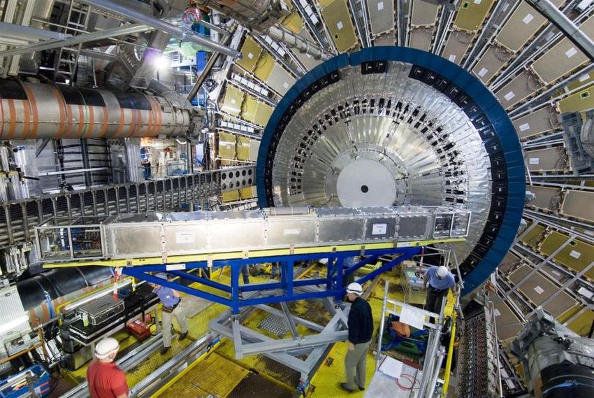
INFN and the National Frascati Laboratories

They are the largest laboratories in the INFN
The main feature is knowing how to build
particle accelerators. This school began in
1957, and continues until today.

 http://www.lnf.infn.it/edu/stagelnf/2009/relazioni_masterclass2009/milardi.pdf

 http://home.infn.it/immagini/picture.php?/552/tags/9-lnf

 Particle accelerators were born to study the constituent elements of matter
 and the laws that regulate their interaction.
INFN - Frascati National Laboratories

Frascati National Laboratories
The first Italian research facility for the study of nuclear and the subnuclear physics
with accelerator machines.

 Elettro sincrotrone (1959)

 AdA (1961) primo acceleratore particelle-
 antiparticelle

 DAFNE (1995)
 ADONE The Big AdA (1969)
Light from accelerators

1887: Joseph Larmor and Alfred Lienard study the case of the radiation emitted by an
electron in motion on a circular trajectory due to a centripetal acceleration.

1947: The visible part of the radiation emitted by
an accelerated electron beam in the small 70
MeV synchrotron of the Schenectady Laboratory
in New York is observed for the first time.

This radiation, for a long time, was
considered as a disturbance for
accelerating machines: the accelerated
particles lost part of their energy in the
form of radiation and this was then
supplied to them.

 1947 General Electric Res. Lab. - 70 MeV Electron
 Synchrotron – N.Y. USA
La Luce di Sincrotrone

 In the second half of the 1950s, synchrotron radiation was
 used in a small number of laboratories around the world to
 conduct pioneering experiments

Today the situation has radically changed:

 Synchrotron light becomes a truly
 unique radiation!

 Thousands of researchers work in
 around 40 laboratories where
 accelerators are designed, built and
 optimized to produce synchrotron
 light.

 ESRF, Grenoble - Francia 6 GeV, C = 844 m
 aperta agli utenti nel 1994

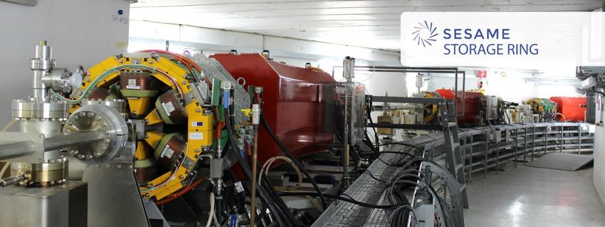
Synchrotron Radiation Sources in the world

Elettra (2.4 GeV) - Trieste DIAMOND (3 GeV) - UK SESAME (2.5 GeV) -
 Jordan

 In construction – Ultimate SR facilities

 Lund (3 GeV) - Svezia Sirius (3 GeV) - Brasile SSRF (3.5 GeV) Shanghai -Cina
Synchrotron radiation properties
Better image quality, using radiation doses similar to or lower than those used in
 conventional radiography.

 The comparison between the two images is clear:

 No other source of electromagnetic radiation presents, all together, the
 multiple and extraordinary characteristics of synchrotron light.
Medical applications

 Esame senologico - TC Tomografia computerizzata Esame senologico -ABI Analyser Based x-ray Imaging, 3D,
 convenzionale produce immagine 3D risoluzione 7 volte migliore.

 It guarantees much better resolutions. Micro calcifications, small deposits of
minerals can clearly be observed, which can indicate the presence of tumors and
 their shape and margins can be more accurately defined.
 A. Bravin - E.S.R.F. Grenoble
La Luce di Sincrotrone in paleobiologia
 Paleontologi dell’ Università di Renne in Francia e Imaging convenzionale
 ricercatori della facility di luce di sincrotrone ESRF a
 Grenoble hanno scoperto la presenza di 356 animaletti,
 inclusi in un pezzo (2 kg) di resina fossile di albero,
 completamente opaco, di 100 milioni di anni fa
 (periodo medio-Cretaceo)

 Imaging a contrasto di fase

 Ambra fossile opaca
M. Lak, D. Neraudeau, A. Nel, P. Cloetens, V. Perrichot and P. Tafforeau, Phase Contrast X-ray Synchrotron Imaging: Opening Access to Fossil Inclusions in Opaque Amber, Microscopy and Microanalysis, (2008), 14:251-259
La Luce di Sincrotrone in paleobiologia

 Sempre ad ESRF usando la microtomografia X a contrasto di
 fase e’ stato possibile effettuare una visualizzazione 3D dei
 microorganismi inclusi nel campione di resina fossile.

 a) Gastropod Ellobiidae; b)
 Myriapod Polyxenidae; c)
 Arachnid; d) Conifer branch
 (Glenrosa); e) Isopod
 crustacean Ligia; f) Insect
 hymenopteran Falciformicidae.

 Scarabeo del
 Cretaceo

M. Lak, D. Neraudeau, A. Nel, P. Cloetens, V. Perrichot and P. Tafforeau, Phase Contrast X-ray Synchrotron Imaging: Opening Access to Fossil
 Inclusions in Opaque Amber, Microscopy and Microanalysis, (2008)
Accelerators, Synchrotron
 radiation and Cultural
 Heritage
Fisica e Beni Culturali

The works of art are complex systems, made up of many different materials that
tend to deteriorate with time and exposure to the environment.

Understanding what a work of art is
composed of is essential for
preserving it

 We must imagine a work of art a bit like
 a "special" patient to whom an accurate
 diagnosis must be made before being
 able to perform “surgery”.
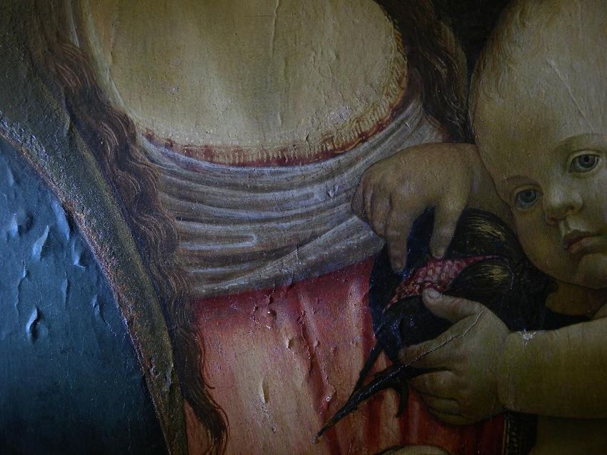
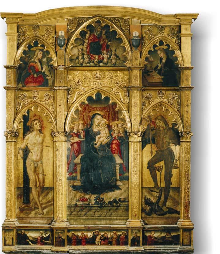
Quali domande ci poniamo davanti ad un’opera da analizzare?

• What are the materials used by the artist?
• What kind of technique did you use?
• Are there materials from later periods?
• Are there synthetic materials?
• Is there degradation?
• Is the cleaning method effective?
• Are the materials used compatible with the
 era or are we facing a fake?

 Niccolo’ Alunno: Madonna con Bambino e Santi (1499)

 Madonna della seggiola
 Dipinto su tavola, Raffello (1513)

 To answer these questions there is a need for a team of scientists, each with different
 diagnostic techniques that work together
CHNet: the INFN network for cultural heritage

The interest for non destructive diagnostic techniques and conservation of the archeological
and artistic heritage has enormously grown. CHNet-Cultural Heritage Network is the INFN
competence network for the study and conservation of artworks, using technologies which
are developed in the field of particle physics.

 CHNet Coordinator: Francesco Taccetti (ftaccetti@fi.infn.it)

 RESEARCH
 TECH-TRANSFER
 TEACHING
 DIAGNOSTIC SERVICES

 http://chnet.infn.it/it/chi-siamo-2/
CHNet: the INFN network for cultural heritage

The mission of INFN-CHNet is to become a reference point on the national
and international scale, for the multidisciplinary community of public and
private sectors that carry out their activities in the field of the study and
diagnosis of cultural heritage.

For decades, the INFN laboratories have not only used the most modern
technologies in this field, but, thanks to specific research projects, they are
developing new ones looking for solutions to the problems posed by operators in
the sector such as, for example, archaeologists, historians , restorers and
conservators.

http://chnet.infn.it/it/home-2/
CHNet: the INFN network for cultural heritage

The different diagnostic techniques provide complementary information that can be
useful for:

- restoration and conservation plans;
- study the materials used and the construction techniques;
- reconstruct the place of origin of the raw materials used for the construction of
the artifacts (so as to be able to reconstruct the trade routes, or to use the original
materials in the case of restoration and consolidation);
- dating artworks or archaeological sites;
- contribute to the authentication of the artworks.

http://chnet.infn.it/it/home-2/
CHNet: the INFN network for cultural heritage

The network is composed of nodes :

✓ INFN structures - level 1 nodes;
✓ Universities and Restoration Centers - level 2 nodes;
✓ Scientific Research Centers Abroad Outside Europe - Level 3 nodes

 Multidisciplinary and international network
Call for proposals: January 27, 2020
https://www.sesame.org.jo/news/sesame-call-proposals-0
CHNet: Le piattaforme operative
The scientific approach to conservation
Material ageing, climate change, atmospheric pollution, anthropic pressure
combined with inappropriate conservation and restoration procedures have also
contributed to degradation of artworks.
The modern approach to conservation requires a deep scientific investigation before
any treatment.

 Sampling techniques

 • Multispectral Imaging (UV, VIS, NIR, SWIR) • Micro-FT-IR Spectroscopy with
 • Micro-photography conventional and Syncrotron radiation
 • Raman Spectroscopy Source
 • FT-IR Spectroscopy • SEM-EDS
 • XRF • Micro-Raman Spectroscopy
 • FT-IR Spectroscopy
Metodi non distruttivi e micro-distruttivi
Imaging Techniques
Multispectral Imaging Techniques
Imaging techniques: are the conversion into visible photographic images of surface
interaction with electromagnetic radiation that cannot be detected by the human eye

 UV FLUORESCECE PHOTOGRAPHY IR REFLECTOGRAPHY
Non-invasive Analysis set up for in situ analyses (UV, VIS, NIR, SWIR)

 UV VIS
 OBJECT OBJECT

 UV LAMP VISIBLE LAMP

 BANDPASS - FILTERS

 REFLEX - CAMERA CONVERTED RELFEX-CAMERA
 OBJECT

 VISIBLE LAMP

 BANDPASS - IR FILTERS

 NIR SWIR
 CONVERTED RELFEX-CAMERA INGAAS-CAMERA
Multispectral imaging techniques

 Stratificazione del dipinto «Ritratto di Giovanna
 Tornabuoni» di Domenico Ghirlandaio :
 DIPINTO 1 preparazione (gesso e colla)
 2 disegno preparatorio
 3 verde di Boemia che fa da base a tutte le parti di
 incarnato
 UV 4-7 diversi strati di colore (poi completati da una mano
 di vernice)

 Dipinto

Lampade UV

 SISTEMA DI ACQUISIZIONE DI IMMAGINI
Multispectral imaging techniques
UV FLUORESCENCE IMAGING QUALITATIVE ANALYSIS
 (Camera acquisition system)
1) Modern restoration interventions (absence of uv fluorescence emission)

 olio su tavola
 «Madonna con
 DIPINTO Bambino» di
 Francesco di Giorgio
 Martini (XV sec.) –
 Accademia di San
 Luca (Roma)

 olio su tavola
 «Madonna con
 Bambino» di scuola
 veneta (XV sec.) –
 Accademia di San
 Luca (Roma)
Multispectral imaging techniques

 UV FLUORESCENCE IMAGING

 DIPINTO

olio su tavola «Madonna con Bambino e San Giovannino» di pittore toscano (Tommaso di Credi?) (inizi secolo XVI) – Palazzo Rosso (Genova)
Multispectral imaging techniques
UV FLUORESCENCE IMAGING

 2) Pictorial materials with UV fluorescence

 DIPINTO

 Olio su tavola «San Giovanni Evangelista, San Zaccaria e una
 Santa» di Francesco Brea (Nizza?, 1512 - 1555) – Palazzo
 Rosso (Genova)
Multispectral imaging techniques
UV FLUORESCENCE IMAGING
 Non solo dipinti….

Cavallo di ceramica della dinastia Han, China. Particolare dell’Afrodite detta "del Fréjus".
 National Gallery of Australia Museo del Louvre
Multispectral imaging techniques
 UV FLUORESCENCE IMAGING Not only paintings….
“Provvisorio 38” - foglio di pergamena del 15 secolo che contiene un frammento del “Filocolo” di Giovanni Boccaccio.

 Immagini multispettrali prese a 470 nm (b), 500 nm (c), 532 nm (d), 600 nm (e), 680 nm (f), 700 nm (g), e 750 nm (h)
Multispectral imaging techniques

 Stratificazione del dipinto «Ritratto di Giovanna
 Tornabuoni» di Domenico Ghirlandaio :
 DIPINTO 1 preparazione (gesso e colla)
 2 disegno preparatorio
 3 verde di Boemia che fa da base a tutte le parti di
 incarnato
 VIS 4-7 diversi strati di colore (poi completati da una mano
 di vernice)

 Dipinto
 LED UV

Lampade visibili

 SISTEMA DI ACQUISIZIONE DI IMMAGINI
Tecniche di imaging multispettrale
FOTOGRAFIA IN LUCE RADENTE
 ✓ Deformazione del supporto in tela/tavola
 ✓ Rigonfiamenti, lacerazioni, strappi, cuciture o integrazioni
 ✓ La presenza di elementi per l’assemblaggio
 ✓ Lo spessore degli strati pittorici e loro sequenza di
 SISTEMA DI
 applicazione
 ACQUISIZIONE ✓ Sollevamenti, cadute e integrazioni del colore
 DI IMMAGINI

 Olio su tavola «Madonna con Bambino e San Giovannino di scuola fiorentina (XV sec.) – Accademia di San Luca (Roma)
CAMERE CCD Multispectral imaging techniques
FOTOGRAFIA SENSORE CCD o CMOS
 matrice di fotodiodi (al silicio) in grado
 di trasformare un segnale luminoso in
 un segnale elettrico (effetto
 fotoelettrico)

 IMMAGINI A COLORI
 filtra RGB o Matrice di Bayer o Color filter array (CFA) o Color filter
 mosaic (CFM)

 R = 700 nm
 G = 546.1 nm
 B = 435.8 nm
 Matrice di Bayer Combinazione RGB
Multispectral imaging techniques

 Pigmenti diversi
 stesi con il tuorlo

 Spettroscopia di riflettanza
➢ Fattore di riflessione spettrale

 ( )
 (λ) = 100
 ( )
Multispectral imaging techniques
Multispectral Reflectance Imaing
 450 nm 500 nm 550 nm 600 nm 650 nm 700 nm

 30 30

 25 25

 20 20

 R (%)
 R (%)

 15 15

 10 10

 5 5

 0 0
 450 500 550 600 650 700 450 500 550 600 650 700

 Wavelegth (nm) Wavelength (nm)
Multispectral imaging techniques

 Stratificazione del dipinto «Ritratto di Giovanna
 Tornabuoni» di Domenico Ghirlandaio :
 DIPINTO 1 preparazione (gesso e colla)
 2 disegno preparatorio
 3 verde di Boemia che fa da base a tutte le parti di
 incarnato
 IR 4-7 diversi strati di colore (poi completati da una mano
 di vernice)
 Dipinto LED UV

Lampade alogene

 Filtro IR

 SISTEMA DI ACQUISIZIONE DI IMMAGINI
 (macchina fotografica modificata)
Multispectral imaging techniques
IR REFLECTOGRAPHY La riflettografia IR viene impiegata principalmente per:

 • Preparatory drawings or hidden tracks
 • “Pentimenti”
 • Identification of some pigments or heterogeneity of the pictorial layout
 • Characterization of mixed inks

 VISIBILE (B/N) RIFLETTOGRAFIA IR

 Olio su tela «Annunciazione» Lorenzo di Credi – Accademia di San Luca (Roma)
Tecniche di imaging multispettrale
IR REFLECTOGRAPHY La riflettografia IR viene impiegata principalmente per:

 • Preparatory drawings or hidden tracks
 • “Pentimenti”
 • Identification of some pigments or heterogeneity of the pictorial layout
 • Characterization of mixed inks

 VISIBILE (B/N) RIFLETTOGRAFIA IR

 Olio su tela «Madonna con Bambino e San Giovannino» Lorenzo di Credi – Galleria Borghese (Roma)
Tecniche di imaging multispettrale
RIFLETTOGRAFIA IR La riflettografia IR viene impiegata principalmente per:

 • Preparatory drawings or hidden tracks
 • “Pentimenti”
 • Identification of some pigments or heterogeneity of the pictorial layout
 • Characterization of mixed inks

 VISIBILE RIFLETTOGRAFIA IR

 Affresco romano «Il Grifo» – Ostia Antica (Roma)
Tecniche di imaging multispettrale
RIFLETTOGRAFIA IR La riflettografia IR viene impiegata principalmente per:

 • Preparatory drawings or hidden tracks
 • “Pentimenti”
 • Identification of some pigments or heterogeneity of the pictorial layout
 • Characterization of mixed inks

 VISIBILE RIFLETTOGRAFIA IR

 Olio su tavola «Madonna col Bambino e i santi Giovanni Battista e Maria Maddalena » Palma il Vecchio – Palazzo Rosso (Genova)
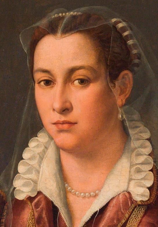
Tecniche di imaging multispettrale
RIFLETTOGRAFIA IR La riflettografia IR viene impiegata principalmente per:

 • Preparatory drawings or hidden tracks
 • “Pentimenti”
 • Identification of some pigments or heterogeneity of the pictorial layout
 • Characterization of mixed inks

 VISIBILE RIFLETTOGRAFIA IR

 Olio su tavola «Ritratto femminile» Angiolo Allori (Bronzino) – Accademia di San Luca (Roma)
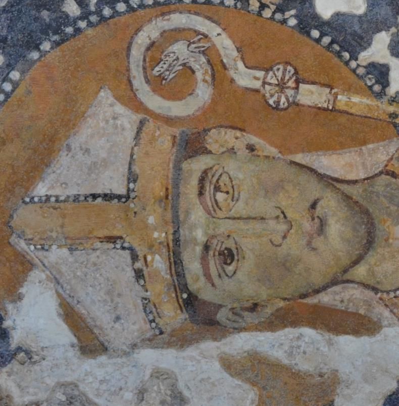
Tecniche di imaging multispettrale
RIFLETTOGRAFIA IR La riflettografia IR viene impiegata principalmente per:

 • Preparatory drawings or hidden tracks
 • “Pentimenti”
 • Identification of some pigments or heterogeneity of the pictorial layout
 • Characterization of mixed inks

 VISIBILE RIFLETTOGRAFIA IR

 Affresco a Santa Luca delle Malve (Matera)
Tecniche di imaging multispettrale
RIFLETTOGRAFIA IR La riflettografia IR viene impiegata principalmente per:

 • Disegni preparatori o tracce nascoste
 • Eventuali pentimenti
 • Identificazione di alcuni pigmenti o eterogeneità della stesura pittorica
 • Caratterizzazione degli inchiostri misti

 Inchiostro ferrogallico

 Inchiostro vegetale

 Inchiostro a base di carbonio

 RIFLETTOGRAFIA IR
Tecniche di imaging multispettrale
 RGB 450 nm 550 nm 600 nm
 VIS
UV

 RGB 600 nm 650 nm 750 nm 1000 nm
Tecniche di imaging multispettrale

 Stratificazione del dipinto «Ritratto di Giovanna
 Tornabuoni» di Domenico Ghirlandaio :
 DIPINTO 1 preparazione (gesso e colla)
 2 disegno preparatorio
 3 verde di Boemia che fa da base a tutte le parti di
 incarnato

Raggi X 4-7 diversi strati di colore (poi completati da una mano
 di vernice)

 Image plate per raggi X

 dipinto

 Tubo a raggi X
Tecniche di imaging multispettrale
RADIOGRAFIA
 La radiografia consente l’analisi della struttura del supporto sia per quanto
 riguarda la realizzazione che lo stato di conservazione e permette di trarre
 informazioni sulla tecnica esecutiva

 VISIBILE RADIOGRAFIA

 Olio su tavola «Madonna con Bambino e San Giovannino» di scuola fiorentina – Accademia di San Luca (Roma)
Tecniche di imaging multispettrale
RADIOGRAFIA

 RADIOGRAFIA
 abete castagno

 VISIBILE

 Olio su tavola «Madonna con Bambino e Santa Caterina d’Alessandria» di Matteo di Giovanni – Accademia di San Luca (Roma)
Tecniche di imaging multispettrale
RADIOGRAFIA Non solo dipinti….

 Foto del pane di terra Asse di Tiberio per il Divo Augusto (14-37 d.C.)
 durante la radiografia Radiografia frontale

 Immagine radiografica Immagine dell’oggetto dopo il
 laterale restauro
 Quadrante di Claudio (41-54 d.C.)
 Ipotesi di
 Immagine visibile Radiografia attribuzione
 Piccolo pane di terra provenienti da Crustumerium , che
 conteneva una fibuletta in ferro. Lo spillo della fibula ed La radiografia consente il recupero dell’impronta del
 il suo gancio sono quasi del tutto scomparsi ed anche il conio anche in situazioni di avanzato stato di corrosione
 corpo della fibula risulta fortemente compromesso. della moneta.
 Inoltre a causa della forte disidratazione della terra si
 erano formate delle fratture
Tecniche di imaging multispettrale
TOMOGRAFIE Non solo dipinti….
 La tomografia consente lo studio dello spessore dello strato di
 metallo e l’identificazione di buchi, deformazioni e riparazioni.

 riparazioni

 Bolle
 d’aria

 Statua romana di bronzo Roman bronzo di
 Cupido (96.AB.53) - Getty Museum
SPECTROSCOPIC TECHNIQUES
Le tecniche spettroscopiche

 Sorgente Campione Rivelatore

 The sample absorbs some components of the incident radiation.
By analyzing the transmitted light we can obtain information on the
 chemical nature of the sample
FT-IR micro spectroscopy and imaging
LA SPETTROSCOPIA INFRAROSSA
Microscopy and Imaging

The objective of mapping and imaging is to generate an image, called a chemical
image, containing spectral information, which can be superimposed on the visible
image.
Depending on the detector used, it is possible to obtain a chemical image:

 • Point by point (raster scan, mapping):
 single point detector MCT (250 μm)
 • In a single shot (using matrix detectors
 FPA: 64x64, 128x128, 256x256, pixel
 40 μm size)
Analisi di sezioni stratigrafiche

 Studio dei processi di degradazione
 dei pigmenti gialli di Van Gogh

 http://www.vangogh.ua.ac.be/
Sezioni stratigrafiche: micro-distruttivo
Studio del degrado dei pigmenti gialli di Van Gogh

Il "Girasoli " di Vincent Van Gogh

Pigmenti gialli a base di cromo
(PbCrO4, PbCrO4 · xPbSO4, o PbCrO4 · xPbO) Non invecchiato Invecchiato
Studio del degrado dei pigmenti gialli di Van Gogh

Il motivo dell’alterazione risiede nel cambiamento del numero di ossidazione del
cromo (da Cr(VI) a Cr(III)) che avvene soprattutto in presenza di solfati e solfuri

 Diminuzione della banda a 1050 cm−1 [ν1(SO42- )]
 e una modifica dell’intensità relativa delle
 bande a 620 e 597 cm−1 [ν4(SO42- ]

 Modifica della struttura dei solfati indica che il
 processo di degrado è in atto
Analisi dei prodotti di degrado

L’ Arco di Settimio Severo
In-situ analysis
ʺPALAZZO CHIGIʺ (Ariccia, Rome)

 Portable Spectrometer ALPHA (Bruker)

 Sanguigna (o Sinopia) di
 Gian Lorenzo Bernini
XRF analyses at Tarquinia (Tomba dei Demoni Azzurri) Ca-K 14 Vestito Floreale Blu Blu Gonna Riga Blu Gonna 12 Grigio Gamba Demone 10 Counts (×103) 8 6 Cu-K Ca-K 4 Fe-K Si-K/ Cu-K Fe-K 2 0 0 2 4 6 8 10 Energy (keV) Pigmento Identificazione Neri Pigmenti Organici Blu Cuprorivaite o Blu Egizio: (CaCuSi4O10 or CaO·CuO·4SiO2) Rossi/Marroni Ocre ed Ossidi di Ferro: (Rossi: ematite-Gialli: goetite) Romani, M., et.al., NON-DESTRUCTIVE CHARACTERIZATION OF FRESCOS IN THE ETRUSCAN TOMB OF BLUE DEMONS (ITALY) BY SPECTROSCOPY TECHNIQUES, Presented at Art'17, 22th-24th November 2017, Torino
Time Gated (TG) Laser Induced Fluorescence (LIF)

 1.0

 Normalized LIF intensity
 0.8

 0.6

 0.4

 0.2

 0.0

 400 500 600 700 800 900 1000
 Wavelength (nm)
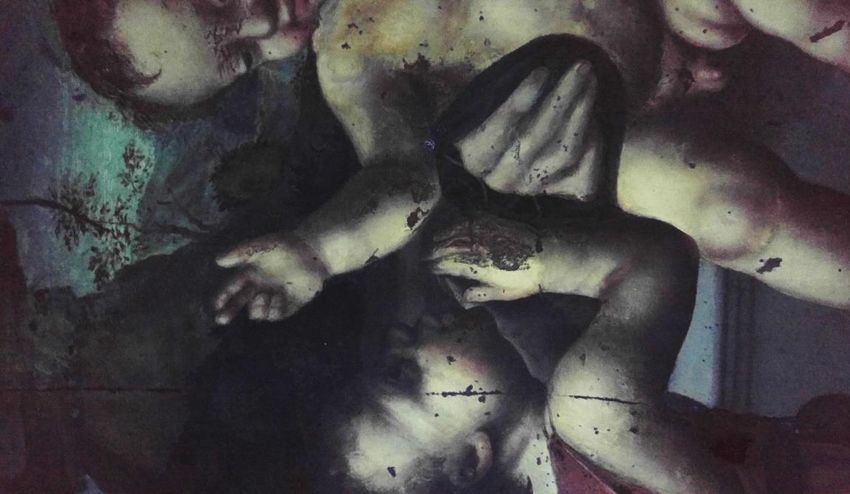
Caratterizzazione di Opere d’Arte Contemporanea
 Nato Frascà, Nascita della forma, 1962, Museo MACRO (Roma)

 Oil

 ?

 A B C
 A. Foto B. Fluorescenza C. Dettaglio
 109
 1010

 108 109
 Opera catalogata Olio su tela
 107
 108
 Pigmenti blu e arancio
 appaiono maggiormente
Counts

 7
 10
 Counts

 106

 5
 No Delay
 30 ns Delay
 106
 No Delay
 degradati
 10
 30 ns Delay
 105

 104
 Identificazione di legante
 PAINT
 104
 REFERENCE acrilico nelle zone più
 103
 250 300 350 400 450 500 550
 103
 250 300 350 400 450 500 550 degradate
 Wavelenght (nm) Wavelength [nm]

 Romani, M., et al., A preliminary study of contemporary binders by TR-LIF spectroscopy for the study of the painting “Nascita della
 Forma” by Nato Frascà, Proceedings of LACONA XI
Importanza di avere un laboratorio di
diagnostica nei Musei
Sviluppo di tecnologie avanzate:
l’acceleratore portatile «MACHINA»

 Corriere della Sera, 25-08-2019
• INFN-LNF:
• M.Cestelli-Guidi, A. Grilli, A. Raco (FTIR and Raman analysis set-
 up)

• INFN-RM2:
• M. Marinelli, G. Verona Rinati (XRF, Multispectral Imaging, TG-LIF
 portable system analysis),

• CHNet: F. Taccetti (National Coordinator), V. Virgili (Latium
 Spokesperson),
• M. Cestelli-Guidi (INFN-LNF Spokesperson)
Follow us :)

 http://w3.lnf.infn.it/

 https://web2.infn.it/Dafne_Light/
 cestelli@lnf.infn.it

http://chnet.infn.it/it/home-2/
You can also read