Thank you for viewing this presentation. We would like to remind you that this material is the property of the author. It is provided to you by ...
←
→
Page content transcription
If your browser does not render page correctly, please read the page content below
Thank you for viewing this presentation.
We would like to remind you that this
material is the property of the author.
It is provided to you by the ERS for your
personal use only, as submitted by the
author.
2016 by the author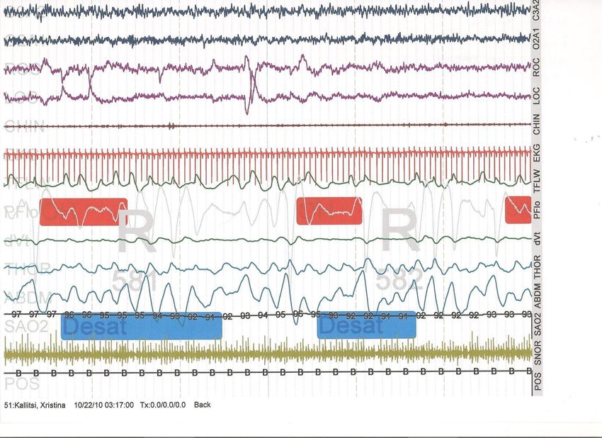
How to interpret sleep
      studies?

           Refika Hamutcu Ersu, MD
              Marmara University
      Division of Paediatric Pulmonology
               Istanbul - Turkey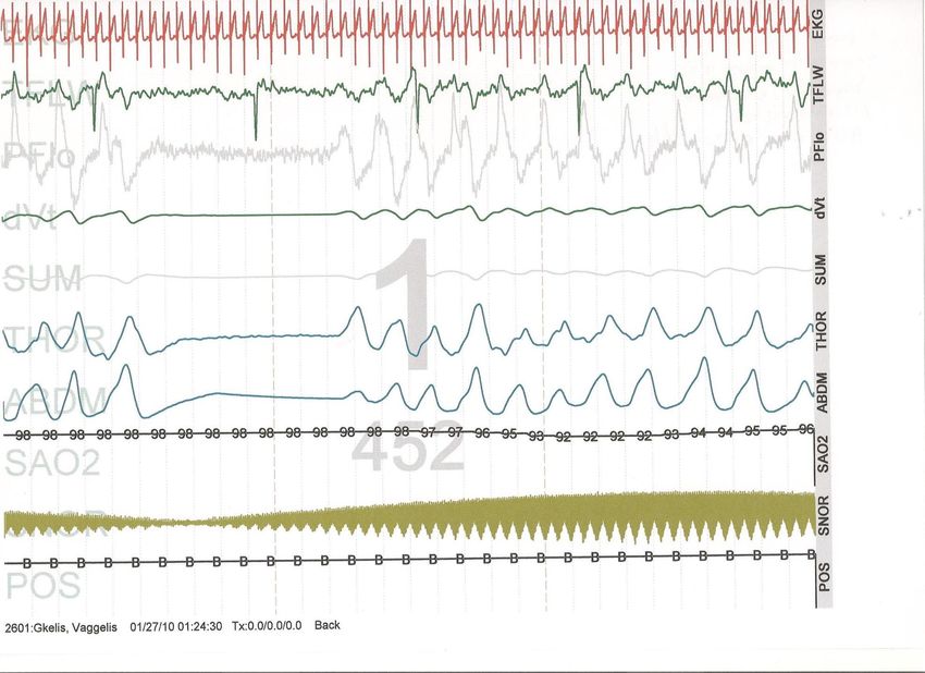
Conflict of interest disclosure I have no real or perceived conflicts of interest that relate to this presentation. This event is accredited for CME credits by EBAP and EACCME and speakers are required to disclose their potential conflict of interest. The intent of this disclosure is not to prevent a speaker with a conflict of interest (any significant financial relationship a speaker has with manufacturers or providers of any commercial products or services relevant to the talk) from making a presentation, but rather to provide listeners with information on which they can make their own judgments. It remains for audience members to determine whether the speaker’s interests, or relationships may influence the presentation. The ERS does not view the existence of these interests or commitments as necessarily implying bias or decreasing the value of the speaker’s presentation. Drug or device advertisement is forbidden.
Laboratory Evaluation
Test to determine diagnosis and
             severity:
       POLYSOMNOGRAPHY
      (Poly-somnus-graphein)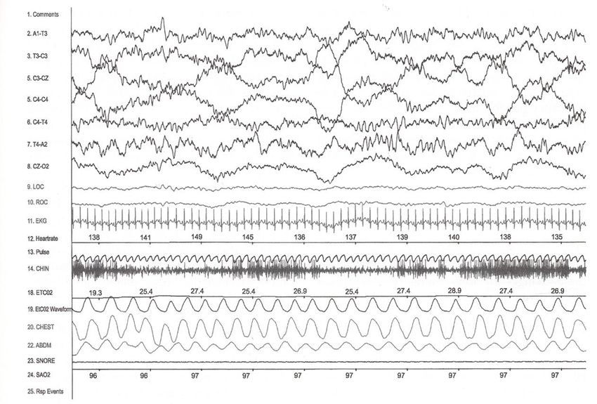
Overnight Polysomnogram
  • Sleep stage analysis
  • Arousal summary
  • Heart rate/rhythm
  • Snoring
  • Leg movements
  • Respiratory events
  • Gas exchange
Is sleep staging needed in PSG?
• REM sleep may be the period of the worst
  obstruction
• Identification of arousal from sleep is
  essential to diagnose UARS
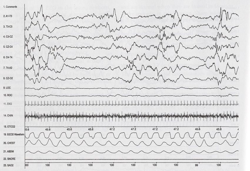
Sleep in Newborns and Infants
Sleep in Newborns and Infants
• REM and NREM sleep states organized
  at third trimester
• 3 sleep states in term newborns:
  active, quiet, indeterminate
• Infants may enter sleep through REM
• Critical sleep reorganization
  at 8-12 wks; more sleep at night
• Development of NREM sleep by 6 mo;
  decreased REM amounts
         Grig-Damberger M, J of Clinical Sleep Medicine 2007;3:201-40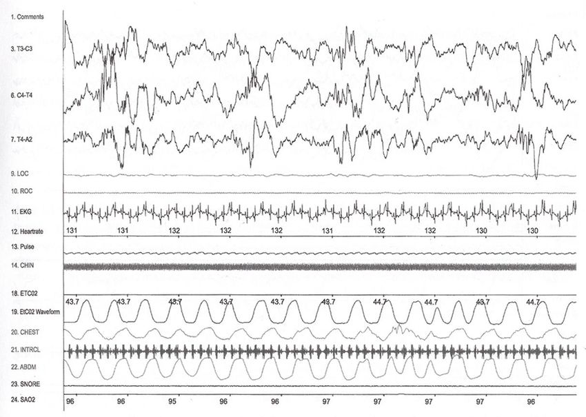
Quiet Sleep in Newborn
Active Sleep in Newborn
Sucking Artefact
Visual Scoring Pediatric Rules

Pediatric sleep scoring rules can be applied to
    children 2 months (post-term) or older
Sleep Stages
•   Stage   W
•   Stage   N1 (NREM 1)
•   Stage   N2 (NREM 2)
•   Stage   N3 (NREM 3)
•   Stage N (NREM)
•   Stage R (REM)
Dominant posterior rhythm
In children:
Dominant Posterior Rhythm replaces- Alpha rhythm
(8-13 Hz).
Dominant Posterior Rhythm (DPR) The dominant
reactive EEG rhythm over the occipital regions in
relaxed wakefulness with eyes closed( slower in
infants and young children) attenuates with eyes
open.
Frequency- 3.5-4.5 hz 3-4 mo post term
5-6 hz 5-6 mo
7.5- 9 hz by age 3 yo, amplitude>50 uVSleep Stage Analysis • Sleep architecture: The analysis of sleep structure throughout the night • Sleep latency: The time from lights out to sleep onset (25 minutes) • REM latency: The time from sleep onset to the first REM sleep period • Sleep efficiency: Calculated by dividing the wake after sleep onset by the total sleep time (>89%) • The sleep stage percentages for the entire recording are also reported
Sleep Cycles
• The first cycle begins by moving from
  wakefulness to non-REM
• The first REM follows the first non-REM
  (70-90 min after sleep onset)
• The 2 sleep states alternate (90-120 min
  intervals)
• Normal sleep usually consists of 4-6 non-
  REM/REM sleep cycles (90-110 min per
  cycle).
• As the night progresses, REM sleep
  periods increase in length

    Kheirandish Gozal L, Pediatr Resp Rev 2006; 75: 550-54
Sleep Architecture in Children w/OSAS
Goh DYT, AJRCCM 2000;162:682-6Stage 1
Moving from wakefulness to non-REM sleep
Rolling eye movements, brain waves >13 Hz dominantStage 2
Relatively low-voltage, mixed frequency EEG background
Sleep spindles (10-13 Hz), K complexesStage 2
Stage 3

      High amplitude delta waves
occupying 20-50% of the epoch-stage 3
 occupying >50% of the epoch-stage 4
Effects of Sleep on Breathing NREM Sleep • Central depression of breathing • Regular timing and amplitude • Decreased chest wall stability • Mild hypoxia and hypercapnia
REM Sleep

  Low voltage, fast frequency brain waves
EMG shows inactivity of all voluntary muscles
REM Sleep
REM Sleep
Effects of Sleep on Breathing

REM Sleep
 •Not controlled by chemoreceptors
 •Irregular timing and amplitude
 •Marked inhibition of skeletal muscle tone
  (except diaphragm, extraocular)
 •Marked hypoxia and hypercapnia
‘Boy are my eyes tired’ had
  REM sleep all night long”
Percentage of apneas in different sleep
                stages

                SWS

               REM

                  Goh DYT, AJRCCM 2000;162:682-6
Sleep Scoring Data • Lights Out • Lights On • Total Sleep Time • Total Recording Time • Sleep Latency • Stage REM Latency • Wake after sleep onset (WASO) • Sleep Efficiency • Time in Each Stage • Percent of TST in each stage
Arousal

Abrupt shift in EEG frequency
At least 3 secons in duration
Arousal Events
• Total number of arousals, and
• The arousal index (number of arousals
  divided by the hours of sleep) should be
  reported.
• Normal children have a mean arousal
  index of 8.8 to 9.5
• Arousals attributed to respiratory events
  give a mesure of sleep distruption
  caused by those respiratory events.
Central Apnea
              >20 sec or 2 breaths w/
               Desat>3% or Arousal

 Flow

Rib Cage

Abdomen

                    Time
Central Apnea
Central Apnea
Periodic Breathing
≥3 respiratory pauses of ≥ 3 sec with less than
 20 sec of normal respiration between pauses
Mixed Apnea
                  Cessation of
               airflow (at least 2
                     breaths)

  Flow

Rib Cage

Abdomen

                   Time
Obstructive Apnea
Cessation of
airflow (at least 2
breaths)
Flow
Rib Cage
Abdomen
TimeHypopnea
Reduction in airflow
>50%, lasting 2 breaths
w/ 3% desat/arousal
Flow
Rib
cage
Abdomen
SpO2
TimeObstructive Apnea-Hypopnea
Respiratory Effort Related Arousal
• When using nasal pressure:
  There is fall in the amplitude of signal from a
  nasal pressure sensor but
Respiratory Events
• Number of Obstructive Apneas
• Number of Mixed Apneas
• Number of Central Apneas
• Number of Hypopneas
• Number of Apneas + Hypopneas
• Apnea Index (AI)
• Hypopnea Index (HI)
• Apnea + Hypopnea Index (AHI)
• Respiratory Effort Related Arousal
• Snoring
Respiratory Events
• Respiratory Effort Related Arousal Index
• Oxygen Desaturations ≥ 3% or ≥ 4%, total
  number
• Oxygen Desaturation Index ≥ 3% or ≥ 4%
• Continuous Oxygen Saturation, mean value
• Minimum Oxygen Saturation during sleep
• Occurrence of hypoventilation (yes/no)
• Occurrence of Cheyne-Stokes breathing
  (yes/no)
Normal Polysomnography Values in
            Children
Movement Events • Number of periodic limb movements of sleep (PLMS): Movements in either or both legs, lasting 0.5 to 5 sec, seperated by 5 to 90 sec, in clusters of 4 • Number of periodic limb movements of sleep (PLMS) with arousals • PLMS index >5 is abnormal • PLMS arousal index (PLMS)
Cardiac Events
• Average heart rate
• Highest heart rate during sleep
• Highest heart rate during recording
• Bradycardia: report highest and lowest
• Asystole: report longest pause observed
• Sinus tachycardia during sleep: report highest heart
  observed
• Narrow complex tachycardia: report highest heart rate
  observed
• Wide complex tachycardia: report highest heart rate
  observed
• Atrial fibrillation
         • Occurrence of other arrhythmias (yes/no)
Polysomnography in Pediatric SDB
• Can be succesfully performed in children
• Should be performed in a lab experienced
with the care of children
• Should include measurement of CO2
• Criteria for scoring and interpretation differ
from those of adults
Schechter, Pediatrics 2002;109:e69Case 1 Airflow Thoraci c Abdomi nal RIP SaO2 (%)
Case 1

• Which respiratory abnormality is observed during this PSG fragment?

   1.   Periodic Breathing
   2.   Central apneas of abnormal duration and associated with desaturation
   3.   Obstructive apneas associated with desaturation
   4.   Obstructive apneas associated with paradoxical breathing movements and
        desaturation
Case 1 • Boy, 9 years old. • Past medical history: recurrent ependymoma located at the posterior fossa. Repeated neurosurgery, chemotherapy, radiotherapy. • Complains of always snoring, fatigue during daytime and recurrent respiratory infections.
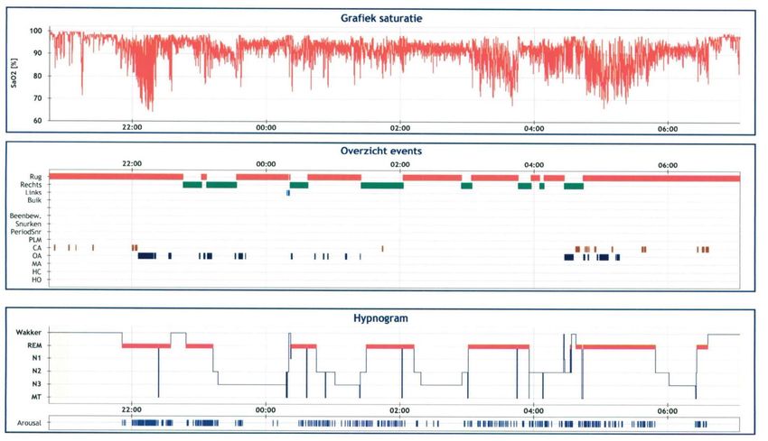
Case 1

• The following pages present the hypnogram and a summary of the
  respiratory parameters of this patient.
Case 1

• What is your diagnosis?

   1.   Central sleep apnea
   2.   Obstructive sleep apnea
   3.   Mixed sleep apnea
   4.   Nocturnal hypoxemia not related to sleep-disordered breathing
Case 2

     • Boy, 8 years old.
     • Referred for snoring and witnessed apneas.
     • Past medical history:
         • Adenotonsillectomy
         • Respiratory allergies – momethasone spray, ceterizine
         • Chronic headache – no causes identified at a local
           hospital including brain MRI
Case 2
Case 2

• Which respiratory abnormality is observed during this PSG fragment?

   1.   Periodic Breathing
   2.   Central apneas
   3.   Obstructive apneas
   4.   Mixed apneas
Case 3

• Girl, 14 years old
• CP
• Epilepsy
• No known respiratory morbidity
• Increasing frequency and severity of snoring and witnessed apneas
  since a few weeks.
Case 3
Case 3: with O2
Case 3: with 02
Case 3: with O2
Case 3
• Which kind of respiratory events are observed and what is the effect
of supplemental oxygen on these events?
1. Obstructive apneas with the occurrence of central apneas during
supplemental oxygen treatment
2. Obstructive apneas with more frequent obstructive apneas during
supplemental oxygen treatment resulting in less severe oxygen
desaturation but increase in CO2 levels.
3. Obstructive apneas with longer obstructive apneas during supplemental
oxygen treatment resulting in increased CO2 levels.
4. Central apneas with longer central apneas during supplemental oxygen
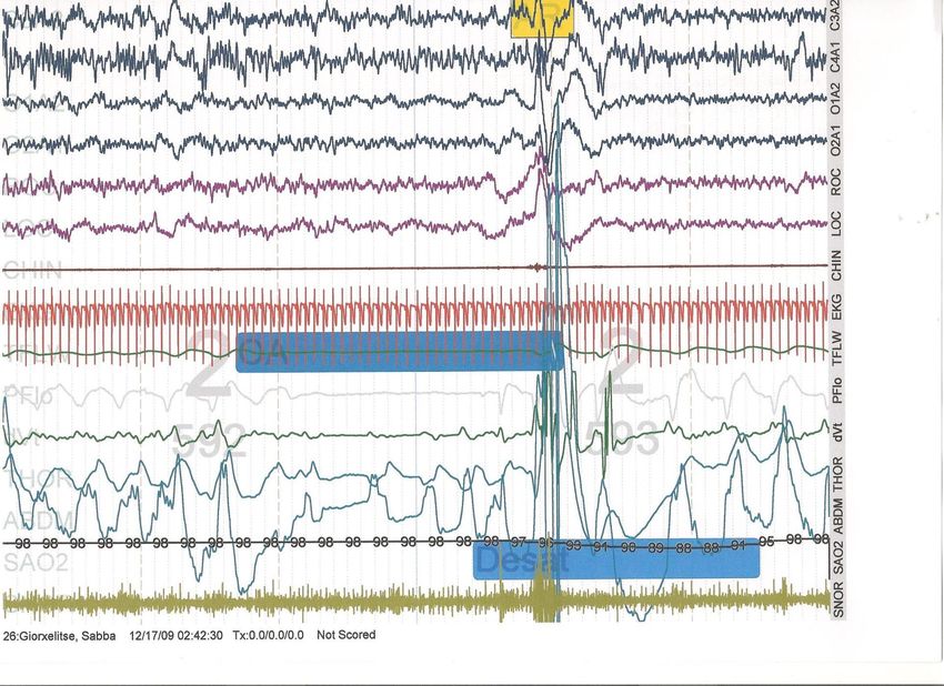
treatment resulting in increased CO2 levels.3 y.o, snoring, mouth-breathing, tonsillar hypertrophy, AHI 4.4 episodes/h
1. What is the sequence of events in the presented epoch?
2. What is the event marked in the EEG recording?
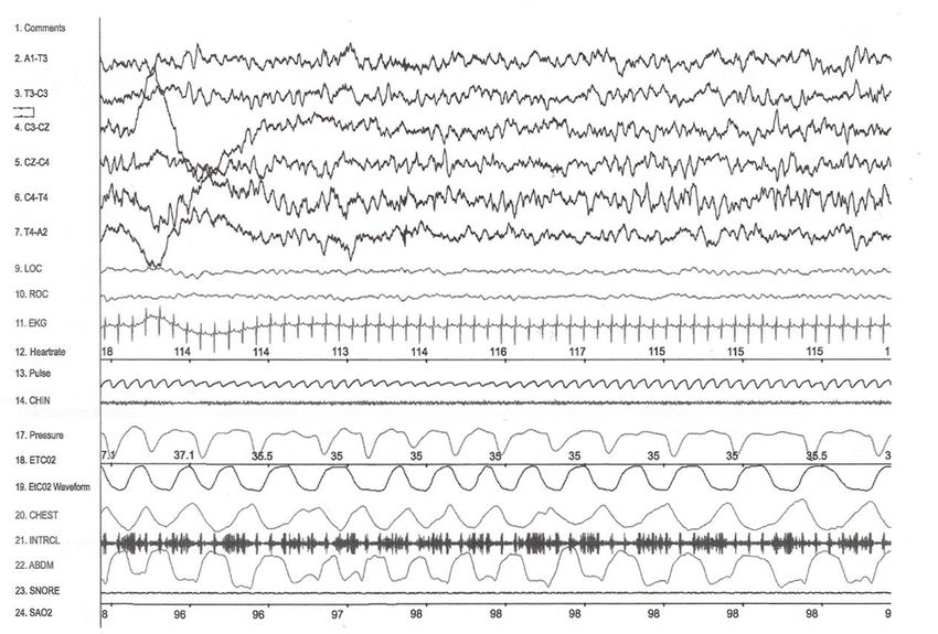
1 y.o., noisy breathing, adenoidal hypertrophy
1. What is the respiratory event in the presented epoch?
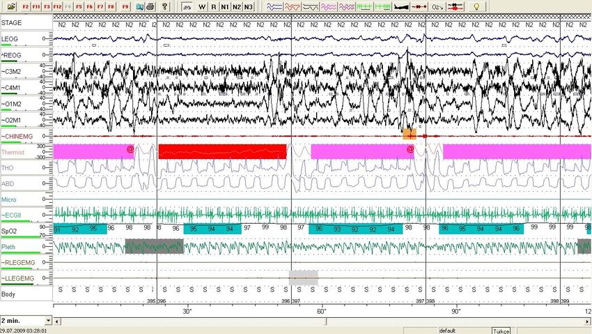
6 y.o, Prader-Willi syndrome
1.   What is the respiratory event in the presented epoch?
2.   What is the stage of sleep?
3.   How would you characterize the chest and abdominal
     wall movements?
Thanks for your attention
You can also read