Ear Abnormalities ARTICLE - BINASSS
←
→
Page content transcription
If your browser does not render page correctly, please read the page content below
ARTICLE

      Ear Abnormalities
      Sana L. Bhatti, MD,*† Lauren T. Daly, MD,‡ Martha Mejia,* Chad Perlyn, MD, PhD*†
      *Division of Plastic Surgery, Nicklaus Children’s Hospital, Miami, FL
      †
       Division of Plastic Surgery, Florida International University College of Medicine, Miami, FL
      ‡
       Division of Plastic Surgery, University of Massachusetts, Worcester, MA

                                                               PRACTICE GAPS

                                                               1. To facilitate prompt treatment for congenital ear abnormalities, pediatric
                                                                  health-care providers should be able to identify common ear defor-
                                                                  mities, some of which can be treated nonsurgically only if recognized
                                                                  within the neonatal period.
                                                               2. Clinicians should know when to refer patients with ear abnormalities to
                                                                  specialists for timely intervention.

                                                               OBJECTIVES After completing this article, readers should be able to:

                                                               1. Understand the normal anatomy of the ear.
                                                               2. Identify common congenital ear abnormalities as they present in the
                                                                  neonatal period.
                                                               3. Recognize the psychosocial impact of ear differences on pediatric
                                                                  patients.
                                                               4. Facilitate prompt diagnosis of congenital ear abnormalities and refer
                                                                  patients to specialists so that nonsurgical treatment can be initiated in
                                                                  the neonatal period.

                                                               ABSTRACT

                                                               Congenital ear abnormalities present an aesthetic and psychosocial concern
                                                               for pediatric patients and their parents. Diagnosis of external ear
                                                               deformities is based on clinical examination and is facilitated by an
                                                               understanding of normal ear anatomy. Ear anomalies can be categorized as
                                                               malformations or deformations. Malformations are characterized by
                                                               absent anatomical structures of the ear (or absence of the ear itself), as
                                                               exemplified by microtia and anotia. Ear deformations are characterized by
                                                               ear anatomical landmarks that are present but are distorted or abnormal,
                                                               with Stahl ear, constricted ear, and prominent ear being common
                                                               presentations. Ear malformations will not improve with growth of the
                                                               patient and uniformly require surgical intervention to recreate an
                                                               anatomically typical ear. Although a small percentage of ear deformations
      AUTHOR DISCLOSURE Drs Bhatti, Daly,
                                                               can self-resolve, most patients with ear deformations will require nonsur-
      and Perlyn and Ms Mejia have no financial
      relationships relevant to this article. This             gical or surgical reconstruction to achieve a normal or more aesthetic ear.
      review does not contain a discussion of an               In recent decades the use of nonsurgical ear splinting or molding has been
      unapproved/investigative use of a                        recognized as a highly effective method in correcting a variety of congenital
      commercial product/device.

180   Pediatrics in Review
                                Downloaded from http://pedsinreview.aappublications.org/ at Swets Blackwell Inc. on April 8, 2021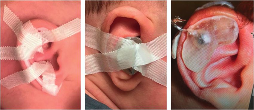
ear deformations when treatment is initiated in the first 8 weeks of life. The urgency in initiating nonsurgical
     treatment of ear deformations at an early age makes prompt recognition of these ear deformations essential
     because surgical correction remains the only viable reconstructive option in older infants and children.

INTRODUCTION                                                            deformations nonsurgically, early referral to a specialist is
Congenital ear abnormalities are the result of an absence or            preferred. Auricular malformation or deformity can cause
malformation of the skin and/or cartilage of the neonatal ear.          significant psychological and social morbidity, including
Auricular anomalies can be categorized as either malfor-                issues with poor self-esteem, social avoidance, anxiety, de-
mations or deformations. Malformations are due to dis-                  pression, and behavioral problems. (6)(7)(8) Fortunately,
rupted embryogenesis, which results in deficient growth of               these symptoms significantly improve after reconstruction of
structures. Examples of malformations include anotia (ab-               the ear anomaly. (6)(7)
sence of external ear), microtia (underdeveloped, usually
malformed ear), cryptotia (ear cartilage partially buried be-           Anatomy
neath the skin), and preauricular sinuses and remnants. (1)             The external ear, or auricle, is the most peripheral compo-
Deformational auricular anomalies have an intact but dis-               nent of the auditory mechanism (Fig 1). The external ear acts
torted chrondrocutaneous framework. Ear deformations are                to funnel sounds to the tympanic membrane in a way that
thought to be due to external forces in utero or ex utero, thus         boosts sound frequencies associated with the human voice
leading to abnormal ear architecture. (1) A variety of different        and aids in differentiating the spatial origin of sounds. (9) In
ear deformations have been described, including Stahl ear,              addition to its role in hearing, the ear is an important
constricted ear, and prominent ear. (2) Along with external             component of craniofacial aesthetics, with minor ear dif-
ear abnormalities there can be hearing loss; this is most               ferences attracting biased visual attention from viewers. (10)
commonly seen with microtia.                                               The external ear is composed of elastic cartilage covered with
                                                                        hairless skin that is tightly adherent anteriorly and more lax
Epidemiology and Pathophysiology                                        posteriorly. The cartilaginous framework of the ear can be viewed
Although the true incidence of congenital ear abnormalities             as a topographical map, with the helix and lobule as the most
is not known, estimates range from 15% to 20% of newborns.              elevated structures, the antihelix and tragus located midlevel, and
(3) Patients with an ear malformation, such as microtia or              the concha presenting as the deepest aspect of the ear. (1) The
anotia, should not be expected to have any spontaneous                  cartilaginous ridges have associated depressions, or scapha (Fig 1).
improvement. On the contrary, up to 30% of patients with a                 Embryologic development of the external ear begins
recognized ear deformation at birth will experience self-               during the fifth week of gestation, with the fetal auricle re-
resolution by 4 to 6 weeks of age. (3)(4)(5) However, be-               sembling the adult ear by the ninth week of gestation. By age
cause of the small window of opportunity to treat these                 3 years, approximately 85% of adult ear growth has been
                                                                        attained. (11)

Figure 1. External ear.                                                 Figure 2. Stahl ear. Left, before. Right, 6 weeks after ear molding.

                                                                                                             Vol. 42 No. 4      APRIL 2021     181
                   Downloaded from http://pedsinreview.aappublications.org/ at Swets Blackwell Inc. on April 8, 2021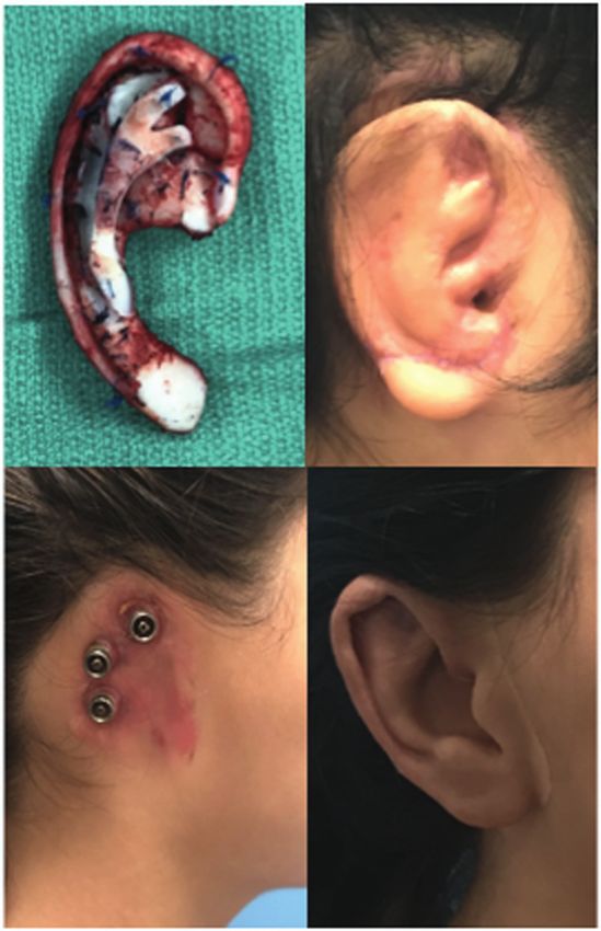
CLINICAL ASPECTS OF EAR DEFORMATIONS from the side of the head and generally has an appreciably large
Stahl Ear surface area. (17) It is estimated that prominent ear is present in
Stahl ear is characterized by an additional abnormal vertical approximately 5% of the white population. (18) Prominent ear
cartilage band crossing the scaphoid fossa from the antihelix can be associated with a large psychological burden for both
to the helix and creating a pointed, elflike appearance to the children and adults. (19)(20)(21) Multiple different operative
ear (Fig 2). (12) The etiology of Stahl ear is unclear, but an techniques, including suture-based techniques, cartilage scor-
anomalous insertion of the transverse auricular muscle has ing, or cartilage excision, have been described to surgically
been implicated. (13) Similar to other congenital ear defor- correct prominent ear. (22)(23) When prominent ear is recog-
mities, Stahl ear that is recognized early can be treated nized and treated at a very young age, ear molding and splinting
successfully in the neonatal period with splinting and are successful methods of reconstruction. (24)
molding. (14)(15) If treated later in childhood, Stahl ear can be
corrected with surgery by resecting or repositioning the Constricted Ear
abnormal vertical cartilage band and reconstructing a more Constricted ear refers to deformities of the superior third of the
normal-appearing antihelical superior crus. (12) auricle; presentation can be diverse (Fig 4). A constricted ear has
been described by several terms, including cup ear, lidded ear, lop
Prominent Ear ear, canoe ear, and cockleshell ear. Cosman (25) described 4 fun-
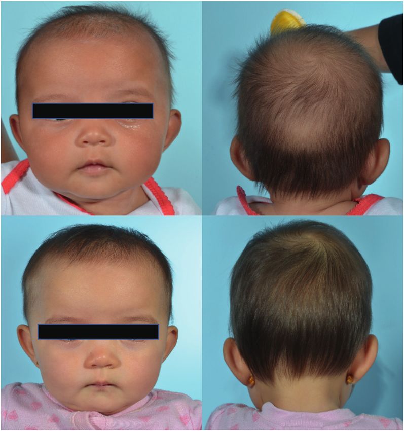
Prominent ear can be caused by an underdeveloped antihelix, damental features of the constricted ear: lidding caused by helical
an overdeveloped conchal bowl, or a combination of the 2 overhang and flattening of the antihelix, protrusion associated
cartilaginous deformations (Fig 3). (16) A prominent ear is with deepened conchal fossa, decreased ear size due to the su-
characterized by an external ear that projects more than 2 cm perior third deficiencies, and low ear position seen in severe cases.
Figure 3. Prominent ear. Top, before.
Bottom, after 6 weeks of ear molding.
182 Pediatrics in Review
Downloaded from http://pedsinreview.aappublications.org/ at Swets Blackwell Inc. on April 8, 2021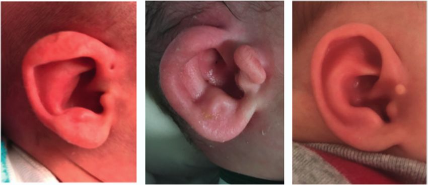
boys (2 or 3:1) and is predominantly unilateral (70%–90%),
                                                                             with a right-left-bilateral ratio of 6:3:1. (26)
                                                                                The cause of microtia is still poorly understood. There is
                                                                             evidence for environmental and genetic causes of microtia.
                                                                             The most common anomalies associated with microtia in-
                                                                             clude vertebral anomalies, macrostomia (a form of lateral
                                                                             facial cleft extending from the corners of the mouth,
                                                                             resulting in a wide oral aperture), oral clefts, facial asym-
                                                                             metry, renal abnormalities, cardiac defects, microphthalmia,
                                                                             holoprosencephaly, and polydactyly. (27)
                                                                                Microtia can present in varying degrees of severity (Fig 5).
                                                                             Although several classification systems of microtia have been
                                                                             described, none are universally used clinically. As such, the
Figure 4. Constricted ear. Left, before. Right, 6 weeks after ear molding.
                                                                             standard should be to document, at the very least, a detailed
                                                                             description of the malformation of the ear, including each
Mild deformities can be treated with nonsurgical molding with ear            anatomical component of the ear, in addition to taking
splints if initiated in the neonatal period. Moderate to severe cases,       photographs.
or cases in older children, require surgical correction.                        Microtia treatment involves restoration of hearing and
                                                                             reconstruction of the external ear, often with a multidisci-
                                                                             plinary approach that includes genetics, otolaryngology,
CLINICAL ASPECTS AND MANAGEMENT OF
                                                                             audiology, and plastic surgery. Hearing screening is neces-
EAR MALFORMATIONS
                                                                             sary for these patients on initial evaluation. An auditory
Microtia                                                                     brainstem response test is recommended soon after birth to
Microtia is the term used for an external ear with absent skin               evaluate both ears for inner ear function. Frequent ear ex-
or cartilage that is small, collapsed, or only has an earlobe                aminations are also advised because these patients have a
present (Fig 5). Microtia can occur as an isolated birth defect              higher risk of ear infections and drainage. Screening with
(the most common presentation), as a part of a spectrum of                   renal ultrasonography is recommended for all patients with
anomalies, or as a component of a syndrome. Treacher                         microtia given the fairly high rate of associated abnormalities
Collins and Goldenhar syndromes are 2 of the most com-                       and high percentage of findings requiring renal follow-up.
monly associated syndromes. In most cases of microtia there                  (28) A study by Koenig et al (28) found that syndromic children
is also agenesis of the external auditory canal. Microtia is                 with microtia demonstrated a higher rate of renal ultrasonog-
most commonly associated with conductive hearing loss,                       raphy abnormalities (22%) than children with isolated microtia
which is due to a malformed middle and external ear. In                      (7%). Of these patients, 69% required specialist follow-up.
unilateral microtia, the contralateral ear has normal hearing.               Varying degrees of renal abnormalities can be found, such as
Microtia prevalence varies geographically and is reported to                 agenesis, hypoplasia, ectopia, hydronephrosis, ureteral abnor-
be 0.83 to 17.4 per 10,000 births. It occurs most frequently in              malities, and vesicoureteral reflux. (28)
                                                                                Early consultation with a surgeon helps develop a trusting
                                                                             relationship with the family as the surgeon guides them
                                                                             through a discussion of treatment options while managing
                                                                             expectations. Ear reconstruction is considered for aesthetic,
                                                                             psychological, and functional reasons. The external ear
                                                                             serves a functional structural purpose and allows children to
                                                                             use glasses, wear earrings, and normalize appearance.
                                                                                The age at which microtia reconstruction should begin
                                                                             depends on psychological and physical considerations. Pa-
                                                                             tients with microtia have a high prevalence of mood disor-
                                                                             ders, with depression in 20.2%, interpersonal sensitivity/
                                                                             social difficulties in 36%, and hostility/aggression in 26.3%.
                                                                             Studies have shown that correction of microtia improves
Figure 5. Microtia.                                                          psychosocial abilities postoperatively. (29)

                                                                                                            Vol. 42 No. 4    APRIL 2021        183
                   Downloaded from http://pedsinreview.aappublications.org/ at Swets Blackwell Inc. on April 8, 2021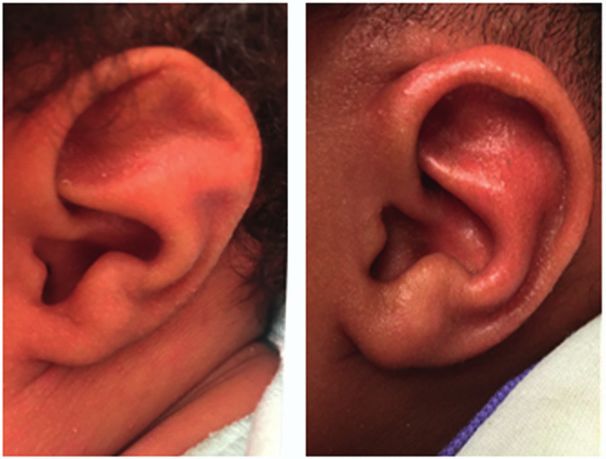
Ear reconstruction would ideally be performed before the           temporal bone region to allow for a stable attachment of a
      child enters school. However, surgery is delayed until rib cartilage   prosthetic ear (Fig 6). Historically for patients with congenital
      is substantial enough to allow for quality auricular framework         microtia, prosthetic ears were often reserved as an alternative to
      construction, which is 6 years and older. Surgery at this time also    failed autologous reconstruction. However, with recent ad-
      allows for the reconstructed ear to be created at adult size. In       vances, the osteointegration process has gained some popularity
      recent years, the preferred age of reconstruction has shifted even     given the relative ease of the procedure for the patient. The
      later, with some surgeons recommending 10 years of age as the          downside is that the prosthesis needs to be replaced frequently,
      ideal time. Worldwide, the predominant method of microtia              and there is no adaption of color to skin temperature or pigment
      reconstruction in children is autologous reconstruction, although      changes due to weather or sun exposure.
      several options exist for reconstruction of microtia.
          Autologous reconstruction uses the patient’s own rib carti-        Cryptotia
      lage. This technique typically requires 3 stages and begins with       Cryptotia, known as hidden ear, is identified as the absence of
      harvest of autologous cartilage rib grafts and creation of the ear     the superior auriculocephalic sulcus, which is due to the su-
      framework. Additional elements include elevation of the                perior third of the auricle being buried underneath the temporal
      framework, creation of a retroauricular sulcus, lobule trans-          skin (Fig 7). Cartilage malformation may also be present.
      position, and tragus formation (Fig 6). Composite recon-               Correction can be attempted with application of a nonsurgical
      struction combines autologous tissues, such as local fascial flaps      molding appliance in the first few weeks of life. Surgical
      and skin grafts, for coverage and alloplastic materials, such as       treatment for older children or more recalcitrant cases involve
      porous polyethylene, for an auricular framework, thus avoiding         division of the abnormally attached skin and placement of a full-
      chest donor site morbidity. Osteointegrated prosthetic recon-          or split-thickness skin graft to create a new sulcus.
      struction involves implanting 2 to 3 magnetic posts into the
                                                                             Preauricular Anomalies
                                                                             Preauricular anomalies include remnants with or without as-
                                                                             sociated ectopic cartilage and sinuses (Fig 8). Screening renal
                                                                             ultrasonography is not indicated for isolated preauricular
                                                                             anomalies. (30) Preauricular remnants do not regress over time,
                                                                             and although there is no physiologic impact, it is a stigmatizing
                                                                             lesion, and, thus, treatment entails surgical excision. Preaur-
                                                                             icular sinuses are typically asymptomatic; however, they can
                                                                             become infected. Infected preauricular sinuses require antibi-
                                                                             otics, and surgical excision is necessary. (31)

                                                                             Management of Ear Deformations
                                                                             Newborn ear molding has been used for nonsurgical cor-
                                                                             rection of auricular deformations since the 1980s. (32)
                                                                             Multiple studies have demonstrated that satisfactory non-
                                                                             surgical correction can be made by forcing the ear into a

      Figure 6. Ear reconstruction.                                          Figure 7. Cryptotia. Left, before treatment. Right, 6 weeks after ear molding.

184   Pediatrics in Review
                              Downloaded from http://pedsinreview.aappublications.org/ at Swets Blackwell Inc. on April 8, 2021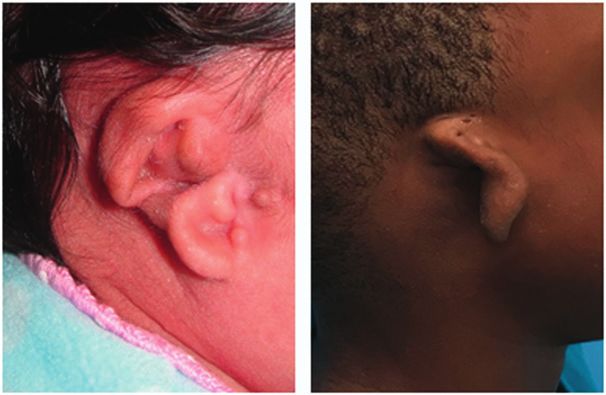
Figure 8. Preauricular anomalies.

proper position and maintaining it there for several weeks.               In ideal circumstances, the infant is seen in a plastic
(32) Permanent results can potentially be achieved if ear              surgery clinic on day 3 after birth and, depending on the
molding is started immediately after birth, preferably before          physician’s preference, either a commercial or a custom
the third day after birth, and can be continued until the infant       molding system can be used. Depending on the auricular
is 3 months old. The impetus to start nonsurgical correction           deformity, a customized mold or prothesis can be fabricated
early is the role of circulating maternal estrogen levels in the       from either plastic or acrylic and held in place with liquid
pliability of ear tissue. It is hypothesized that retained cir-        adhesive and tape (Fig 9). Average treatment duration is 4 to
culating maternal estrogen decreases the structural density            8 weeks, with biweekly follow-up visits for reevaluation
of collagen. Cartilage elasticity is dependent on the con-             and any necessary splint adjustments. Newborns are fre-
centration of proteoglycan aggregate, of which hyaluronic              quently seen by their pediatrician during this period, and
acid is a major component, and the presence of hyaluronic              coordination of appointments for families is encouraged.
acid is increased by estrogens. Maternal estrogen levels peak          Complications associated with nonsurgical correction are
in neonates at 3 days of life, then taper significantly and             rare and usually minor and can include minor superficial
normalize at approximately 6 weeks of age. (33)                        excoriations and skin loss due to pressure necrosis. (32)

Figure 9. Custom molding system.

                                                                                                         Vol. 42 No. 4   APRIL 2021    185
                  Downloaded from http://pedsinreview.aappublications.org/ at Swets Blackwell Inc. on April 8, 2021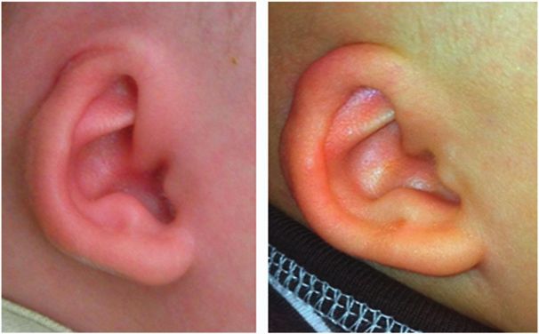
Table. Ear Malformations and Deformations: Recommended Diagnostics and Treatment Options
                                    DIAGNOSTIC                                                                        NONSURGICAL
          EAR ABNORMALITY           HEARING    RENAL ULTRASONOGRAPHY                SURGERY                           CORRECTION
          Malformations
            Microtia                +             +                                 +                                 –
            Cryptotia               –             –                                 Possible                          +
            Preauricular remnant    –             –                                 +                                 –
            Preauricular sinus      –             –                                 If infected                       –
          Deformations
            Prominent ear           –             –                                 If not corrected with molding     +
            Stahl ear               –             –                                 If not corrected with molding     +
            Constricted ear         –             –                                 If not corrected with molding     +

                                                                                           •   Based on strong research evidence, early referral is
      Summary                                                                                  mandatory for nonsurgical correction in the
      •   Based on strong research evidence, the external ear is a                             neonatal period for most congenital ear anomalies.
          complex 3-dimensional anatomical landmark with                                       (3)(4)(15)(32)(33)
          raised cartilaginous crura and depressed scapha.                                 •   Based on strong research evidence, if recognized
          (9)(10)                                                                              in the neonatal period, most congenital ear
      •   Based on some research evidence as well as consensus,                                anomalies can be treated successfully with
          ear differences are associated with psychosocial stresses                            nonsurgical intervention. However, if diagnosis is
          and self-awareness in school-age children and adults.                                delayed, surgical intervention is required to
          (20)(21)                                                                             reconstruct congenital ear deformities (Table). (16)(18)
                                                                                           •   Based on some research evidence as well as
      •   Based primarily on consensus due to lack of
          relevant clinical studies, ear anomalies can                                         consensus, hearing screening and renal
          be divided into either malformations or                                              ultrasonography are typically not required except
          deformations. Malformations include anotia,                                          for cases of microtia. (28)(30)
          microtia, cryptotia, and preauricular anomalies.
          Deformations include Stahl, prominent, and
          constricted ears. (1)
                                                                                              References for this article can be found at
                                                                                    http://pedsinreview.aappublications.org/content/42/No. 4/180.

186   Pediatrics in Review
                                 Downloaded from http://pedsinreview.aappublications.org/ at Swets Blackwell Inc. on April 8, 2021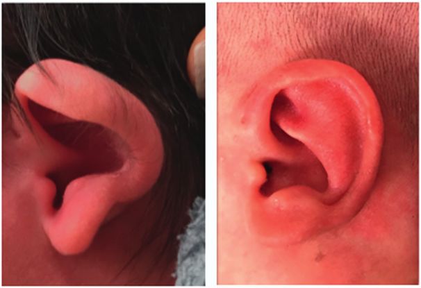
PIR QUIZ

                     Individual CME quizzes are available via the blue CME link under the article title in the online Table of Contents
                     of any issue. To learn how to claim MOC points, go to: http://www.aappublications.org/content/moc-credit.

1. You are making rounds in the newborn nursery. As you perform the physical
   examination of a term male newborn you notice an ear deformity possibly
   consistent with a Stahl ear. The remainder of the physical examination findings
   are normal. The baby is feeding well and is ready for discharge. Which of the
   following is the best next step in management of the ear deformity?
     A. Advise the parents to tape both ears backward.
     B. Follow up with his primary care provider at his 2-month health supervision
        visit.
     C. No follow-up is needed as this will spontaneously resolve.                                   REQUIREMENTS: Learners can
                                                                                                     take Pediatrics in Review quizzes
     D. Recommend referral to plastic surgery if no improvement by 6 months of age.
                                                                                                     and claim credit online only at:
     E. Schedule him an outpatient appointment with plastic surgery in 48 hours.                     http://pedsinreview.org.
2. A term female newborn was noted on physical examination in the newborn
                                                                                                     To successfully complete 2021
   nursery to have an isolated microtia on the left side. The remainder of the
                                                                                                     Pediatrics in Review articles for
   physical examination findings are normal, with no evidence of dysmorphic                           AMA PRA Category 1 Credit™,
   features. Which of the following is the most appropriate immediate next step in                   learners must demonstrate
   testing in this patient?                                                                          a minimum performance
                                                                                                     level of 60% or higher on this
     A.   Auditory brainstem response test.                                                          assessment. If you score less
     B.   Chromosomal microarray.                                                                    than 60% on the assessment,
     C.   Echocardiography.                                                                          you will be given additional
     D.   Magnetic resonance imaging of the brain.                                                   opportunities to answer
                                                                                                     questions until an overall 60%
     E.   Upper endoscopy.                                                                           or greater score is achieved.
3. A 2-year-old boy with unilateral microtia is followed in your practice. The patient
                                                                                                     This journal-based CME activity
   has been followed by plastic surgery since birth. The parents are seeking your
                                                                                                     is available through Dec. 31,
   opinion as to when their child’s surgical reconstruction of the ear should be                     2023, however, credit will be
   performed. They have read on the Internet conflicting information. Which of the                    recorded in the year in which
   following is the optimal age to complete the surgical intervention in this patient?               the learner completes the quiz.

     A. As soon as possible.
     B. At 4 years of age, before starting school to avoid him being subject to
        bullying.
     C. At 10 years of age.
     D. At mid-adolescence after the child goes through his growth spurt.
                                                                                                     2021 Pediatrics in Review is
     E. The timing is variable with every child depending on the child’s size.                       approved for a total of 30
                                                                                                     Maintenance of Certification
4. A 4-year-old boy is brought to the clinic for a health supervision visit. The patient
                                                                                                     (MOC) Part 2 credits by the
   is healthy and has had normal growth and development. He has no recent                            American Board of Pediatrics
   illnesses. On physical examination he is noted to have a preauricular tag on the                  (ABP) through the AAP MOC
   right. Otherwise his physical examination findings are normal. The parents                         Portfolio Program. Pediatrics in
   inquire about the prognosis of the tag. Which of the following is the best                        Review subscribers can claim
                                                                                                     up to 30 ABP MOC Part 2 points
   approach to the management of this patient?
                                                                                                     upon passing 30
     A. Renal ultrasonography because preauricular tags are usually associated                       quizzes (and claiming full
        with renal anomalies.                                                                        credit for each quiz) per year.
                                                                                                     Subscribers can start claiming
     B. Excision and antibiotics if preauricular tags become infected.
                                                                                                     MOC credits as early as October
     C. No intervention required because such tags regress overtime.                                 2021. To learn how to claim
     D. No surgical excision required because the tag does not have an associated                    MOC points, go to: https://www.
        preauricular sinus tract.                                                                    aappublications.
     E. Surgical excision for cosmetic reasons because preauricular tags can                         org/content/moc-credit.
        potentially grow over time.

                                                                                                        Vol. 42 No. 4       APRIL 2021    187
                 Downloaded from http://pedsinreview.aappublications.org/ at Swets Blackwell Inc. on April 8, 2021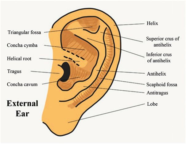
5. Because primary care clinicians diagnose auricular deformations on initial
         physical examination in the newborn nursery, the success of ear molding
         nonsurgical correction in achieving permanent results depends on which
         of the following factors?
          A.   Absence of associated renal anomalies.
          B.   Absence of family history of ear deformations.
          C.   Early intervention, before the third day after birth.
          D.   Female sex.
          E.   First born.

188   Pediatrics in Review
                            Downloaded from http://pedsinreview.aappublications.org/ at Swets Blackwell Inc. on April 8, 2021
Ear Abnormalities
Sana L. Bhatti, Lauren T. Daly, Martha Mejia and Chad Perlyn
Pediatrics in Review 2021;42;180
DOI: 10.1542/pir.2019-0167
Updated Information & including high resolution figures, can be found at:
Services http://pedsinreview.aappublications.org/content/42/4/180
References This article cites 32 articles, 2 of which you can access for free at:
http://pedsinreview.aappublications.org/content/42/4/180.full#ref-list
-1
Subspecialty Collections This article, along with others on similar topics, appears in the
following collection(s):
Surgery
http://classic.pedsinreview.aappublications.org/cgi/collection/surgery
_sub
Plastic Surgery
http://classic.pedsinreview.aappublications.org/cgi/collection/plastic
_surgery_sub
Permissions & Licensing Information about reproducing this article in parts (figures, tables) or
in its entirety can be found online at:
https://shop.aap.org/licensing-permissions/
Reprints Information about ordering reprints can be found online:
http://classic.pedsinreview.aappublications.org/content/reprints
Downloaded from http://pedsinreview.aappublications.org/ at Swets Blackwell Inc. on April 8, 2021Ear Abnormalities
Sana L. Bhatti, Lauren T. Daly, Martha Mejia and Chad Perlyn
Pediatrics in Review 2021;42;180
DOI: 10.1542/pir.2019-0167
The online version of this article, along with updated information and services, is
located on the World Wide Web at:
http://pedsinreview.aappublications.org/content/42/4/180
Pediatrics in Review is the official journal of the American Academy of Pediatrics. A monthly
publication, it has been published continuously since 1979. Pediatrics in Review is owned,
published, and trademarked by the American Academy of Pediatrics, 345 Park Avenue, Itasca,
Illinois, 60143. Copyright © 2021 by the American Academy of Pediatrics. All rights reserved.
Print ISSN: 0191-9601.
Downloaded from http://pedsinreview.aappublications.org/ at Swets Blackwell Inc. on April 8, 2021You can also read