Effect of C reactive protein on the sodium calcium exchanger 1 in cardiomyocytes
←
→
Page content transcription
If your browser does not render page correctly, please read the page content below
EXPERIMENTAL AND THERAPEUTIC MEDICINE 22: 815, 2021

                   Effect of C reactive protein on the sodium‑calcium
                             exchanger 1 in cardiomyocytes
     YONG XIE1,2*, QIAN LI3*, HAI‑FENG ZHANG1,2*, TU‑CHENG HUANG1,2, YING YANG1,2, YONG‑QING LIN1,2,
      JING‑TING MAI1,2, ZHU‑ZHI WEN1,2, WO‑LIANG YUAN1,2, JING‑FENG WANG1,2 and YANG‑XIN CHEN1,2

            1
            Department of Cardiology and 2Guangdong Province Key Laboratory of Arrhythmia and Electrophysiology,
     Sun Yat‑sen Memorial Hospital of Sun Yat‑sen University, Guangzhou, Guangdong 510120; 3Department of Dermatology,
                 Nanfang Hospital of Southern Medical University, Guangzhou, Guangdong 515110, P.R. China

                                    Received September 23, 2020; Accepted March 26, 2021

                                                    DOI: 10.3892/etm.2021.10247

Abstract. Numerous previous studies have found that                Introduction
C‑reactive protein (CRP) is associated with cardiac arrhythmia
and cardiac remodeling. However, the underlying mecha‑             C‑reactive protein (CRP), which is an acute‑phase protein is
nisms of this association remain unclear. Sodium‑calcium           mainly synthesized in the liver and serves important roles in
exchanger 1 (NCX1) serves an important role in the regula‑         cardiovascular diseases (1‑3). Over the past several decades,
tion of intracellular calcium concentration, which is closely      several studies have found that CRP may be an important
related with cardiac arrhythmia and cardiac remodeling. The        risk factor for a number of cardiovascular diseases, such as
present study aimed to evaluate the effects of CRP on NCX1         coronary heart disease (CHD), dilated cardiomyopathy and
and intracellular calcium concentration in cardiomyocytes.         atrial fibrillation (AF) (1,4). A high‑sensitivity hsCRP level
Primary neonatal mouse ventricular cardiomyocytes were             >3 mg/l was independently associated with a 60% excess risk
cultured and treated with varying concentrations of CRP (0,        in incident CHD as compared with levels
2 XIE et al: EFFECTS OF CRP ON NCX1 IN CARDIOMYOCYTES
resulting in early afterdepolarization and arrhythmia (17,18). Treatment with human CRP. Following incubation for 24 h
Hence, calcium imbalance has been identified as a treatment in DMEM/F12 with 10% fetal bovine serum and 100 U/ml
target to manage arrhythmia in the clinical setting. penicillin/streptomycin, the cardiomyocytes were maintained
Although higher CRP levels can increase the risk of cardiac in serum‑free DMEM/F12 for 24 h in 37˚C and then treated
arrhythmia, the mechanisms involved in are not clear. The with human recombinant CRP (purity, >98%; Merck KGaA).
present study aimed to evaluate the effects of CRP on NCX1 CRP purity was confirmed by 12% SDS‑PAGE. The endotoxin
and intracellular calcium concentration in cardiomyocytes level as 0.0005 EU/µg for CRP preparation was determined
and explore the potential underlying mechanism. It is hoped by the Limulus amebocyte lysate assay (Pyrotell® ‑T; cat.
that this would assist in the development of anti‑inflammatory no. T0051; Associates of Cape Cod, Inc.). The cardiomyo‑
therapies for patients with heart disease and infection. cytes were cultured with PBS and CRP at clinically relevant
concentrations at 5, 10, 20, 30 and 40 µg/ml for 24 h in 37˚C.
Materials and methods The NF‑κ B specific inhibitor PDTC (10 µM; Sigma‑Aldrich;
Merck KGaA) was added to cells for 1 h prior to being stimu‑
Animal ethics. A total of 200 neonatal (1‑2 days) C57BL/6J lated with CRP (40 µg/ml) for 24 h at 37˚C (20). The NF‑κ B
mice, weighing 1.8±0.32 g, were used in this investigation pathway was tested at 0, 10, 30 and 60 min after CRP stimula‑
and this was approved by the Institutional Animal Care and tion with the cardiomyocytes (24,25).
Use Committee of Sun Yat‑sen Memorial Hospital of Sun
Yan‑Sen University (approval no. 175). Animal use and care Reverse transcription‑quantitative (RT‑q) PCR. Total RNA was
were in accordance with the animal care guidelines, which extracted from cardiomyocytes by RNAiso plus (Takara Bio, Inc.)
conformed to the Guide for the Care and Use of Laboratory and reverse transcribed (RT) to cDNA at 37˚C for 15 min and
Animals published by the US National Institutes of Health 85˚C for 5 sec using the PrimeScript™ RT Master Mix (Perfect
(NIH publication no. 85‑23; revised 1996) (19). All animals Real Time; cat. no. RR036B; Takara Bio, Inc.). Quantification
were purchased from The Animal Research Center Of Sun of NCX1 transcript levels was performed by amplification of
Yat‑Sen University. Mice were housed under a temperature at cDNA prepared from the isolated RNA with the TB Green®
22˚C and the humidity at 50‑60% with 12‑h light/dark cycle Premix Ex Taq™ (Tli RNase H Plus; cat. no. RR420A; Takara
and had free access to rodent chow and tap water. Bio, Inc.) and primers specific for NCX1 (forward 5'‑AGGCCA
GAAATAG GAG CCATC‑3' and reverse, 5'‑AGTGTGCCT
Culturing of neonatal mice cardiomyocytes. Cardiac myocytes GTCCCCCTAAA‑3'); and glyceraldehyde‑3‑phosphate dehy‑
were prepared from the ventricles of 1‑2 day old C57BL/6J drogenase (GAPDH) as the internal control (forward, 5'‑TGT
mice as described by a previous study (18). Briefly, neonatal GTCCGTCGTGGATCTGA‑3' and reverse, 5'‑TTGCTGTTG
mice were anesthetized with isoflurane intermittently; the AAGTCGCAGGAG‑3'). The thermocycling conditions were:
induction and maintenance dose of isoflurane were 5 and Pre‑denaturation at 95˚C for 5 min, followed by 40 cycles of 95˚C
1%, respectively, and the hearts were extracted after cervical for 5 sec, annealing at 62˚C for 25 sec and extension at 72˚C for
dislocation was performed under anesthesia. The atrial and 20 sec. Results were presented as fold difference for each gene
vascular tissues were removed and the ventricles were enzy‑ against GAPDH by use of 2‑ΔΔCq method (26). Melting curves
matically digested in 0.125% trypsin (Gibco; Thermo Fisher were used to confirm that only a single product was present.
Scientific Inc.) for 5 min in 37˚C, followed by 0.06% collage‑
nase II (MP Biomedicals, LLC) for 2 h in a thermostat shaker Western blotting. Total proteins were extracted from cultured
at 37˚C and a speed of 62 rpm. The supernatant was collected cardiomyocytes using RIPA lysis buffer (Cell Signaling
and the resuspended digested cardiac tissues was centrifuged Technology Inc.), and protein concentration was measured
at 0.5 x g for 5 min in 4˚C. The pellet containing the cells with a bicinchoninic acid protein assay kit. Protein samples
was collected and resuspended with Dulbecco's Modified (25‑100 µg) were separated on 10‑13% SDS‑PAGE gels and
Eagle medium/Ham's F‑12 medium (DMEM/F12) containing transferred onto 0.22‑µm PVDF membranes. After blocking
10% fetal bovine serum (FBS) and 100 U/ml penicillin/ with 5% non‑fat skimmed milk at room temperature for 1 h,
streptomycin (all Thermo Fisher Scientific, Inc.). The cells the membranes were incubated overnight at 4˚C with primary
were plated in tissue culture dishes and maintained at 37˚C in antibodies (all Abcam) to NCX1 (cat. no. ab177952, 1:1,000),
a 5% CO2 incubator for 30‑35 min to remove non‑myocytes. NF‑κ B (cat. no. ab32536,1:2,000) and inhibitor of NF‑κ Bα
The supernatants were transferred to new culture dishes (Iκ Bα; cat. no. ab32518; 1:2,000) and GAPDH (cat. no. 5174;
and cultured in DMEM/F12 with 10% FBS, 100 U/ml peni‑ 1,1000; Cell Signaling Technology Inc.). After washing 5 times
cillin/streptomycin and 1% 5‑Bromo‑2'‑deoxyuridine (BrdU; with TBST which include 2% tween‑20 (5 min washes each),
Sigma‑Aldrich; Merck KGaA) at 37˚C which could inhibit the the membranes were incubated with a horseradish peroxidase
growth of non‑myocardial cell (20‑22). (HRP)‑conjugated secondary antibody (cat. no. 98164; goat
anti‑rabbit IgG; 1:5,000; Cell signaling technology, Inc.) for
Cell viability assay. Cell viability was assessed using 1 h at room temperature. Membranes were developed using
the MTS assay. Cardiomyocytes seeded in 96‑well plates the Immobilon™ Western Chemiluminescent HRP substrate
(5x10³ cells/well) were incubated with CRP (0, 5, 10, 20, 40 (EMD Millipore) and visualized using the Gel Documentation
and 100 µg/ml) for 24, 48 and 72 h. MTS (20 µl) was added and Analysis System (G‑Box; Syngene, Europe). Band
into each well and co‑cultured for 4 h. The absorbance at intensities were quantified by scanning densitometry and the
490 nm measured by the microplate reader presented the cell densitometry ratios of the target proteins to GAPDH were
viability (23). determined by Image J software (National Institutes of Health).EXPERIMENTAL AND THERAPEUTIC MEDICINE 22: 815, 2021 3 Figure 1. NCX1 protein and mRNA expression in cardiomyocytes following treatment with CRP. (A) There was a significantly higher expression of NCX1 mRNA and (B) protein following 40 µg/ml CRP stimulation compare with this in the control group and 5, 10 and 20 µg/ml groups, (C) which was quantified. Data are presented as mean ± SEM, significance was determined using one way ANOVA with the post hoc Tukey's test. #P
4                                          XIE et al: EFFECTS OF CRP ON NCX1 IN CARDIOMYOCYTES

Figure 3. NF‑κ B pathway may be involved in the regulation roles of CRP on NCX1. (A) The NF‑κ B inhibitor PDTC (10 µM) attenuated the effects of CRP
on NCX1 compared with those in the CRP and control groups (treat with PBS), (B) which is quantified. Data are presented as mean ± SEM. Significance was
determined using a two sided one way ANOVA with Tukey test. #P
EXPERIMENTAL AND THERAPEUTIC MEDICINE 22: 815, 2021 5
(triggered by delayed afterdepolarizations) in animal models of Authors' contributions
heart failure (39). In addition, KB‑R7943, a selective inhibitor
for the reverse mode of NCX1 (25) and the NF‑κ B inhibitor JFW and YXC designed the study. YX cultured the primary
PDTC (18) may serve roles in the anti‑arrhythmia in patients cardiomyocytes and performed the experiments to find the
with cardiac diseases and inflammation. suitable stimulation concentration of CRP. YX and TCH
It is well‑established that NCX1 is an important player in were responsible for confirming the raw data authenticity. QL
calcium balance (16). In the present study, it was observed that performed the RT‑qPCR and western blotting experiments.
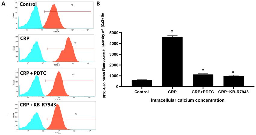
CRP upregulated the expression of NCX1 and increased the HFZ performed the statistical analysis and drafted the manu‑
[Ca2+]in, which was significantly attenuated by NF‑κ B specific script. TCH, YY, QL, JTM, ZZW and WLY revised the paper
inhibitor PDTC. In addition in the present study, CRP also for important intellectual content. TCH provided advice for
increased the expression of NF‑κ B and decreased the expres‑ this study and collected the experiments data. YY ,MJT, ZZW
sion of Iκ Bα, suggesting that CRP‑induced changes of NCX1 and WLY participated in the research design. All authors have
and [Ca 2+]in, were regulated by the NF‑κ B pathway. It was read and approved the manuscript.
previously reported in our previous study, that CRP reduced
the expression of K+ channel interacting proteins 2 (KChIP2) Ethics approval and consent to participate
in murine cardiomyocytes (23). KChIP2 is a member of the
Ca 2+‑binding protein, which is only expressed in the heart This study was approved by the Institutional Animal Care
and interacts with Kv4.2 or Kv4.3 to form transient outward and Use Committee of Sun Yat‑sen Memorial Hospital of Sun
currents and participates in the regulation of the early repolar‑ Yan‑Sen University (approval no. 175; Guangzhou, China).
ization and the QTc interval of heart (40). The data presented Animal use and care were in accordance with the animal care
herein, along with previous studies, suggested that CRP serves guidelines, which conformed to the Guide for the Care and
a role in the KChIP2 in the heart. Use of Laboratory Animals published by the US National
CRP has also been demonstrated to mediate cardiovas‑ Institutes of Health (NIH Publication No. 85‑23, revised 1996).
cular diseases, such as coronary heart disease (41). It has been
reported that CRP is associated with vascular and ventricular Patient consent for publication
remodeling (42). Notably, it has been suggested that the
upregulation of NCX1 and higher [Ca 2+]in were involved in the Not applicable.
structural and electronic remodeling of ventricles in cardiac
diseases, such as heart failure and cardiomyopathy (43). Competing interests
Hence, the results from the present study suggested that CRP
may be a risk factor of arrhythmia due to the myocardial The authors declare that they have no competing interests.
remolding and electronic remodeling. However, a limitation
of the present study is a lack of in vivo experiments. References
In conclusion, the present study demonstrated that CRP
1. Li Y, Zhong X, Cheng G, Zhao C, Zhang L, Hong Y, Wan Q,
increases NCX1 expression and [Ca2+]in in cardiomyocytes and He R and Wang Z: Hs‑CRP and all‑cause, cardiovascular, and
that the NF‑κ B pathway is involved in the regulation process. cancer mortality risk: A meta‑analysis. Atherosclerosis 259:
The findings of the present study suggested that CRP can act 75‑82, 2017.
2. Ghazizadeh H, Rezaei M, Avan A, Fazilati M, Pasdar A,
as a predictor of ventricular arrhythmia due to the activation Tavallaie S, Kazemi E, Seyedi SMR, Ferns GA, Azimi‑Nezhad M
of the reverse mode of NCX1 function and [Ca2+]in increase. and Ghayour‑Mobarhan M: Association between serum cell
adhesion molecules with hs‑CRP, uric acid and VEGF genetic
polymorphisms in subjects with metabolic syndrome. Mol Biol
Acknowledgements Rep 47: 867‑875, 2020.
3. Han K, Lu Q, Zhu WJ, Wang TZ, Du Y and Bai L: Correlations of
degree of coronary artery stenosis with blood lipid, CRP, Hcy, GGT,
Not applicable. SCD36 and fibrinogen levels in elderly patients with coronary heart
disease. Eur Rev Med Pharmacol Sci 23: 9582‑9589, 2019.
Funding 4. Amorim S, Campelo M, Moura B, Martins E, Rodrigues J,
Barroso I, Faria M, Guimaraes T, Macedo F, Silva‑Cardoso J and
Maciel MJ: The role of biomarkers in dilated cardiomyopathy:
This work was supported by the National Natural Science Assessment of clinical severity and reverse remodeling. Rev Port
Foundation of China (grant nos. 81870170, 81870334, Cardiol 36: 709‑716, 2017 (In English, Portuguese).
5. Buckley DI, Fu R, Freeman M, Rogers K and Helfand M:
81770229, 81570329, 81970200, 81700215 and 81703098), the C‑reactive protein as a risk factor for coronary heart disease:
Guangdong Province Natural Science Fund‑Vertical collabo‑ A systematic review and meta‑analyses for the U.S. preventive
ration doctor initiative program (grant no. 2017A030310446) services task force. Ann Intern Med 151: 483‑495, 2009.
6. Lee Y, Park HC, Shin JH, Lim YH, Shin J and Park JK: Single
and Science and Technology Program of Guangdong Province and persistent elevation of C‑reactive protein levels and the risk
of China (grant no. 2015B010131010), the Science and of atrial fibrillation in a general population: The ansan‑ansung
cohort of the Korean genome and epidemiology study. Int J
Technology Program of Guangzhou City of China (grant Cardiol 277: 240‑246, 2019.
nos. 201803040010 and 201707010206). 7. Kazumi T, Kawaguchi A, Hirano T and Yoshino G: C‑reactive
protein in young, apparently healthy men: Associations with serum
leptin, QTc interval, and high‑density lipoprotein‑cholesterol.
Availability of data and materials Metabolism 52: 1113‑1116, 2003.
8. Hodzic E, Drakovac A and Begic E: Troponin and CRP as
The datasets used and/or analyzed during the current study are indicators of possible ventricular arrhythmias in myocardial
infarction of the anterior and inferior walls of the heart. Mater
available from the corresponding author on reasonable request. Sociomed 30: 185‑188, 2018.6 XIE et al: EFFECTS OF CRP ON NCX1 IN CARDIOMYOCYTES
9. Li C, Jia L, Wang Z, Niu L and An X: The efficacy of radiofre‑ 27. McElnea EM, Quill B, Docherty NG, Irnaten M, Siah WF,
quency ablation in the treatment of pediatric arrhythmia and its Clark AF, O'Brien CJ and Wallace DM: Oxidative stress, mito‑
effects on serum IL‑6 and hs‑CRP. Exp Ther Med 14: 3563‑3568, chondrial dysfunction and calcium overload in human lamina
2017. cribrosa cells from glaucoma donors. Mol Vis 17: 1182‑1191,
10. Nagai T, Anzai T, Kaneko H, Anzai A, Mano Y, Nagatomo Y, 2011.
Kohsaka S, Maekawa Y, Kawamura A, Yoshikawa T and 28. Shen JB, Yang R, Pappano A and Liang BT: Cardiac P2X
Ogawa S: Impact of systemic acidosis on the development of purinergic receptors as a new pathway for increasing Na+ entry
malignant ventricular arrhythmias after reperfusion therapy in cardiac myocytes. Am J Physiol Heart Circ Physiol 307:
for ST‑elevation myocardial infarction. Circ J 74: 1808‑1814, H1469‑H1477, 2014.
2010. 29. Xie Y, Gu ZJ, Wu MX, Huang TC, Ou JS, Ni HS, Lin MH,
11. Wu KC, Gerstenblith G, Guallar E, Marine JE, Dalal D, Cheng A, Yuan WL, Wang JF and Chen YX: Disruption of calcium
Marbán E, Lima JA, Tomaselli GF and Weiss RG: Combined homeostasis by cardiac‑specific over‑expression of PPAR‑ γ
cardiac magnetic resonance imaging and C‑reactive protein in mice: A role in ventricular arrhythmia. Life Sci 167: 12‑21,
levels identify a cohort at low risk for defibrillator firings and 2016.
death. Circ Cardiovasc Imaging 5: 178‑186, 2012. 30. LaRocca TJ, Fabris F, Chen J, Benhayon D, Zhang S,
12. Saxon LA, Bristow MR, Boehmer J, Krueger S, Kass DA, McCollum L, Schecter AD, Cheung JY, Sobie EA, Hajjar RJ
De Marco T, Carson P, DiCarlo L, Feldman AM, Galle E and and Lebeche D: Na+/Ca2+ exchanger‑1 protects against systolic
Ecklund F: Predictors of sudden cardiac death and appropriate failure in the Akitains2 model of diabetic cardiomyopathy via a
shock in the comparison of medical therapy, pacing, and defi‑ CXCR4/NF‑κ B pathway. Am J Physiol Heart Circ Physiol 303:
brillation in heart failure (COMPANION) trial. Circulation 114: H353‑H367, 2012.
2766‑2772, 2006. 31. Balasubramaniam SL, Gopalakrishnapillai A, Gangadharan V,
13. Theuns DA, Smith T, Szili‑Torok T, Muskens‑Heemskerk A, Duncan RL and Barwe SP: Sodium‑calcium exchanger 1
Janse P and Jordaens L: Prognostic role of high‑sensitivity regulates epithelial cell migration via calcium‑dependent extra‑
C‑reactive protein and B‑type natriuretic peptide in implantable cellular signal‑regulated kinase signaling. J Biol Chem 290:
cardioverter‑defibrillator patients. Pacing Clin Electrophysiol 35: 12463‑12473, 2015.
275‑282, 2012. 32. Kobayashi Y, Tanno K, Ueno A, Fukamizu S, Murata H,
14. Streitner F, Kuschyk J, Veltmann C, Ratay D, Schoene N, Watanabe N, Sasaki T, Yamamoto T, Takayama M and Nagao K:
Streitner I, Brueckmann M, Schumacher B, Borggrefe M and In‑hospital electrical storm in acute myocardial infarction‑clin‑
Wolpert C: Role of proinflammatory markers and NT‑proBNP ical background and mechanism of the electrical instability. Circ
in patients with an implantable cardioverter‑defibrillator and an J 83: 91‑100, 2018.
electrical storm. Cytokine 47: 166‑172, 2009. 33. Nortamo S, Ukkola O, Lepojärvi S, Kenttä T, Kiviniemi A,
15. Vianello E, Dozio E, Barassi A, Sammarco G, Tacchini L, Junttila J, Huikuri H and Perkiömäki J: Association of sST2
Marrocco‑Trischitta MM, Trimarchi S and Corsi Romanelli MM: and hs‑CRP levels with new‑onset atrial fibrillation in coronary
A pilot observational study on magnesium and calcium imbal‑ artery disease. Int J Cardiol 248: 173‑178, 2017.
ance in elderly patients with acute aortic dissection. Immun 34. Hamilton S and Terentyev D: Altered intracellular calcium
Ageing 14: 1, 2017. homeostasis and arrhythmogenesis in the aged heart. Int J Mol
16. Shattock MJ, Ottolia M, Bers DM, Blaustein MP, Boguslavskyi A, Sci 20: 2386, 2019.
Bossuyt J, Bridge JH, Chen‑Izu Y, Clancy CE, Edwards A, et al: 35. Davlouros PA, Gkizas V, Vogiatzi C, Giannopoulos G,
Na+/Ca2+ exchange and Na+/K+‑ATPase in the heart. J Physiol 593: Alexopoulos D and Deftereos S: Calcium homeostasis and
1361‑1382, 2015. kinetics in heart failure. Med Chem 12: 151‑161, 2016.
17. Hamilton S and Terentyev D: Proarrhythmic remodeling of 36. Kim JJ, Němec J, Papp R, Strongin R, Abramson JJ and
calcium homeostasis in cardiac disease; implications for diabetes Salama G: Bradycardia alters Ca(2+) dynamics enhancing
and obesity. Front Physiol 9: 1517, 2018. dispersion of repolarization and arrhythmia risk. Am J Physiol
18. Ma HJ, Li Q, Ma HJ, Guan Y, Shi M, Yang J, Li DP and Heart Circ Physiol 304: H848‑H860, 2013.
Zhang Y: Chronic intermittent hypobaric hypoxia ameliorates 37. Song J, Gao E, Wang J, Zhang XQ, Chan TO, Koch WJ,
ischemia/reperfusion‑induced calcium overload in heart via Shang X, Joseph JI, Peterson BZ, Feldman AM and Cheung JY:
Na/Ca2+ exchanger in developing rats. Cell Physiol Biochem 34: Constitutive overexpression of phosphomimetic phospho‑
313‑324, 2014. lemman S68E mutant results in arrhythmias, early mortality,
19. Guide for the Care and Use of Laboratory Animals published by and heart failure: Potential involvement of Na+/Ca 2+ exchanger.
the US National Institutes of Health, NIH publication no. 85‑23, Am J Physiol Heart Circ Physiol 302: H770‑H781, 2012.
revised 1996. 38. Javidanpour S, Dianat M, Badavi M and Mard SA: The inhibi‑
20. Battiprolu PK, Hojayev B, Jiang N, Wang ZV, Luo X, Iglewski M, tory effect of rosmarinic acid on overexpression of NCX1 and
Shelton JM, Gerard RD, Rothermel BA, Gillette TG, et al: stretch‑induced arrhythmias after acute myocardial infarction in
Metabolic stress‑induced activation of FoxO1 triggers diabetic rats. Biomed Pharmacother 102: 884‑893, 2018.
cardiomyopathy in mice. J Clin Invest 122: 1109‑1118, 2012. 39. Jordan MC, Henderson SA, Han T, Fishbein MC, Philipson KD
21. Simpson P and Savion S: Differentiation of rat myocytes in single and Roos KP: Myocardial function with reduced expression of
cell cultures with and without proliferating nonmyocardial cells. the sodium‑calcium exchanger. J Card Fail 16: 786‑796, 2010.
Cross‑striations, ultrastructure, and chronotropic response to 40. Murthy A, Workman SW, Jiang M, Hu J, Sifa I, Bernas T,
isoproterenol. Circ Res 50: 101‑116, 1982. Tang W, Deschenes I and Tseng GN: Dynamic palmitoylation
22. Lokuta A, Kirby MS, Gaa ST, Lederer WJ and Rogers TB: On regulates trafficking of K channel interacting protein 2 (KChIP2)
establishing primary cultures of neonatal rat ventricular myocytes across multiple subcellular compartments in cardiac myocytes.
for analysis over long periods. J Cardiovasc Electrophysiol 5: J Mol Cell Cardiol 135: 1‑9, 2019.
50‑62, 1994. 41. Liu J, Yuen J and Kang S: Sleep duration, C‑reactive protein
23. Xie Y, Mai JT, Wang F, Lin YQ, Yuan WL, Luo NS, Fang MC, and risk of incident coronary heart disease‑results from the
Wang JF and Chen YX: Effects of C‑reactive protein on K(+) Framingham offspring study. Nutr Metab Cardiovasc Dis 24:
channel interaction protein 2 in cardiomyocytes. Am J Transl 600‑605, 2014.
Res 7: 922‑931, 2015. 42. Anzai T: Inflammatory mechanisms of cardiovascular remod‑
24. Li M, Ye J, Zhao G, Hong G, Hu X, Cao K, Wu Y and Lu Z: Gas6 eling. Circ J 82: 629‑635, 2018.
attenuates lipopolysaccharide‑induced TNF‑ α expression and 43. Ujihara Y, Iwasaki K, Takatsu S, Hashimoto K, Naruse K,
apoptosis in H9C2 cells through NF‑κ B and MAPK inhibition Mohri S and Katanosaka Y: Induced NCX1 overexpression
via the Axl/PI3K/Akt pathway. Int J Mol Med 44: 982‑994, 2019. attenuates pressure overload‑induced pathological cardiac
25. Martin TP, McCluskey C, Cunningham MR, Beattie J, Paul A remodelling. Cardiovasc Res 111: 348‑361, 2016.
and Currie S: CaMKIIδ interacts directly with IKK β and
modulates NF‑κ B signalling in adult cardiac fibroblasts. Cell This work is licensed under a Creative Commons
Signal 51: 166‑175, 2018. Attribution-NonCommercial-NoDerivatives 4.0
26. Livak KJ and Schmittgen TD: Analysis of relative gene expres‑ International (CC BY-NC-ND 4.0) License.
sion data using real‑time quantitative PCR and the 2(‑Delta Delta
C(T)) method. Methods 25: 402‑408, 2001.You can also read