Enhancing polyp detection: technological advances in colonoscopy imaging - Translational Gastroenterology ...
←
→
Page content transcription
If your browser does not render page correctly, please read the page content below
Review Article

Enhancing polyp detection: technological advances in
colonoscopy imaging
Antonio Lee, Nicholas Tutticci

Endoscopy Unit, Queen Elizabeth II Jubilee Hospital, Brisbane, Australia
Contributions: (I) Conception and design: None; (II) Administrative support: None; (III) Provision of study materials or patients: None; (IV)
Collection and assembly of data: None; (V) Data analysis and interpretation: None; (VI) Manuscript writing: All authors; (VII) Final approval of
manuscript: All authors.
Correspondence to: Dr. Nicholas Tutticci, BSc, MBBS, FRACP. Endoscopy Unit, Queen Elizabeth II Jubilee Hospital, Corner Kessels and Troughton
Road, Coopers Plains, Brisbane 4108, Australia. Email: Nicholas.Tutticci@health.qld.gov.au.

                 Abstract: The detection and removal of polyps at colonoscopy is core to the current colorectal cancer
                 (CRC) prevention strategy. However, colonoscopy is flawed with a well described miss rate and variability in
                 detection rates associated with incomplete protection from CRC. Consequently, there is significant interest
                 in techniques and technologies which increase polyp detection with the aim to remedy colonoscopy’s ills.
                 Technologic advances in colonoscope imaging are numerous and include; increased definition of imaging,
                 widening field of view, virtual technologies to supplant conventional chromocolonoscopy (CC) and now
                 computer assisted detection. However, despite nearly two decades of technologic advances, data on gains in
                 detection from individual technologies have been modest at best and heterogenous and conflicted as a rule.
                 This state of detection technology science is exacerbated by use of relatively blunt metrics of improvement
                 without consensus, the myopic search for gains over single generations of technology improvement and an
                 unhealthy focus on adenomatous lesions. Yet there remains cause for optimism as detection gains from new
                 technology, while small, may still improve CRC prevention. The technologies are also readily available in
                 current generation colonoscopes and have roles beyond simply detection such as lesion characterization,
                 further improving their worth. Coupled with the imminent expansion of computer assisted detection the
                 detection future from colonoscope imaging advances looks bright. This review aims to cover the major
                 imaging advances and evidence for improvement in polyp detection.

                 Keywords: Colonoscopy; polyp; detection; imaging; high definition (HD); virtual chromoendoscopy; AI; advances

                 Received: 06 December, 2019; Accepted: 17 January, 2020.
                 doi: 10.21037/tgh.2020.02.05
                 View this article at: http://dx.doi.org/10.21037/tgh.2020.02.05

Introduction                                                                 improve detection and this prevailing narrative has driven
                                                                             the development and evolution of multiple technologies
The colonoscopic detection and removal of polyps is
                                                                             such that the major colonoscope manufacturers all currently
central to the current paradigm of colorectal cancer
(CRC) prevention. Despite its key role, colonoscopy                          produce high definition (HD) imaging systems with one
is imperfect in this task. Tandem colonoscopy studies                        or more integrated detection technologies (Olympus,
consistently show up to 25% of adenomas are missed and                       Fujfilm and Pentax, Tokyo, Japan) (5). New colonoscope
substantial variability in adenoma detection rates between                   systems have even been developed specifically around new
Colonoscopists is the norm. Alarmingly, lower detection                      detection technology such as widening field of view (FUSE
rates are associated with higher risk for interval CRC (1-4).                EndoChoice Inc., Alpharetta, GA, USA) (6). Despite
Intuitively the remedy to colonoscopy’s imperfection is to                   imaging advances gathering pace since the early 2000s the

© Translational Gastroenterology and Hepatology. All rights reserved.       Transl Gastroenterol Hepatol 2020 | http://dx.doi.org/10.21037/tgh.2020.02.05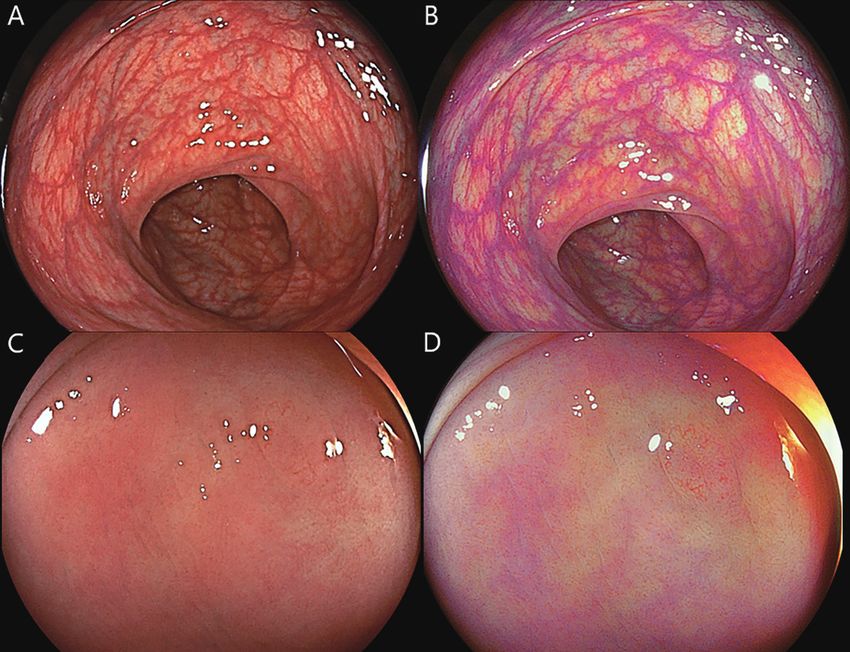
Page 2 of 11                                                                         Translational Gastroenterology and Hepatology, 2020

impact on detection has been disappointing. Improvements                     One explanation for the generally meagre improvements
from individual technology advances are marginal or                      in detection seen with imaging technology advances is that
modest in positive studies but balanced by large numbers                 imaging represents only a single step in the multi-step
of heterogenous or negative results. This is reflected in                process from colonoscopy preparation through to detection
recent guidelines which suggest technologic advancements                 and polyp removal for histopathologic assessment. Ideal
can be used but fall short of recommending routine use in                bowel preparation, optimal colonoscopy technique and
average risk colonoscopy (7). The increasingly vast number               the aide of distal attachments culminate in the exposure of
of detection technology studies suffer from common                       colonic mucosa to the imaging technology and each of these
limitations; the central two being an overly narrow focus                have been demonstrated to individually impact on detection
on adenomatous polyp detection and secondly reporting                    (10-12). In a recent network-meta-analysis, Facciorusso
only averaged detection rates from multiple Colonoscopists.              et al. concluded that while there were detection gains for
Both are problems. Firstly, detection of sessile serrated                most technologies (distal caps or imaging enhancements)
lesions (SSLs) in addition to adenomas is of critical                    there was limited evidence for superiority of any one single
importance as they, like adenomas, are variably detected.                technology (13).
More importantly SSLs are precursors in a CRC pathway                        The conclusion in the current ESGE guideline is
which appears overrepresented in interval CRC (8,9). A                   that most of the current technologies provide only
hypothetical technology which improves adenoma detection                 “marginal detection gains” however we believe there is
yet has no, or worse, a negative impact on serrated lesion               still a case for optimism (7). Most of the imaging advances
detection may fail to achieve optimal CRC prevention.                    covered in this review are already available on current
The fixation on adenomas is compounded by the focus on                   generation colonoscopes, so Colonoscopists already have
the adenoma detection rate (ADR), whilst undoubtedly                     an opportunity to gain experience day to day. These
established and important, may be too blunt to evaluate                  technologies also have established benefits beyond
subtle detection gains in new technologies. Common                       detection such as characterization and real time prediction
throughout the detection literature are studies without                  of polyp histology and the assessment of polypectomy scars
significant difference in ADR but significant improvements               at surveillance (7,14). When a technology has multiple
in diminutive or flat adenoma detection or reduction in                  practical benefits its inclusion, and use, is more likely and
adenoma miss rates. There is no consensus on what metrics                incorporation into successive colonoscope systems more
should be used and what represents a clinically meaningful               readily justified. Perhaps more importantly, technology
detection gain. The second core issue is that the provision of           advances are still arriving. The current paradigm of
averaged detection rates may obscure assessment of the real              detection imaging technology has recently been disrupted
value of new technologies. A generally accepted paradigm is              by the advent of computer aided detection (CAD) systems
that interventions which will improve the detection rate of              which have capacity to detect polyps and relay this to the
the lowest detectors are likely to be of more overall clinical           Colonoscopist. In this review we aim to cover the major
value than improving that of high detectors. Studies which               areas of colonoscope imaging advances with a focus on the
                                                                         evidence base for detection gains for each technology.
do not routinely report whether detection gains are seen in
this former group limit the practical remedial value of that
technology. Furthermore, the inclusion of both high and                  Colonoscope imaging definition and field of view
low detectors in a technology study may blunt averaged
                                                                         HD
detection gains if high detectors, already operating nearer
the boundaries of detection, are not similarly aided by the              Detection of mucosal abnormalities in the gastrointestinal
study technology. Adding to this a myriad of additional                  tract is primarily dependent on the image seen by the
factors require consideration when reviewing the literature,             endoscopist and the ability to recognize subtle differences
such as; heterogeneity in reporting on training programs                 in mucosal characteristics. The ability to discriminate these
and learning curves in new technologies, the combination                 fine details was greatly advanced with the advent of HD
multiple generations of imaging technology in meta-                      endoscopes in the early 2000s. Prior to HD endoscopes,
analyses and the inherent limitation that while imaging                  the image resolution achieved by standard definition
studies may be randomized, blinding is usually not possible.             endoscopes was approximately 370,000 pixels (15).

© Translational Gastroenterology and Hepatology. All rights reserved.   Transl Gastroenterol Hepatol 2020 | http://dx.doi.org/10.21037/tgh.2020.02.05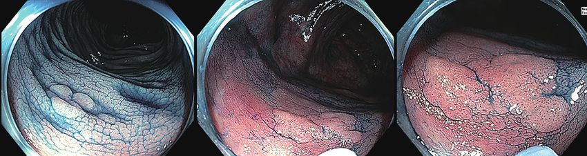
Translational Gastroenterology and Hepatology, 2020                                                                                   Page 3 of 11

With improvements in charge-coupled device chips and                     ADR (43.8% vs. 36.5%; P=0.03) than SD (20). Interestingly
complementary metal oxide semi-conductor (CMOS)                          in contrast to adenomas, no difference in SSL detection rate
technology, HD endoscopes now have image resolutions                     or missed SSLs was demonstrated (5.8% vs. 4.5%; P=0.361,
ranging from 850,000 pixels to more than 1 million pixels.               2.1% vs. 1.2% P=0.308) though this should be interpreted
Disappointingly these advances in definition have only                   within the context of a relatively low overall SSL detection
translated into marginal gains in detection. Data from early             rate. A second randomized control trial (RCT) without
studies of early HD systems were presented in a 2011 meta-               tandem colonoscopy of 1,221 average risk screening patients
analysis which included 4,422 patients and demonstrated an               however failed to demonstrate a significant difference in
increased adenoma diagnostic yield of 3.5% for HD over                   ADR (HD 32% vs. SD 28%; P=0.01) though HD did detect
SD (16). Study heterogeneity, non-randomized studies and                 an overall greater number of diminutive adenomas in the
technological limitations of early HD systems may have                   cohort (135 vs. 97; P=0.002) (21). Counterintuitively a lower
contributed to the limited detection difference. However,                overall detection of SSLs was seen the HD compared to the
optimism that randomized studies of more recent HD                       SD group (62 vs. 81; P=0.005) (21).
technology would demonstrate more impressive detection                      Overall HD colonoscopy appears to be associated with
gains are yet to materialize. A multicentre trial in the                 a marginal increase in adenoma detection which may be
United Kingdom of 262 patients in the UK National Bowel                  more marked when comparing over multiple generations of
Cancer Screening Program randomized patients to either                   colonoscope imaging technology. There is no clear data to
SD (Olympus Lucera System, Toyko, Japan) or the HD                       support HD improving SSL detection.
(Pentax HiLine system, Tokyo, Japan) did not demonstrate
a difference in polyp or adenoma detection, though HD was
                                                                         Field of view
superior in the detection of flat adenomas (18.6% vs. 5.2%;
P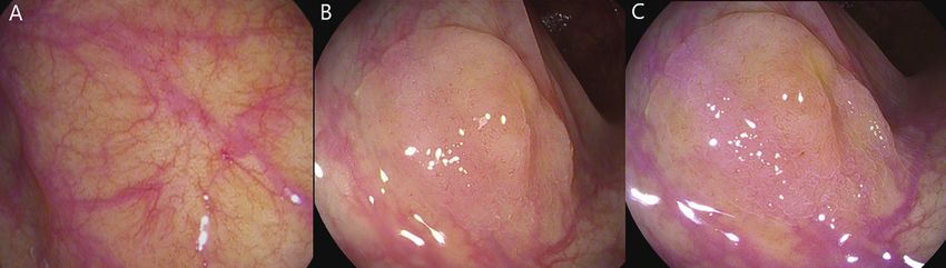
Page 4 of 11 Translational Gastroenterology and Hepatology, 2020
A B C
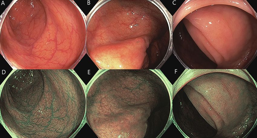
Figure 1 Flat adenomatous polyps and background colonic mucosa highlighted with chromocolonoscopy (A) 5 mm flat (Paris 0-IIa)
adenoma. A 7 mm flat (Paris 0-IIa) adenoma detected with chromocolonoscopy (B) and viewed with near focus magnification (C).
which is reflected in limited inclusion into routine practice. P=0.12). The higher CC ADR (55.5% vs. 48.4%) was not
More study in this area may be required. statistically significant however CC did detect a higher
proportion of flat adenomas, diminutive adenomas and a
higher number of polyps per patient (31).
Chromocolonoscopy (CC) and VCC
In addition to improved adenoma detection, increased
CC proximal serrated lesion detection has also been
demonstrated in a RCT where CC was compared with SD
CC was first described in 1977 and has consistently been
WL colonoscopy (12% vs. 6%; OR 1.96; P=0.012) though
associated with improved detection in the majority of
again the CC mean procedure time was significantly longer
studies with increases in overall polyp detection, adenoma
detection and serrated polyp detection rates (27-30). Whilst (36.8 vs. 30.6 minutes) (32).
not a universally considered a technologic imaging advance, Despite the benefits of CC, an increase in detection
a brief review of CC is warranted as virtual CC technologies of advanced adenomas has not been demonstrated and
aim to replicate and supplant CC, thus it remains the guidelines have fallen short of recommending CC use in
yardstick by VCC technologies should be judged. routine average-risk colonoscopy (28). In addition to the
CC involves colonic inspection after the application costs of procurement and preparation of the dye and the
of a contrast dye, indigo-carmine or methylene blue. CC equipment for delivery, CC has consistently been associated
highlights the topography and microtopography of the with a time cost of longer withdrawal times, though it is
colonic mucosa, reflected in increased flat and diminutive possible these longer times are an inherent component
adenoma detection (29,31) (Figure 1). A 2016 Cochrane of the improved detection of CC rather than simply an
review of seven randomized controlled trials concluded, unwanted negative consequence (7,28,29).
despite heterogeneity in patient cohorts, that CC increased It is possible that overcoming the logistic hurdles in
both the mean number of polyps detected as well as the delivering CC could reduce the barrier to widespread CC
number of neoplastic lesions found; OR detection of at use. This was studied in multi-centre, placebo controlled
least one polyp 1.87 (95% CI: 1.51 to 2.3) (29). With the randomised double blind trial of methylene blue colonic
widespread introduction of HD imaging which was already release tablets (MB-MMX) (33). Methylene blue was
associated with detection gains the question of whether combined with a pH and time dependent multimatrix
CC still conferred additional detection was assessed by structure designed to release the dye in the colon. A total
Kahi et al. in 2010 (31). Six hundred and sixty patients were of 1,249 patients were randomised in the intention to treat
randomised to HD CC or white light (WL). Consistent analysis with an increase in the detection of at least one
with previous studies the CC mean procedure time was adenoma, traditional serrated adenoma or SSL in the MB-
significantly longer (30.6 vs. 21.9 min; PTranslational Gastroenterology and Hepatology, 2020 Page 5 of 11 A B C D E F Figure 2 Normal colonic mucosa viewed with high definition white light (HD WL) (A) and narrow band imaging (NBI) (D) Diminutive flat (Paris 0-IIa) adenoma viewed with HD WL (B) and NBI (E). Large flat sessile serrated lesion viewed with HD WL (C) and NBI (F). continues to have a role in detection particularly in high mixed or limited in effect (7). However current generation risk patient groups. However it is the juxtaposition these VCC does appear superior to initial VCC technology benefits with the challenges in implementation that has giving a glimmer of hope to a future role of VCC in routine facilitated interest and research in VCC. colonoscopy (34). VCC Narrow band imaging (NBI) Push button VCC is now routinely incorporated into HD Conventional white light colonoscopy has three optical colonoscope systems either as light filters (NBI, Olympus filters covering the visible wavelength range from 400 Tokyo Japan) narrow wavelength laser (BLI/LCI Fujifilm, to 800 nm. NBI is achieved by filtering out the red light Tokyo, Japan) or post-image acquisition processing bandwidth, leaving a narrowed spectrum of light, at 415 (I-Scan Pentax, FICE Fujifilm, Tokyo, Japan). In principle and 540 nm, to penetrate the mucosa less deeply, resulting VCC aims to replicate the enhanced surface contrast and in increased contrast for surface vessels and structures (5) microtopography advantages of CC with simple push button (Figure 2). technique rather than the physical spraying of dye in CC. A recent meta-analysis of data from 4,491 individual In addition VCC aims to exaggerate the mucosal capillary patients in eleven randomised controlled trials compared vascular networks which is hypothesized to improve the efficacy of NBI to white light colonoscopy (34). Overall, detection through the absence of the normal background the ADR was 42.3% for HD-WL vs. 45.2% for NBI (OR vascular network and, for adenomatous lesions, highlight for adenoma detection of 1.14 NBI compared to HD- denser neoplastic vascular network (34). These conceptual WLE; 95% CI: 1.01–1.29; P=0.04). Despite heterogeneity advantages of VCC have unfortunately not translated into in bowel prep scores, NBI was found to be superior to WL clear detection gains with evidence of increased detection when bowel prep grade was best compared to only adequate © Translational Gastroenterology and Hepatology. All rights reserved. Transl Gastroenterol Hepatol 2020 | http://dx.doi.org/10.21037/tgh.2020.02.05
Page 6 of 11                                                                         Translational Gastroenterology and Hepatology, 2020

(OR 1.30; 95% CI: 1.04–1.62; P=0.02). ADR was also found                 are generated by two lasers with wavelengths of 410 and
to be higher only with newer generation “bright” NBI than                450 nm (40). White light endoscopy is achieved through
WLE (OR 1.28; 95% CI: 1.05–1.56; P=0.02), but not in                     irradiation of phosphor by the 450 nm-wavelenght laser.
initial generation NBI. Non-adenomatous polyps (OR 1.24;                 BLI is proposed to facilitate characterization of mucosal
95% CI: 1.06–1.44; P=0.008), right sided non-adenomatous                 vessels and structures and LCI to increase detection by
polyps (OR 1.35; 1.05–1.74; P=0.02) and flat polyps (OR                  improving contrast between red coloured lesions and white
1.24; 95% CI: 1.02–1.51; P=0.03) were also more likely                   mucosa (Figure 3).
to be detected with NBI than WL. SSLs are likely to                         There have been several randomised control trials for
comprise a substantial proportion of non-adenomatous                     the utility of BLI and LCI in polyp detection (41-44). BLI
right sided polyps. SSL detection was specifically assessed              was associated with a higher number of adenomas per
in recent meta-analysis where the only VCC technology                    patient than WL and in a tandem colonoscopy study BLI
demonstrating gains was NBI (SSL detection rate RR 2.04;                 followed by WL, had a lower polyp miss rate of 1.6% vs.
95% CI: 1.18–3.54; P=0.01 and mean SSLs per patient 0.03                 10.0% miss rate in the WLE-BLI group (42). More recent
vs. 0.024; P
Translational Gastroenterology and Hepatology, 2020                                                                                   Page 7 of 11

                    A                                                   B

                    C                                                   D

Figure 3 Normal colonic mucosa viewed with high definition white light (HD WL) (A) and linked colour imaging (LCI) (B). Diminutive
3 mm flat (Paris 0-IIa) adenoma visualized with HD WL (C) and LCI (D).

A                                                   B                                           C

Figure 4 Normal colonic mucosal vascular pattern viewed with I-Scan OE mode 2 (A). A large flat (Paris 0-IIa) sessile serrated lesion with
disruption of background colonic mucosal vascular pattern viewed with high definition white light (B) and I-Scan OE mode 2 (C). OE,
optical enhancement.

colonoscopy (49). Polyp detection rates (67.9% vs. 48.1%,                   the capacity to determine the relative benefit of the VCC
P≤0.0001) and flat polyp detection (27.8% vs. 9.9%;                         technology. A more recent tandem RCT compared HD
P=0.04) were higher in the HD i-Scan group (50). However                    i-Scan and HD WL colonoscopy in 740 patients with an
combining HD with VCC and comparing to SD limits                            i-Scan demonstrating a higher ADR for i-Scan (47.2% vs.

© Translational Gastroenterology and Hepatology. All rights reserved.   Transl Gastroenterol Hepatol 2020 | http://dx.doi.org/10.21037/tgh.2020.02.05
Page 8 of 11 Translational Gastroenterology and Hepatology, 2020 37.7% P=0.01) (51). No difference in SSL detection was low rate of false positive findings at 7% (58,59). However seen. In a tandem RCT involving a Lynch patient cohort these studies assessed CAD colonoscopy video recordings. i-Scan demonstrated substantially lower adenoma miss rate A recent RCT of CAD during real-time HD colonoscopy than HD WL (12% vs. 62%, P=0.007) without difference in in 1,058 patients demonstrated a significantly greater ADR ADR (52). (29.1% vs. 20.3%; P90%) has Coast University Hospital, AUSTRALIA for providing the been demonstrated in deep learning CAD studies with a images in Figure 3. © Translational Gastroenterology and Hepatology. All rights reserved. Transl Gastroenterol Hepatol 2020 | http://dx.doi.org/10.21037/tgh.2020.02.05
Translational Gastroenterology and Hepatology, 2020                                                                                   Page 9 of 11

Footnote                                                                       performance: a pilot study. Am J Gastroenterol
                                                                               2010;105:2312-7.
Conflicts of Interest: The authors have no conflicts of interest
                                                                         12.   Facciorusso A, Del Prete V, Buccino RV, et al. Comparative
to declare.
                                                                               Efficacy of Colonoscope Distal Attachment Devices in
                                                                               Increasing Rates of Adenoma Detection: A Network Meta-
Ethical Statement: The authors are accountable for all
                                                                               analysis. Clin Gastroenterol Hepatol 2018;16:1209-
aspects of the work in ensuring that questions related
                                                                               1219.e9.
to the accuracy or integrity of any part of the work are
                                                                         13.   Facciorusso A, Triantafyllou K, Murad MH, et al.
appropriately investigated and resolved.
                                                                               Compared Abilities of Endoscopic Techniques to Increase
                                                                               Colon Adenoma Detection Rates: A Network Meta-
References                                                                     analysis. Clin Gastroenterol Hepatol 2019;17:2439-
                                                                               2454.e25.
1.  Rex DK, Cutler CS, Lemmel GT, et al. Colonoscopic
                                                                         14.   Desomer L, Tutticci N, Tate DJ, et al. A standardized
    miss rates of adenomas determined by back-to-back
                                                                               imaging protocol is accurate in detecting recurrence after
    colonoscopies. Gastroenterology 1997;112:24-8.
                                                                               EMR. Gastrointest Endosc 2017;85:518-26.
2. van Rijn JC, Reitsma JB, Stoker J, et al. Polyp miss rate
    determined by tandem colonoscopy: a systematic review.               15.   ASGE Technology Committee. High-definition and high-
    Am J Gastroenterol 2006;101:343-50.                                        magnification endoscopes. Gastrointestinal Endoscopy
3. Kaminski MF, Regula J, Kraszewska E, et al. Quality                         2014;80:919-27.
    indicators for colonoscopy and the risk of interval cancer.          16.   Subramanian V, Mannath J, Hawkey CJ, et al. High
    N Engl J Med 2010;362:1795-803.                                            definition colonoscopy vs. standard video endoscopy
4. Corley DA, Jensen CD, Marks AR, et al. Adenoma                              for the detection of colonic polyps: a meta-analysis.
    detection rate and risk of colorectal cancer and death. N                  Endoscopy 2011;43:499-505.
    Engl J Med 2014;370:1298-306.                                        17.   Di Caro S, Fini L, Vega R, et al. Multicentre randomised
5. East JE, Vleugels JL, Roelandt P, et al. Advanced                           controlled trial comparing standard and high resolution
    endoscopic imaging: European Society of Gastrointestinal                   optical technologies in colorectal cancer screening.
    Endoscopy (ESGE) Technology Review. Endoscopy                              Frontline Gastroenterology 2019;10:244-52.
    2016;48:1029-45.                                                     18.   Roelandt P, Demedts I, Willekens H, et al. Impact
6. Kudo T, Saito Y, Ikematsu H, et al. New-generation                          of endoscopy system, high definition, and virtual
    full-spectrum endoscopy versus standard forward-                           chromoendoscopy in daily routine colonoscopy: a
    viewing colonoscopy: a multicenter, randomized, tandem                     randomized trial. Endoscopy 2019;51:237-43.
    colonoscopy trial (J-FUSE Study). Gastrointest Endosc                19.   Adler A, Wegscheider K, Lieberman D, et al. Factors
    2018;88:854-64.                                                            determining the quality of screening colonoscopy: a
7. Bisschops R, East JE, Hassan C, et al. Advanced imaging                     prospective study on adenoma detection rates, from 12,134
    for detection and differentiation of colorectal neoplasia:                 examinations (Berlin colonoscopy project 3, BECOP-3).
    European Society of Gastrointestinal Endoscopy (ESGE)                      Gut 2013;62:236-41.
    Guideline - Update 2019. Endoscopy 2019;51:1155-79.                  20.   Pioche M, Denis A, Allescher HD, et al. Impact of 2
8. Hetzel JT, Huang CS, Coukos JA, et al. Variation                            generational improvements in colonoscopes on adenoma
    in the detection of serrated polyps in an average risk                     miss rates: results of a prospective randomized multicenter
    colorectal cancer screening cohort. Am J Gastroenterol                     tandem study. Gastrointest Endosc 2018;88:107-16.
    2010;105:2656-64.                                                    21.   Zimmermann-Fraedrich K, Groth S, Sehner S, et al.
9. Kahi CJ. How does the serrated polyp pathway alter CRC                      Effects of two instrument-generation changes on adenoma
    screening and surveillance? Dig Dis Sci 2015;60:773-80.                    detection rate during screening colonoscopy: results from
10. Rex DK, Imperiale TF, Latinovich DR, et al. Impact of                      a prospective randomized comparative study. Endoscopy
    bowel preparation on efficiency and cost of colonoscopy.                   2018;50:878-85.
    Am J Gastroenterol 2002;97:1696-700.                                 22.   Deenadayalu VP, Chadalawada V, Rex DK. 170 degrees
11. Rex DK, Hewett DG, Raghavendra M, et al. The                               wide-angle colonoscope: effect on efficiency and miss
    impact of videorecording on the quality of colonoscopy                     rates. Am J Gastroenterol 2004;99:2138-42.

© Translational Gastroenterology and Hepatology. All rights reserved.   Transl Gastroenterol Hepatol 2020 | http://dx.doi.org/10.21037/tgh.2020.02.05
Page 10 of 11 Translational Gastroenterology and Hepatology, 2020
23. Fatima H, Rex DK, Rothstein R, et al. Cecal insertion adenoma detection rate in the colon by electronic
and withdrawal times with wide-angle versus standard chromoendoscopy and distal attachment: systematic
colonoscopes: a randomized controlled trial. Clin review and meta-analysis. Gastrointest Endosc
Gastroenterol Hepatol 2008;6:109-14. 2019;90:721-731.e1.
24. Núñez-Rodríguez H, Diez-Redondo P, Perez-Miranda 36. Giardiello FM, Allen JI, Axilbund JE, et al. Guidelines
M, et al. Role of Full-spectrum Endoscopy in Colorectal on genetic evaluation and management of Lynch
Cancer Screening: Randomized Trial. J Clin Gastroenterol syndrome: a consensus statement by the US Multi-society
2019;53:191-6. Task Force on colorectal cancer. Am J Gastroenterol
25. Facciorusso A, Del Prete V, Buccino V, et al. Full- 2014;109:1159-79.
spectrum versus standard colonoscopy for improving polyp 37. East JE, Suzuki N, Stavrinidis M, et al. Narrow band
detection rate: A systematic review and meta-analysis. J imaging for colonoscopic surveillance in hereditary non-
Gastroenterol Hepatol 2018;33:340-6. polyposis colorectal cancer. Gut 2008;57:65-70.
26. Jovani M, Campbell EJ, Hur C, et al. Effect of video 38. Cellier C, Perrod G, Colas C, et al. Back-to-
monitor size on polyp detection: a prospective, Back Comparison of Colonoscopy With Virtual
randomized, controlled trial. Gastrointest Endosc Chromoendoscopy Using a Third-Generation Narrow-
2019;90:254-258.e2. Band Imaging System to Chromoendoscopy With
27. Tada M, Katoh S, Kohli Y, et al. On the dye spraying Indigo Carmine in Patients With Lynch Syndrome. Am J
method in colonofiberscopy. Endoscopy 1977;8:70-4. Gastroenterol 2019;114:1665-70.
28. Kamiński MF, Hassan C, Bisschops R, et al. Advanced 39. Omata F, Ohde S, Deshpande GA, et al. Image-enhanced,
imaging for detection and differentiation of colorectal chromo, and cap-assisted colonoscopy for improving
neoplasia: European Society of Gastrointestinal Endoscopy adenoma/neoplasia detection rate: a systematic review and
(ESGE) Guideline. Endoscopy 2014;46:435-49. meta-analysis. Scand J Gastroenterol 2014;49:222-37.
29. Brown SR, Baraza W, Din S, et al. Chromoscopy versus 40. Yoshida N, Dohi O, Inoue K, et al. Blue Laser Imaging,
conventional endoscopy for the detection of polyps in Blue Light Imaging, and Linked Color Imaging for the
the colon and rectum. Cochrane Database Syst Rev Detection and Characterization of Colorectal Tumors.
2016;4:CD006439. Gut Liver 2019;13:140-8.
30. Kahi CJ, Li X, Eckert GJ, et al. High colonoscopic 41. Ikematsu H, Sakamoto T, Togashi K, et al. Detectability
prevalence of proximal colon serrated polyps in average- of colorectal neoplastic lesions using a novel endoscopic
risk men and women. Gastrointest Endosc 2012;75:515-20. system with blue laser imaging: a multicenter randomized
31. Kahi CJ, Anderson JC, Waxman I, et al. High-definition controlled trial. Gastrointest Endosc 2017;86:386-94.
chromocolonoscopy vs. high-definition white light 42. Shimoda R, Sakata Y, Fujise T, et al. The adenoma miss
colonoscopy for average-risk colorectal cancer screening. rate of blue-laser imaging vs. white-light imaging during
Am J Gastroenterol 2010;105:1301-7. colonoscopy: a randomized tandem trial. Endoscopy
32. Hurt C, Ramaraj R, Farr A, et al. Feasibility and economic 2017;49:186-90.
assessment of chromocolonoscopy for detection of 43. Min M, Deng P, Zhang W, et al. Comparison of linked
proximal serrated neoplasia within a population-based color imaging and white-light colonoscopy for detection
colorectal cancer screening programme (CONSCOP): an of colorectal polyps: a multicenter, randomized, crossover
open-label, randomised controlled non-inferiority trial. trial. Gastrointest Endosc 2017;86:724-30.
Lancet Gastroenterol Hepatol 2019;4:364-75. 44. Paggi S, Mogavero G, Amato A, et al. Linked color
33. Repici A, Wallace MB, East JE, et al. Efficacy of Per-oral imaging reduces the miss rate of neoplastic lesions in the
Methylene Blue Formulation for Screening Colonoscopy. right colon: a randomized tandem colonoscopy study.
Gastroenterology 2019;156:2198-2207.e1. Endoscopy 2018;50:396-402.
34. Atkinson NSS, Ket S, Bassett P, et al. Narrow-Band 45. Yoshida N, Hisabe T, Ikematsu H, et al. Comparison
Imaging for Detection of Neoplasia at Colonoscopy: Between Linked Color Imaging and Blue Laser Imaging
A Meta-analysis of Data From Individual Patients for Improving the Visibility of Flat Colorectal Polyps: A
in Randomized Controlled Trials. Gastroenterology Multicenter Pilot Study. Dig Dis Sci 2019. [Epub ahead
2019;157:462-71. of print].
35. Aziz M, Desai M, Hassan S, et al. Improving serrated 46. Oliveira Dos Santos CE, Malaman D, Pereira-Lima JC, et
© Translational Gastroenterology and Hepatology. All rights reserved. Transl Gastroenterol Hepatol 2020 | http://dx.doi.org/10.21037/tgh.2020.02.05Translational Gastroenterology and Hepatology, 2020 Page 11 of 11
al. Impact of linked-color imaging on colorectal adenoma of autofluorescence imaging of adenomatous and polypoid
detection. Gastrointest Endosc 2019;90:826-34. lesions during colonoscopy: a systematic review and meta-
47. Fujimoto D, Muguruma N, Okamoto K, et al. Linked analysis. Endosc Int Open 2015;3:E226-35.
color imaging enhances endoscopic detection of 54. Takeuchi Y, Sawaya M, Oka S, et al. Efficacy of
sessile serrated adenoma/polyps. Endosc Int Open autofluorescence imaging for flat neoplasm detection: a
2018;6:E322-E334. multicenter randomized controlled trial (A-FLAT trial).
48. Leung WK, Guo CG, Ko MKL, et al. Linked color Gastrointest Endosc 2019;89:460-9.
imaging versus narrow-band imaging for colorectal polyp 55. Moriichi K, Fujiya M, Okumura T. The efficacy of
detection: a prospective randomized tandem colonoscopy autofluorescence imaging in the diagnosis of colorectal
study. Gastrointest Endosc 2020;91:104-112.e5. diseases. Clin J Gastroenterol 2016;9:175-83.
49. Bhandari P, Thayalasekaran S, Keisslich R, et al. Detection 56. Vinsard DG, Mori Y, Misawa M, et al. Quality assurance
and characterization of colorectal polyps using high- of computer-aided detection and diagnosis in colonoscopy.
definition white light and i-Scan: Evidence-based Gastrointest Endosc 2019;90:55-63.
consensus recommendations using a modified Delphi 57. Liedlgruber M, Uhl A. Computer-aided decision support
process. United European Gastroenterol J 2018;6:748-54. systems for endoscopy in the gastrointestinal tract: a
50. Testoni PA, Notaristefano C, Vailati C, et al. High- review. IEEE Rev Biomed Eng 2011;4:73-88.
definition colonoscopy with i-Scan: better diagnosis for 58. Urban G, Tripathi P, Alkayali T, et al. Deep Learning
small polyps and flat adenomas. World J Gastroenterol Localizes and Identifies Polyps in Real Time With 96%
2012;18:5231-9. Accuracy in Screening Colonoscopy. Gastroenterology
51. Kidambi TD, Terdiman JP, El-Nachef N, et al. Effect 2018;155:1069-1078.e8.
of I-scan Electronic Chromoendoscopy on Detection 59. Misawa M, Kudo SE, Mori Y, et al. Artificial Intelligence-
of Adenomas During Colonoscopy. Clin Gastroenterol Assisted Polyp Detection for Colonoscopy: Initial
Hepatol 2019;17:701-708.e1. Experience. Gastroenterology 2018;154:2027-2029.e3.
52. Bisschops R, Tejpar S, Willekens H, et al. Virtual 60. Wang P, Berzin TM, Glissen Brown JR, et al. Real-time
chromoendoscopy (I-SCAN) detects more polyps in automatic detection system increases colonoscopic polyp
patients with Lynch syndrome: a randomized controlled and adenoma detection rates: a prospective randomised
crossover trial. Endoscopy 2017;49:342-50. controlled study. Gut 2019;68:1813.
53. Zhao ZY, Guan YG, Li BR, et al. Detection and miss rates
doi: 10.21037/tgh.2020.02.05
Cite this article as: Lee A, Tutticci N. Enhancing polyp
detection: technological advances in colonoscopy imaging.
Transl Gastroenterol Hepatol 2020.
© Translational Gastroenterology and Hepatology. All rights reserved. Transl Gastroenterol Hepatol 2020 | http://dx.doi.org/10.21037/tgh.2020.02.05You can also read