Extremity Fracture Management - Joint Trauma System Emergency War Surgery Course - Army ...
←
→
Page content transcription
If your browser does not render page correctly, please read the page content below
Emergency War Surgery Course
Joint Trauma System

         Extremity Fracture
              Management

       Joint Trauma System Battlefield Trauma Educational Program

                                                                1
EWS Extremity Fractures
Scenario

  A 22‐year‐old female sailor arrives to you after her left leg
  was hit by a piece of flying debris on the deck of an aircraft
  carrier. She has an obvious open fracture of her proximal left
  calf and no palpable pulse. On plain film, she has a severely
  comminuted tibial plateau fracture. On angiogram, she has a
  popliteal artery injury.
  1. How do you reduce and stabilize the fracture?
  2. Should the person get fasciotomies? If so, how?

2020, v1.1                                                         2
EWS Extremity Fractures Objectives Describe the initial evaluation and management of extremity fractures of upper and lower extremities. Debride non‐viable tissue in open fractures. Spare viable tissue in open fractures. Use early antibiotics with cephazolin. Give tetanus toxoid as soon as possible. Describe the diagnosis and management of compartment syndrome. 2020, v1.1 3

EWS Extremity Fractures
General Considerations
∎ Perform early debridement & irrigation.
∎ Administer early antibiotics for
contaminated injuries – cefazolin
1‐2 gm q8 – and tetanus toxoid ASAP.
∎ Stabilize the bone.
∎ Preserve viable bone fragments.
Viable soft‐tissue attachment or
constitute a large portion of the joint.
∎ Preserve maximal amount of viable Open lower extremity fracture
soft tissue. with stabilization. Requires
Ensures greatest reconstruction further debridement.
options at Role 3 and 4 facilities.
2020, v1.1 4
EWS Extremity Fractures
General Considerations

 ∎ Avoid internal fixation.
 ∎ Casts should not be used
   – may act as tourniquets.
                                           1                      2
 ∎ Always evaluate the neurovascular
   status of an extremity and check
   repeatedly.

 ∎ Check for compartment syndrome.
                                           3                          4

                                       Source: Emergency War Surgery, 5th U.S. Edition

2020, v1.1                                                                        5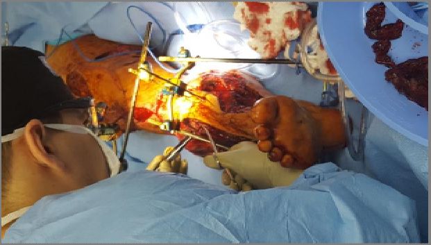
EWS Extremity Fractures
Fracture Stabilization
Splints and External Fixation
Splinting may be the only far‐forward option.
Most appropriate for low energy and closed fractures.
In general, intended to limit further injury and
not to definitively treat.
Complete reduction may not be possible.
Neuro‐vascular exam should be conducted post‐splinting.
Must be suitable for mode of transportation.
2020, v1.1 6
EWS Extremity Fractures
Fracture Stabilization
Splints and External Fixation (continued)
Use caution against constrictive/circumferential splints (avoid
compartment syndrome).
Splint should immobilize the joint above and below the fracture.
Apply adequate padding at pressure points to prevent injury.
Address open wounds first.
Document date and time of most recent debridement and
irrigation on the splint itself.
2020, v1.1 7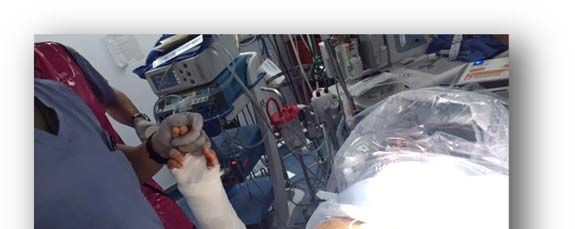
EWS Extremity Fractures
External Fixation
External Fixation over Internal Fixation
∎ Internal fixation at definitive care
location only.
∎ External fixation
Minimizes further soft tissue trauma.
Provides access for wound care and
re‐evaluation of compartments.
Early fracture stabilization blunts
inflammatory mediators.
External fixation of left lower
Benefit of pain control and ease of extremity fractures
transport.
2020, v1.1 8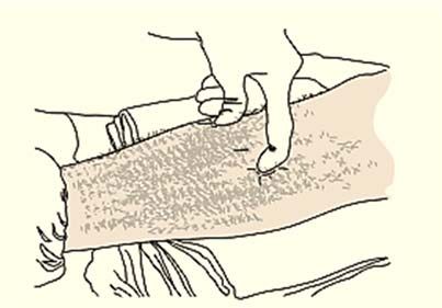
EWS Extremity Fractures
External Fixation
External Fixation General Principles
An understanding of anatomy for safe insertion of pins is critical.
May be done without use of plain films or fluoroscopy.
Can be done without power instruments.
Place enough pins to adequately stabilize fracture for transport.
Usually 2 pins per multipin clamp, but 3 may be required.
In austere environment (without fluoroscopy)
Surgeons should make small longitudinal incisions at pin sites and spread
down bluntly to avoid neurovascular bundles.
Stop pin advancement once purchasing the far cortex.
Place pins both near and far from the fracture on both bone fragments.
Pins may be placed to span joints if fracture extends to articular surface.
2020, v1.1 9
EWS Extremity Fractures
 External Fixation

 External Fixation General Principles

Pins should be a minimum        Bicortical placement of pins                   Addition of cross‐bar and two bar
of 2‐3 fingerbreadths from                                                     clamps. Apply longitudinal
fracture.                                                                      traction to reduce fracture and
                                                                               tighten the frame in alignment.

                             Source: Emergency War Surgery, 5th U.S. Edition

 2020, v1.1                                                                                                 10
EWS Extremity Fractures
Upper Extremity
∎ Splinting preferred in most situations
due to risk of nerve injury.
Humerus and shoulder fractures:
sling and swath or coaptation splint
Forearm and elbow injures:
long arm posterior splint or double
sugar‐tong splint
∎ If fixation required, pins should be Preparing a right upper
placed using an open technique. extremity splint
2020, v1.1 11EWS Extremity Fractures
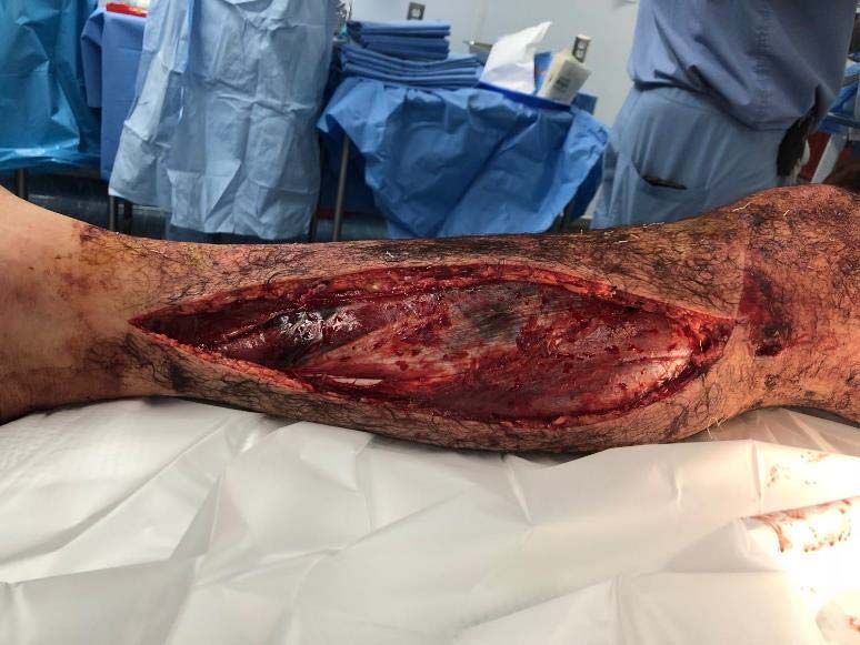
External Fixation
External Fixation of the Femur
1. Placing a towel underneath the thigh helps reproduce the bow of the femur.
2. Place a 1 cm longitudinal incision in line with mid‐lateral axis of the femur.
3. Place pin outside of fracture hematoma and at least 3 fingerbreadths away from
fracture.
4. Pins can be placed at any point along the anterolateral aspect of the femur.
5. Use multipin clamp as a guide to place second pin.
6. Repeat for distal fragment of the fracture.
7. Connect the two clamps with elbows, bar‐to‐bar clamps and two longitudinal bars
placed parallel to each other.
Source: Emergency War Surgery, 5th U.S. Edition
2020, v1.1 12EWS Extremity Fractures
External Fixation
External Fixation of the Tibia
External fixations similar to femur, although pins placed over
the anteromedial face of the tibia.
Anteromedial surface is the safest location for pins.
Source: Emergency War Surgery, 5th U.S. Edition
2020, v1.1 13EWS Extremity Fractures
External Fixation
External Fixation to Span Knee
∎ Indication: Proximal tibia fracture, distal femur fracture,
extensive knee injuries or vascular repairs in popliteal fossa.
∎ Pins placed anteromedial on the proximal tibia and
anterolateral on the distal femur.
Slight 10°‐15° flexion at the knee
2020, v1.1 14EWS Extremity Fractures
External Fixation
External Fixation to Span Ankle
∎ Indication: open distal tibia fractures
and open ankle wounds.
∎ Pins should be inserted on the
anteromedial surface of the tibia and
medial aspect of the calcaneus.
Make a longitudinal incision over the
calcaneus and dissect to the bone, avoiding
the posterior neurovascular structures.
Check distal vascular status before and after –
mark where the posterior tibial and dorsalis
Ankle external fixation
pedis artery pulses can be felt. with fasciotomies
2020, v1.1 15EWS Extremity Fractures
Compartment Syndrome
∎ Can occur with any injury to any fascial compartment.
Fascial defect, if penetrating, may not be adequate to fully
decompress the compartment.
∎ Mechanisms of Injury
Open fractures
Closed Fractures
Penetrating wounds
Crush Injuries
Vascular Injuries
And more…
2020, v1.1 16EWS Extremity Fractures
Compartment Syndrome
Clinical Signs of Compartment Syndrome
∎ Early: Pain out of proportion,
pain with passive stretch,
tense/swollen compartment.
∎ Late: Paresthesias,
pulselessness, pallor,
paralysis.
Medial fasciotomy incision
2020, v1.1 17EWS Extremity Fractures
Compartment Syndrome Risks
Risks for Acute Traumatic Compartment Syndrome
Decreased 1. Tight cast or dressing, closure of prior fasciotomy, excess traction
Compartment 2. External limb compression or crush particularly in obtunded or incapacitated casualty
Volume 3. Frostbite, burns or electric injury (may include escharotomy)
Increased 1. Edema accumulation: embolism, intravascular thrombosis, replantation, venous tourniquet,
Compartment injections, extravasation, infiltration, ergotamine ingestion, ischemia‐reperfusion, swelling,
Contents artery injury or spasm, revascularization procedures, prolonged arterial tourniquet use,
shock hypoperfusion, angiography and catheterization, limbs positioned well above heart,
mal‐positioned joints (ankle dorsiflexion,) or stretched muscles
2. Prolonged immobilization and limb compression particularly with obtunded or drugged
casualty, some surgical positioning
3. Hemorrhage, hemophilia, coagulopathy, anticoagulation, vessel injury
4. Large volume crystalloid resuscitation
5. Fractures particularly tibia fractures in adults, supracondylar humerus fractures in children
displaced, comminuted, or open fractures increase hemorrhage, swelling, and CS risk
6. Popliteal cyst, long leg brace
Source: JTS Acute Extremity Compartment Syndrome (CS) and the Role of Fasciotomy in Extremity War Wounds, 25 Jul 2016
2020, v1.1 18EWS Extremity Fractures
Compartment Syndrome
∎ Clinical diagnosis
Formal measurement is not necessary.
Measurements may be helpful in obtunded patients.
ΔPEWS Extremity Fractures
CS Risk Assessment
Risk Assessment for Extremity Compartment Syndrome
YES Established NO
Compartment
Syndrome?
Warm
Close monitoring: YES ischemia NO Therapeutic
Periodic reassessments duration fasciotomy
>12 hours?
Capacity to
YES NO
closely Prophylactic
monitor fasciotomy
over time?
Source: JTS Acute Extremity Compartment Syndrome (CS) and the Role of Fasciotomy in Extremity War Wounds, 25 Jul 2016
2020, v1.1 20EWS Extremity Fractures
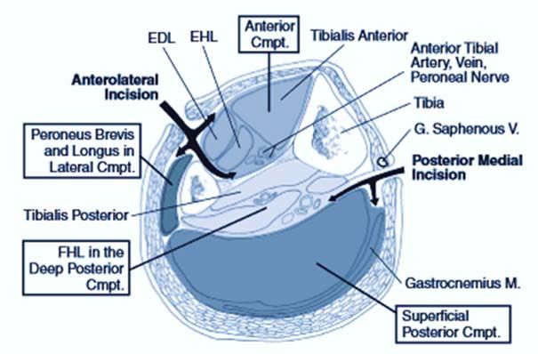
Compartment Releases
∎ All named compartments on Compartment Syndrome CPG.
∎ Leg: Release all 4 compartments (medial and lateral “H” incisions)
∎ Thigh: Release anterior and posterior, +/‐ medial compartment
∎ Gluteal: Release gluteus maximus
Volar Compartment
Tibialis
Anterior Anterior
EDL EHL Anterior Tibial
Cmpt.
Artery, Vein,
Anterolateral Peroneal Nerve
Mobile Wad Incision Tibia
Peroneus Brevis G. Saphenous V.
Medial Lateral & Longus in
Lateral Cmpt. Posterior Medial Incision
Tibialis Posterior
FHL in the Deep
Posterior Cmpt. Gastrocnemius M.
Dorsal Compartment
Superficial Posterior Cmpt.
Upper extremity Calf
2020, v1.1 21EWS Extremity Fractures Compartment Releases ∎ Forearm: Release volar and brachioradialis (a.k.a. mobile wad) +/‐ dorsal compartment ∎ Arm: Release anterior and posterior ∎ Shoulder: Release shoulder ∎ Hand: Release carpal tunnel and 11 compartments (you may need to refer to a book or Google) ∎ Foot: Controversial and not routinely recommended. Reviewed in ASSET. 2020, v1.1 22
EWS Extremity Fractures
Compartment Releases
Delayed Diagnosis of Compartment Syndrome
∎ Casualties with > 12 hours of warm ischemia
time should NOT receive fasciotomies.
∎ Medical treatment for rhabdomyolysis
Appropriate resuscitation (goal
UOP 75‐100 mL/hr) and intensive support
Role of amputation is currently unclear;
use medical judgement.
Right forearm fasciotomy
2020, v1.1 23EWS Extremity Fractures Exercise A 22‐year‐old female sailor arrives to you after her left leg was hit by a piece of flying debris on the deck of an aircraft carrier. She has an obvious open fracture of her proximal left calf and no palpable pulse. On plain film, she has a severely comminuted tibial plateau fracture. On angiogram, she has a popliteal artery injury. 1. How do you reduce and stabilize the fracture? 2. Should the person get fasciotomies? If so, how? 2020, v1.1 24
EWS Extremity Fractures
References
Joint Trauma System (JTS), Orthopedic Trauma: Extremity Fractures Clinical
Practice Guideline (CPG), 26 Feb 2020.
https://jts.amedd.army.mil/assets/docs/cpgs/JTS_Clinical_Practice_Guidelines_(CPGs)/Orthopaedic_Trau
ma_Extremity_Fractures_26_Feb_2020_ID56.pdf
JTS, Acute Extremity Compartment Syndrome (CS) and the Role of Fasciotomy in
Extremity War Wounds CPG, 25 Jun 2016.
https://jts.amedd.army.mil/assets/docs/cpgs/JTS_Clinical_Practice_Guidelines_(CPGs)/Extremity_Compa
rtment_Syndrome‐Fasciotomy_Extremity_War_Wounds_25_Jul_2016_ID17.pdf
The Office of The Surgeon General, Borden Institute. Emergency War Surgery, 5th
U.S. Edition, 2018. Chap 22, 34
https://www.cs.amedd.army.mil/Portlet.aspx?ID=cb88853d‐5b33‐4b3f‐968c‐2cd95f7b7809
Photos are part of the JTS image library unless otherwise noted.
2020, v1.1 25You can also read