Focal Active Colitis Presented With Chronic Diarrhea - Cureus
←
→
Page content transcription
If your browser does not render page correctly, please read the page content below
Open Access Case
                                          Report                                                  DOI: 10.7759/cureus.8140

                                          Focal Active Colitis Presented With Chronic
                                          Diarrhea
                                          Bayarmaa Mandzhieva 1 , John Taylor 2 , Hammad Zafar 3 , Mamoon Ur Rashid 1 , Abu H. Khan 4

                                          1. Internal Medicine, AdventHealth Orlando, Orlando, USA 2. Internal Medicine, AdventHealth, Orlando,
                                          USA 3. Internal Medicine, Advent Health, Orlando, USA 4. Gastroenterology, AdventHealth, Orlando,
                                          USA

                                          Corresponding author: John Taylor, john.taylor.do@adventhealth.com

                                          Abstract
                                          There are various etiologies of colonic injury and inflammation. The most commonly described
                                          colitides in clinical practice are associated with infection, inflammatory bowel disease,
                                          ischemia, radiation and medications. The colonic wall has a limited set of responses to different
                                          types of injury; therefore, there is overlap between many of these disorders. Focal active colitis
                                          is characterized by isolated neutrophilic cryptitis with the background mucosa displaying
                                          normal crypt architecture. This inflammatory pattern can be easily unnoticed by pathologists
                                          because on low-power examination the mucosa may have almost normal appearance. General
                                          practitioners also may not be familiar with this term, underlying etiologies, associated risk
                                          factors, course, available therapies and follow up.

                                          We present a case of an 82-year-old female with chronic diarrhea and weight loss. She had a
                                          negative infectious workup and normal radiology series. She subsequently underwent
                                          endoscopic evaluation in lieu of persistent and debilitating symptoms which revealed
                                          nonspecific macroscopic findings with pathology noting focal active colitis. She was empirically
                                          treated with a 14-day course of Xifaxan and responded well to management with almost
                                          complete resolution of her symptoms and no recurrence on six-month follow-up.

                                          Categories: Internal Medicine, Gastroenterology
                                          Keywords: chronic diarrhea, focal active colitis, cryptitis, xifaxan, irritable bowel syndrome, infectious
                                          colitis, inflammatory bowel disease.

                                          Introduction
                                          Focal active colitis (FAC) is a histologic term that describes the isolated finding of focal
                                          infiltration of the colonic crypts by neutrophils. Significant controversy is still present around
                                          the clinical implication of the diagnosis of FAC, which may or may not be clinically relevant.
Received 04/02/2020
Review began 05/01/2020
Review ended 05/01/2020                   Prior studies have shown that the incidence of Crohn’s disease (CD) in adults presenting with
Published 05/15/2020
                                          FAC was relatively low and has varied between 0% and 13%, whereas the incidence of
© Copyright 2020                          infectious-type colitis has been demonstrated to be nearly 50% with FAC appearing to be an
Mandzhieva et al. This is an open         incidental finding without clinical relevance in about 25% of the patients [1,2]. In addition,
access article distributed under the
                                          based on clinical presentation and outcomes, a greater number of FAC cases in adults have
terms of the Creative Commons
                                          been found to have an infectious etiology, although specific infectious pathogens were not
Attribution License CC-BY 4.0., which
permits unrestricted use, distribution,   detected in the majority of cases [3]. This can be partially explained by the fact that some of
and reproduction in any medium,           them are notoriously fastidious and may be difficult to recover. FAC is also encountered in
provided the original author and          patients with irritable bowel syndrome (IBS) and drug-induced colonic injury. The aim of this
source are credited.
                                          article is to present manifestations of FAC with chronic diarrhea and weight loss which is not a

                                          How to cite this article
                                          Mandzhieva B, Taylor J, Zafar H, et al. (May 15, 2020) Focal Active Colitis Presented With Chronic
                                          Diarrhea. Cureus 12(5): e8140. DOI 10.7759/cureus.8140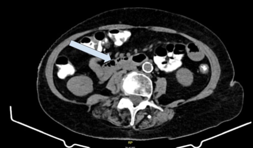
frequently encountered problem for general physicians. Our case also demonstrates effective
                                   treatment of FAC with Xifaxan, which is quite rarely reported upon extensive review of medical
                                   literature. Xifaxan could be one of the potential therapies for this condition, and our case
                                   emphasizes the necessity to conduct further research on underlying pathogenesis of FAC and its
                                   therapeutic options given the lack of strong recommendations and standardized approach
                                   available currently.

                                   Case Presentation
                                   An 82-year-old female presented to the gastroenterology office with complaints of diarrhea,
                                   nausea, abdominal cramping, increased flatulence and anorexia for the past three months. She
                                   also estimated that she had lost nearly 20 lbs in the past six months unintentionally.

                                   She described the diarrhea as explosive and the amount of diarrhea correlating to the amount of
                                   food she was eating, but not the type of food. She reported taking a recent course of Flagyl for
                                   three days with no symptom improvement and no other recent exposure to antibiotics. She had
                                   tried Imodium without relief. She denied fever, chills, emesis, constipation, jaundice, melena,
                                   hematochezia, mucus in the stools, tenesmus, chest pain, respiratory or urinary symptoms.

                                   She denied similar episodes in the past, any recent changes to her medications, chronic
                                   nonsteroidal anti-inflammatory drugs (NSAIDs) use, sick contacts or recent international
                                   travel. She had a history of giardia infection more than 10 years ago and had a normal
                                   colonoscopy in 2007.

                                   Her past medical history was significant for intraductal carcinoma in situ of left breast,
                                   coronary artery disease, hypertension, hyperlipidemia, thyrotoxicosis, peripheral artery disease
                                   and osteopenia. Her past surgical history included left breast lumpectomy, hysterectomy,
                                   cholecystectomy, left renal mass removal and appendectomy. Family history was negative for
                                   inflammatory bowel disease (IBD) and gastrointestinal tract malignancies. She had a remote
                                   history of smoking and denied alcohol or illicit drug use.

                                   In the office at the initial presentation, vital signs were within normal limits and physical
                                   examination was unremarkable. A complete blood count, comprehensive metabolic panel and
                                   thyroid-stimulating hormone were normal. She had negative stool studies for Clostridium
                                   difficile and negative stool cultures for Vibrio, Yersinia, Shigella, Salmonella, Campylobacter and
                                   Escherichia coli 0157:H7.

                                   She underwent a CT of the abdomen and pelvis with contrast prior to her initial office visit,
                                   ordered by her urologist for evaluation of resolved flank pain, which was negative for acute
                                   intra-abdominal abnormalities (Figure 1).

2020 Mandzhieva et al. Cureus 12(5): e8140. DOI 10.7759/cureus.8140                                                                 2 of 7
FIGURE 1: CT of the abdomen/pelvis with contrast
No bowel wall thickening or obstruction. No mesenteric stranding or free fluid.
She was recommended to complete stool studies with giardia antigen and ova and parasite
microscopic exam, and she was scheduled for a diagnostic colonoscopy with biopsies. Stool
studies returned negative for infectious etiologies. Given significant unintentional weight loss
in an elderly patient, esophagogastroduodenoscopy (EGD) was ordered as well.
She was empirically treated with a trial of Xifaxan three times daily for 14 days for possible IBS
with diarrhea (IBS-D) given previously failed treatment with Imodium and Flagyl. She was also
given FDgard two tablets twice daily for symptomatic management of abdominal cramps.
She completed bowel preparation for colonoscopy with SUPREP® Bowel Prep Kit (Braintree
Laboratories, Braintree, MA) (sodium sulfate, potassium sulfate and magnesium sulfate).
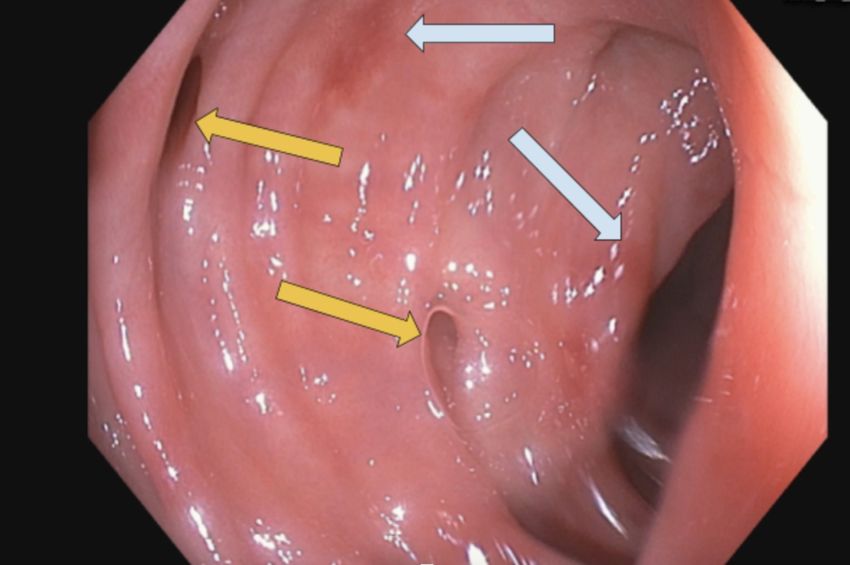
EGD and colonoscopy with good bowel preparation noted mild Helicobacter pylori negative
gastritis, patchy erythema in the sigmoid colon (Figure 2) with pathology noting FAC, 5-mm
hyperplastic polyp in the sigmoid, diverticulosis and medium sized internal and external
hemorrhoids. Small bowel biopsies ruled out celiac disease and multiple random biopsies from
each segment of the colon were negative for microscopic colitis. A small 5-mm sessile polyp in
the sigmoid colon was completely removed with cold snare. Two endoclips were deployed
prophylactically at the polypectomy site.
2020 Mandzhieva et al. Cureus 12(5): e8140. DOI 10.7759/cureus.8140 3 of 7FIGURE 2: Colonoscopy with good bowel preparation
Mild patchy mucosal erythema involving sigmoid colon (gray arrows). Few scattered diverticula
(yellow arrows).
Given colonoscopy findings, she was diagnosed with acute focal colitis and a decision was made
to complete the course of Xifaxan and to consider switching to ciprofloxacin and Flagyl in the
event of treatment failure. On the two-week follow-up appointment, she reported significant
improvement in her symptoms (mainly diarrhea, abdominal cramps and flatulence) after the
14-day course of Xifaxan. The patient was advised to follow a high-fiber diet with over-the-
counter Benefiber or Metamucil two to three times per day and low FODMAP (Fermentable
Oligosaccharides, Disaccharides, Monosaccharides and Polyols) diet.
Discussion
FAC pattern is characterized by a single focus or multiple foci of neutrophilic infiltration of the
crypts with an otherwise unremarkable colonic mucosa. The background crypts are well
preserved with no evidence of chronic injury [4].
Multiple etiologic entities can present as FAC on pathology reports. Most common clinical
scenarios include acute self-limited infectious colitis, IBD early in its course, ischemic colitis,
IBS, Clostridium difficile colitis, medication or chemical injury and bowel preparation artifact
[4]. Medications associated with FAC are NSAIDs, mycophenolate acid, and ipilimumab, among
others [5]. Although diarrhea is the most frequent reason for workup in the majority of these
patients, FAC can also be an incidental finding as a result of Phospho-soda bowel preparation in
asymptomatic patients undergoing screening colonoscopy [6]. Ozdil et al. found that 6.6% of
IBS patients had FAC, and it may be eligible to take a routine biopsy in female patients and in
patients aged above 50 years [7].
Osmond et al. in their retrospective review of cases of FAC in pediatric patients reported the
following presenting clinical symptoms: diarrhea, rectal bleeding, abdominal pain, vomiting
and others [8].
2020 Mandzhieva et al. Cureus 12(5): e8140. DOI 10.7759/cureus.8140 4 of 7Of these patients, most presented as an infectious or acute self-limited colitis with a significant
subset of the patients being immunocompromised. During the subsequent follow-up, none of
the patients were later found to have either ulcerative colitis or CD [1].
Chronic diarrhea sometimes can be a very challenging clinical scenario due to multitude of
differential diagnoses to consider. The majority of the macroscopic findings during endoscopic
evaluation are unremarkable or nonspecific [9]. Greenson et al. in their study on adult
population reported that endoscopic findings of FAC varied. Some of the patients had
completely normal endoscopic findings, whereas others had scattered aphthous ulcers on
normal background mucosa or patchy erythema [1]. Thus, the patient's past medical history,
medication review and clinical correlation are crucial.
FAC is a histopathological diagnosis. The hallmark is neutrophilic cryptitis which involves
several adjacent crypts on a single biopsy fragment. The lamina propria is often hypercellular
with normal architecture of the background mucosa and no Paneth cell metaplasia. The
presence of apoptotic crypt eplithelial cells is variable [10].
Features on histology cannot distinguish between incidental and clinically significant
FAC. Hence, it remains a diagnostic challenge in which features on pathology must
appropriately fit the correct clinical presentation.
Management of FAC itself is not well described. Therefore, physicians generally use their
clinical judgment and treatment available for presumed underlying etiology, such as IBD, IBS or
acute infectious colitis.
The clinicopathological features of CD and certain enteric pathogens can mimic each other to
some degree and can all present with an endoscopic picture of focal/segmental enterocolitis.
Most notable microorganisms in this regard are Campylobacter, Salmonella, Shigella, Yersinia
infections, Escherichia coli serotype O157:H7, Chlamydia trachomatis, Mycobacterium
tuberculosis, Actinomyces spp, Entamoeba histolytica, Giardia lamblia and others [11]. Normal gut
flora might become pathogenic under certain circumstances as well.
There is evidence that supports the notion that bacteria act as the driving force for both active
and chronic intestinal inflammation. One hypothesis is that chronic inflammation may be due
to abnormal immune responses to the normal gut flora. These notions support the use of
antibiotics in the case of active intestinal inflammation and also brings up the idea of
manipulating the enteric flora as a possible therapeutic for the more chronic cases [12].
IBS has been attributed to many triggers with numerous studies suggesting that up to one-third
of IBS-D cases result from a prior bacterial infection [13].
The use of oral antibiotics has likely therapeutic utility in multiple enteric conditions with
these notions in mind. However, several barriers exist to antibiotic use such as concerns for
growing bacteria resistance, interactions with other drugs and antibiotic side effects. The use of
minimally absorbed antibiotics, which are more gut targeted, may provide a solution to these
concerns [14].
Rifaximin (Xifaxan) is a safe, rifamycin-derived antibiotic with a wide range of antimicrobial
spectrum covering most Gram-positive and Gram-negative bacteria, including aerobes and
anaerobes . It has very poor systemic absorption after oral administration and complete fecal
excretion as unchanged drug [12].
2020 Mandzhieva et al. Cureus 12(5): e8140. DOI 10.7759/cureus.8140 5 of 7Rifaximin has several indications, which include select cases of infectious diarrhea, hepatic
encephalopathy and surgical prophylaxis. The drug has also found use in the prevention of
small intestinal bacterial overgrowth, Clostridium difficile associated colitis, diverticular disease
and infectious diarrhea. It may also have benefit in other conditions such as IBD and IBS [14].
Different studies suggested that it could be beneficial in the treatment of active IBD refractory
to the standard treatment. For example, Shafran et al. presented recently an open-label study
on the efficacy and safety of rifaximin 600 mg/day for 16 weeks in the treatment of mildly to
moderately active CD. At the end of the study, 59% of patients were in remission [15].
Rifaximin proved more effective than placebo for global symptoms and bloating in IBS patients.
The modest therapeutic gain was similar to that yielded by other currently available therapies
for IBS [13].
Banaag et al. in their multicenter retrospective study have shown that there are approximately
25% of patients presenting with diarrhea, abdominal pain and IBS with the histological finding
of FAC without a definitive diagnosis. More than 50% of patients who had diarrhoea had FAC,
and of these 44% responded to 5-aminosalicylic acid (5ASA), evident by resolution of
symptoms within two to six weeks [16].
In summary, FAC remains a diagnostic challenge. One must incorporate the patient's presenting
history, medications and findings on endoscopy to help narrow the differential
diagnosis. Histology can be helpful; however, there is no specific finding or feature that can
distinguish between incidental and clinically significant FAC (such as a minimum neutrophil
count).
As such, further studies should be conducted to determine patterns of histological disease
and/or a threshold of inflammation so that one can predict disease trajectory and allow for
better patient stratification.
We presented the case of FAC being effectively treated with rifaximin.
Rifaximin is a very promising agent with an excellent safety profile and more experimental
studies with rifaximin in clinical practice will help to further define the role of this antibiotic in
gastroenterology.
Conclusions
Our case is unique because infectious pathogens were not identified and she did not have risk
factors commonly associated with FAC in the literature such as IBD, NSAIDs use or bowel
preparation with Phospho-soda. She achieved significant resolution of her diarrhea within two
weeks of Xifaxan treatment with no recurrence for at least six months. In summary, FAC should
be strongly considered in the differential diagnosis in patients presenting with diarrhea and
macroscopically normal mucosa. The case also highlights the necessity of gaining more
experience in treatment of FAC with Xifaxan.
Additional Information
Disclosures
Human subjects: Consent was obtained by all participants in this study. Conflicts of interest:
In compliance with the ICMJE uniform disclosure form, all authors declare the following:
Payment/services info: All authors have declared that no financial support was received from
any organization for the submitted work. Financial relationships: All authors have declared
2020 Mandzhieva et al. Cureus 12(5): e8140. DOI 10.7759/cureus.8140 6 of 7that they have no financial relationships at present or within the previous three years with any
organizations that might have an interest in the submitted work. Other relationships: All
authors have declared that there are no other relationships or activities that could appear to
have influenced the submitted work.
References
1. Greenson JK, Stern RA, Carpenter SL, Barnett JL: The clinical significance of focal active
colitis. Hum Pathol. 1997, 28:729-733. 10.1016/s0046-8177(97)90183-0
2. Volk EE, Shapiro BD, Easley KA, Goldblum JR: The clinical significance of a biopsy-based
diagnosis of focal active colitis: a clinicopathologic study of 31 cases. Mod Pathol. 1998,
11:789-794.
3. Schneider EN, Havens JM, Scott MA, et al.: Molecular diagnosis of Campylobacter jejuni
infection in cases of focal active colitis. Am J Surg Pathol. 2006, 30:782-785.
10.1097/00000478-200606000-00017
4. Assarzadegan N, Montgomery E, Pezhouh MK: Colitides: diagnostic challenges and a pattern
based approach to differential diagnosis. Diagn Histopathol. 2017, 23:536-543.
10.1016/j.mpdhp.2017.11.006
5. Marginean EC: The ever-changing landscape of drug-induced injury of the lower
gastrointestinal tract. Arch Pathol Lab Med. 2016, 140:748-758. 10.5858/arpa.2015-0451-RA
6. Driman DK, Preiksaitis HG: Colorectal inflammation and increased cell proliferation
associated with oral sodium phosphate bowel preparation solution. Hum Pathol. 1998,
29:972-978. 10.1016/S0046-8177(98)90203-9
7. Ozdil K, Sahin A, Calhan T, et al.: The frequency of microscopic and focal active colitis in
patients with irritable bowel syndrome. BMC Gastroenterol. 2011, 11:96. 10.1186/1471-230X-
11-96
8. Osmond A, Ashok D, Francoeur CA, Miller M, Walsh JC: Is focal active colitis of greater
clinical significance in pediatric patients? A retrospective review of 68 cases with clinical
correlation. Hum Pathol. 2018, 74:164-169. 10.1016/j.humpath.2018.01.012
9. Lin W-C, Chang C-W, Chen M-J, et al.: Challenges in the diagnosis of ulcerative colitis with
concomitant bacterial infections and chronic infectious colitis. PLoS One. 2017, 12:e0189377.
10.1371/journal.pone.0189377
10. Jessurun J: The differential diagnosis of acute colitis: clues to a specific diagnosis . Surg Pathol
Clin. 2017, 10:863-885. 10.1016/j.path.2017.07.008
11. Hertogh GD, Aerssens J, Geboes KP, Geboes K: Evidence for the involvement of infectious
agents in the pathogenesis of Crohn’s disease. World J Gastroenterol. 2008, 14:845-852.
12. Gionchetti P, Rizzello F, Morselli C, Romagnoli R, Campieri M: Management of inflammatory
bowel disease: does rifaximin offer any promise?. Chemotherapy. 2005, 51:96-102.
10.1159/000081995
13. Menees SB, Maneerattannaporn M, Kim HM, Chey WD: The efficacy and safety of rifaximin for
the irritable bowel syndrome: a systematic review and meta-analysis. Am J Gastroenterol.
2012, 107:28-35. 10.1038/ajg.2011.355
14. Su CG, Aberra F, Lichtenstein GR: Utility of the nonabsorbed (You can also read