Functional Connectivity of the Anterior Nucleus of the Thalamus in Pediatric Focal Epilepsy
←
→
Page content transcription
If your browser does not render page correctly, please read the page content below
ORIGINAL RESEARCH
                                                                                                                                                  published: 02 August 2021
                                                                                                                                            doi: 10.3389/fneur.2021.670881

                                               Functional Connectivity of the
                                               Anterior Nucleus of the Thalamus in
                                               Pediatric Focal Epilepsy
                                               Rory J. Piper 1,2,3*, Chayanin Tangwiriyasakul 4,5 , Elhum A. Shamshiri 6,7,8 , Maria Centeno 9 ,
                                               Xiaosong He 10 , Mark P. Richardson 4 , Martin M. Tisdall 2 and David W. Carmichael 3
                                               1
                                                 Department of Neurosurgery, John Radcliffe Hospital, Oxford, United Kingdom, 2 Department of Neurosurgery, Great
                                               Ormond Street Hospital for Children, UCL Great Ormond Street Institute of Child Health, London, United Kingdom,
                                               3
                                                 Wellcome EPSRC Centre for Medical Imaging, Department of Biomedical Engineering, King’s College London, London,
                                               United Kingdom, 4 Department of Basic and Clinical Neuroscience, Institute of Psychiatry Psychology and Neuroscience,
                                               King’s College London, London, United Kingdom, 5 School of Biomedical Engineering and Imaging Sciences, King’s College
                                               London, London, United Kingdom, 6 San Francisco Veterans Affairs Health Care System (SFVAHCS), San Francisco, CA,
                                               United States, 7 Department of Psychiatry, University of California, San Francisco, San Francisco, CA, United States, 8 Sierra
                                               Pacific Mental Illness Research Education and Clinical Centers, San Francisco, CA, United States, 9 Epilepsy Unit, Neurology
                                               Department, Hospital Clinic, Barcelona, Spain, 10 Department of Psychology, University of Science and Technology of China,
                            Edited by:
                                               Hefei, China
                         Jan Kassubek,
           University of Ulm, Germany

                        Reviewed by:           Objective: Whilst stimulation of the anterior nucleus of the thalamus has shown efficacy
                            Yann Quidé,        for reducing seizure frequency in adults, alterations in thalamic connectivity have not
              University of New South
                       Wales, Australia
                                               been explored in children. We tested the hypotheses that (a) the anterior thalamus has
                Eelco Van Duinkerken,          increased functional connectivity in children with focal epilepsy, and (b) this alteration
Hospital Universitário Gaffrée e Guinle,
                                               in the connectome is a persistent effect of the disease rather than due to transient
                                  Brazil
                                               epileptiform activity.
                  *Correspondence:
                        Rory J. Piper          Methods: Data from 35 children (7–18 years) with focal, drug-resistant epilepsy and 20
             rorypiper@doctors.org.uk
                                               healthy children (7–17 years) were analyzed. All subjects underwent functional magnetic
                    Specialty section:         resonance imaging (fMRI) whilst resting and were simultaneously monitored with scalp
          This article was submitted to        electroencephalography (EEG). The fMRI timeseries were extracted for each Automated
                Applied Neuroimaging,
                a section of the journal
                                               Anatomical Labeling brain region and thalamic subregion. Graph theory metrics [degree
                 Frontiers in Neurology        (DC) and eigenvector (EC) centrality] were used to summarize the connectivity profile of
         Received: 22 February 2021            the ipsilateral thalamus, and its thalamic parcellations. The effect of interictal epileptiform
            Accepted: 27 May 2021              discharges (IEDs) captured on EEG was used to determine their effect on DC and EC.
          Published: 02 August 2021

                              Citation:
                                               Results: DC was significantly higher in the anterior nucleus (p = 0.04) of the thalamus
          Piper RJ, Tangwiriyasakul C,         ipsilateral to the epileptogenic zone in children with epilepsy compared to controls. On
    Shamshiri EA, Centeno M, He X,
                                               exploratory analyses, we similarly found a higher DC in the lateral dorsal nucleus (p =
      Richardson MP, Tisdall MM and
   Carmichael DW (2021) Functional             0.02), but not any other thalamic subregion. No differences in EC measures were found
Connectivity of the Anterior Nucleus of        between patients and controls. We did not find any significant difference in DC or EC in
       the Thalamus in Pediatric Focal
  Epilepsy. Front. Neurol. 12:670881.
                                               any thalamic subregion when comparing the results of children with epilepsy before, and
    doi: 10.3389/fneur.2021.670881             after the removal of the effects of IEDs.

Frontiers in Neurology | www.frontiersin.org                                           1                                           August 2021 | Volume 12 | Article 670881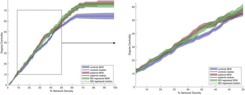
Piper et al. Thalamic Connectivity in Pediatric Epilepsy
Conclusions: Our data suggest that the anterior and lateral dorsal nuclei of the
thalamus are more highly functionally connected in children with poorly controlled focal
epilepsy. We did not detect a convincing change in thalamic connectivity caused by
transient epileptiform activity, suggesting that it represents a persistent alteration to
network dynamics.
Keywords: epilepsy, focal epilepsies, childhood epilepsies, deep brain stimulation, functional magnetic resonance
imaging, electroencephaloagraphy, connectivity, thalamus
INTRODUCTION seem paradoxical to findings from functional studies, this still
strengthens the association of the abnormal connectivity between
Focal epilepsy is increasingly recognized as a disorder of thalamus and the epileptogenic zone (17, 18). What is not clear
brain connectivity (1–4), and both structural and functional from these imaging studies, however, is whether this altered
connectome studies suggest that large-scale network alterations thalamic connectivity is a result of the persistent effect of epilepsy
associate with epilepsy (5, 6). The thalamus has been implicated or is instead due to transient interictal epileptiform activity.
as a major “hub” in epilepsy since it integrates information across Interictal epileptiform discharges (IEDs) have been associated
multiple functional cortical networks (7). Thalamo-cortical with significant thalamic or basal ganglia involvement in children
connectivity has long been implicated in the network-based with focal epilepsy (19, 20). This raises a question as to whether
pathogenesis of epileptic seizures (8–10), and has been shown to the differences in the functional connectivity seen in other studies
be a mechanism of interhemispheric seizure propagation, both in may be driven by the transient epileptiform activity.
generalized (11, 12) and focal epilepsies (13). There has been notably less work on establishing the
Deep brain stimulation (DBS) has therefore become a significance of the thalamus in the functional connectome in
potential surgical treatment for epilepsy and the SANTE trial childhood focal epilepsies, particular in extratemporal epilepsy
in 2010 showed that bilateral stimulation of the anterior nuclei which has recently been reported to constitute 37.9% of all
of the thalamus reduced seizure frequency in adults with both pediatric epilepsy surgeries (21). We therefore set out to test the
medically refractory, temporal lobe, and extratemporal lobe hypothesis that the anterior nucleus of the thalamus has a higher
epilepsy (14). Mechanistic understanding of the alterations to level of functional connectivity in children with focal epilepsy
thalamic connectivity that may be targeted by this therapy are than in those without epilepsy. We aimed to do this by using
invaluable for increasing, and potentially predicting efficacy. graph theory to measure connectiveness (degree and eigenvector
Thalamic DBS could be therapeutic in children but thus far has centrality) of the anterior thalamus, as performed in prior studies
not yet been accepted as a therapeutic option to the same degree (16). We additionally wished to test the hypothesis that these
as in adults. In part this may be explained by the paucity of data changes are not predominantly due to transient effects, but rather
on therapeutic outcomes of DBS in this group (15). persisting alterations in the network.
There have been a number of human neuroimaging studies
in temporal lobe epilepsy (TLE) in adults that examined the
METHODS
significance of the thalamus in the functional and structural
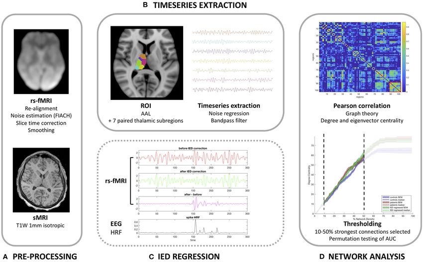
connectomes. Magnetic resonance imaging (MRI) currently Our methodology and imaging pipeline is summarized in
offers the only non-invasive method of examining the role Figure 1.
of thalamic connectivity within the whole-brain connectome.
Graph theory has been increasingly used to analyze these Patients
structural and functional brain networks to understand the This study examined data obtained from 35 children with
alterations posed by disease states such as epilepsy. Measures drug-resistant, focal epilepsy undergoing evaluation for
such as degree centrality (number of functional connections with epilepsy surgery at Great Ormond Street Hospital (London,
other brain regions) and eigenvector centrality (“influence” of a United Kingdom) (20/35 female, median age 15 years, age range
node within a network) have commonly been used. 7–18 years), previously described in the work by Centeno et al.
Studies from functional MRI (fMRI) have shown that the (19, 22). We excluded patients in whom there were large lesions
thalamus is a hub of functional connectivity in patients with that deformed brain architecture to ensure a reliable parcellation
TLE when compared to controls (16). The study by He et al. of brain regions would be possible in all subjects. The putative
(16) demonstrated that the thalami of patients with TLE who epileptogenic zone (determined by the epilepsy multidisciplinary
were not seizure-free following temporal lobe resection had team by means of clinical, neurophysiological, and neuroimaging
a higher degree and eigenvector centrality than compared to data) was most commonly the frontal lobe (21/35 cases). The
patients rendered seizure free and healthy controls. Studies putative epileptogenic zone was lateralized to the left-side in
of connectivity using diffusion tensor imaging (DTI) have 20/35 children. We summarize the patient details individually
shown findings suggestive of decreased structural connectivity in Table 1 and at a group level in Table 2. Our control group
between medial thalamic and temporal regions. Whilst this may consisted of twenty children without epilepsy (13/20 female,
Frontiers in Neurology | www.frontiersin.org 2 August 2021 | Volume 12 | Article 670881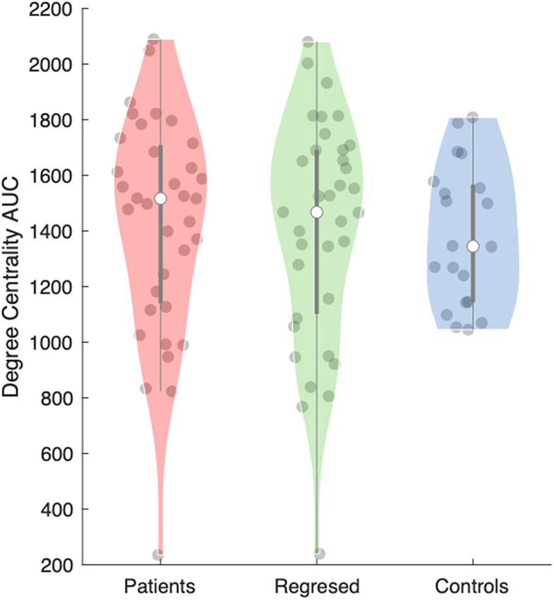
Piper et al.                                                                                                                    Thalamic Connectivity in Pediatric Epilepsy

  FIGURE 1 | Imaging analysis pipeline. (A) Pre-processing pipeline. (B) Timeseries extraction. (C) Demonstration of the correction method for interictal epileptiform
  discharges (IEDs) in the anterior nucleus of the thalamus for patients 13 (one spike type) as detailed in Table 1. (D) Functional network analysis.

median age 10 years, age range 7–17 years). Ethical approval was                         which children were asked to rest with their eyes closed, since
given by the NRES Committee London: Surrey Borders Research                              this session had reduced motion compared to the second on
Ethics Committee London Center (REC reference: 11/LO/1421).                              average (22).
The guardian of each participant provided informed and written
consent on the participant’s behalf.
                                                                                         Image Pre-processing
Magnetic Resonance Imaging Acquisition                                                   Image pre-processing was performed using Statistical Parametric
All subjects underwent simultaneous EEG-fMRI according to                                Mapping (SPM; https://www.fil.ion.ucl.ac.uk/spm/software/
the protocol previously published Centeno et al. (22). Briefly,                          spm12/). fMRI data was re-aligned and then the Functional
subjects were scanned at Great Ormond Street Hospital (London,                           Image Artifact Correction Heuristic (FIACH) method (23)
United Kingdom) in a 1.5T Siemens Avanto scanner using a 12                              was applied. This removes biophysically implausible signal
channel receive head coil. One cubic millimeter isotropic T1-                            jumps, and creates a parsimonious noise model from brain
weighted images were acquired using a Fast Low Angle Single                              regions exhibiting high noise levels. This method has been
Shot (FLASH) gradient echo sequence. The fMRI acquisition                                shown to be highly effective when compared to a number of
consisted of echo planar imaging (EPI) with 3.3×3.3×4 mm                                 alternative pipelines (23, 24). The six noise regressors estimated
resolution with a field of view of 210 mm, TR of 2,160 ms, TE                            by FIACH were added to the six re-alignment parameters
of 30 ms and flip angle of 75 . There were 30 contiguous slices in                       generated by SPM. The fMRI data was then corrected for
each volume with a slice thickness of 3 mm, slice gap of 1 mm,                           slice-timing and then registered to the 1 mm isotropic T1W
and matrix of 64 × 64. There were 300 volumes per session                                volume. Image registration was visually verified for each subject.
but the first five volumes in each session were omitted. Each                            Each patient’s T1W volumes were normalized to standard space
subject underwent up-to four fMRI sessions (each for 10 min and                          (MNI 152 template) and then the fMRI was normalized using
48 s), based on their ability to tolerate all of the sessions. In two                    the derived transform. The fMRI volumes were then converted
randomly allocated sessions the child watched a cartoon, and in                          to a 2 mm isotropic resolution following the normalization
the other two sessions the child was asked to rest with their eyes                       step. A Gaussian kernel was used to smooth fMRI 8 mm in
closed. We chose to include the first resting fMRI session, in                           each direction.

Frontiers in Neurology | www.frontiersin.org                                         3                                           August 2021 | Volume 12 | Article 670881
Piper et al. Thalamic Connectivity in Pediatric Epilepsy
TABLE 1 | Demographic, clinical, neurophysiological, and neuroradiological descriptions of each child.
ID Age* Age of Sex Laterality Location of EZ MRI features Focal vs. No. of IEDs Medications
(years) onset** of EZ multifocal
(years)
1 8 Female Left Temporal Tuberous sclerosis Focal 2 NZP and ZNS
2 14 4 Female Left Frontal Cryptogenic Focal 2 LCM and LVT
3 11 0.25 Male Left Hypothalamus/temporal Hypothalamic hamartoma Focal 59 LVT
4 15 10 Male Left Posterior quadrant Cryptogenic Multifocal 31, 10 CBZ
5 17 Male Right Parietal Focal cortical dysplasia Multifocal 51, 37, 71, 28 LVT, CBZ, and VPA
6 15 10 Male Right Frontal-central Cryptogenic Focal 15 CBZ
7 17 3 Female Left Temporal Cryptogenic Multifocal 175, 30, 131 LVT
8 14 2.5 Female Right Temporal Focal cortical dysplasia Multifocal 206, 16 LVT and TOP
9 11 6 Female Right Frontal-temporal Cryptogenic Focal 132 CBZ and LTG
10 11 7 Female Right Frontal Focal cortical dysplasia Focal 141 OXC
11 17 10 Female Right Frontal Cryptogenic Focal 34 LTG and LEV
12 16 6 Female Left Frontal Cryptogenic Focal 7 VPA and CBZ
13 16 13 Female Left Insula Focal cortical dysplasia Focal 76 TPM and CBZ
14 11 3 Male Right Frontal Cryptogenic Multifocal 128, 29 CBZ
15 11 8 Male Right Frontal Cryptogenic Multifocal 25, 1 LVT and VPA
16 16 2 Female Left Temporal-parietal-occipital Polymicrogyria Multifocal 236, 62, 39 LEV and CLNZ
17 15 9 Male Left Temporo-occipital Hippocampal sclerosis Multifocal 83, 21 LMT 575 Zonasimne 200
18 15 8 Male Left Fronto-temporal Focal cortical dysplasia Focal 112 OXZC 1200 LVT
19 17 5 Female Left Frontal Cryptogenic Focal 82 OXC
20 8 4 Female Left Frontal Middle cerebral artery stroke Focal 150 VPA 1200, LEV 600, ETHX
1000
21 16 Male Left Frontal Cryptogenic Multifocal 129, 77 PMP
22 13 3 Male Left Frontal Focal cortical dysplasia Focal 26 OXC and CLBZ
23 10 3 Female Left Frontal Cryptogenic Multifocal 47, 4 LVT and CBZ
24 11 6 Male Right Posterior quadrant Cryptogenic Multifocal 25, 23, 5 OXC
25 17 8 Male Left Frontal Cryptogenic Multifocal 35, 13 LVT, OXC, and CLBZ
26 17 Male Right Occipital Ischaemic perinatal insult Focal 21 OXC, LMT, and LVT 2000
27 18 Female Right Frontal Focal cortical dysplasia Multifocal 148, 6 LTG
28 17 5 Female Right Fronto-temporal Bilateral polymicrogyria Multifocal 242, 43 LVT and VPA
29 11 Female Right Parietal Cryptogenic Focal 81 OXC, PHE, and CLBZ
30 17 5 Male Left Frontal Cryptogenic Focal 67 LVT and LAC
31 11 5 Female Right Parietal Focal cortical dysplasia Focal 450 OXC, CLBZ, and VPA
32 17 12 Female Left Frontal Cryptogenic Multifocal 62, 1, 75 VPA
33 13 7 Female Left Frontal Focal cortical dysplasia Multifocal 128, 23 VPA
34 15 Female Right Posterior cingulate Dysembryoplastic Focal 26 LVT and LTG
neuroepithelial tumor
35 7 Male Left Frontal Cryptogenic Focal 145 LVT, OXC, and CLBZ
The number of interictal epileptiform discharges (IEDs) are listed for each IED type within each patient. EZ, epileptogenic zone; IED, interictal epileptiform discharge; *Age rounded to
nearest integer. **Age-of-onset (if known) is an approximate. CBZ, Carbamazepine; CLBZ, Clobazam; GAB, Gabapentin; LCM, Lacosamide; LTG, Lamotrigine; LVT, Levetiracetam; NZP,
nitrazepam; OXC, Oxcarbazepine; PHE, Phenobarbital; PGB, Pregabalin; PMP, Perampanel; RUF, Rufinamide; TPM, Topiramate; VPA, Valproate; ZNS, Zonisamide.
Electroencephalography (EEG) Acquisition the standard canonical hemodynamic response, and its temporal
and Analysis and dispersion derivatives as implemented in SPM8.
Simultaneous scalp EEG data was acquired using a 64-channel,
MRI-compatible cap (EASYCAP, Brain Products, Munich, Timeseries Analyses
Germany). The full EEG acquisition protocol is available in Determination of the fMRI timeseries for each region within
prior work (22). The onsets and durations of IEDs for each the brain had the following processing steps applied using
session were identified by a neurologist and neurophysiologist, MATLAB (MathWorks, Natick, MA, United States). This script
as described in prior work (19, 22). Events onsets and durations is freely available to download (https://github.com/roryjpiper/rs-
were used to generate a temporal regressor by convolution with fMRI.git). A general linear model was applied to control noise
Frontiers in Neurology | www.frontiersin.org 4 August 2021 | Volume 12 | Article 670881Piper et al. Thalamic Connectivity in Pediatric Epilepsy
TABLE 2 | Summaries of the patient and control cohorts. Statistical Analyses
For the purpose of statistical analysis, we selected a range of 10–
Patients Controls
50% of the highest correlations found in the adjacency matrix.
Median age (range) 15 (7–18) years 10 (7–17) years Fifty percent was used as the upper limit since after this point the
Male:female ratio 15:20 7:13 number of connections (defined as positive correlation values)
Median age of seizure onset (range)* 6 (0.25–13) years – did not increase any further in some subjects. To determine the
Median duration of epilepsy* 7 (3–14) years –
difference in DC and EC between groups that are relatively robust
to our choice of network density and thresholds, we compared
*Data missing for eight patients. results derived from the thalamus ipsilateral to hemisphere of
the epileptogenic focus in patients to the median results derived
from the left and right thalami of controls. Using a MATLAB-
according to the six re-alignment parameters aforementioned. based permutation test (30), we statistically compared the median
We then took the mean timeseries signal from the voxels of area-under-the-curve (AUC) between 10 and 50% of the network
each region of the brain according to the Automated Anatomical density for each graph theory metric (see Figure 2) between the
Labeling (AAL) template (25). The cerebellar regions were results for these subject groups. Statistical testing for measures
excluded to leave 90 cerebral regions. We investigated thalamic of the anterior nucleus of the thalamus were performed with
nuclei by removing the left and right thalami from the AAL an established priori hypothesis, whereas those performed on
atlas and replaced these with seven paired thalamic subregions, the remaining thalamic subregions were exploratory, and the
which instead parcellated the cerebrum into 102 regions. The reported p-values should be interpreted as descriptive. Our
sub-parcellations of the thalamus used here are described in statistical testing used 10,000 permutations and outputted a two-
previously published work by He et al. (26). These thalamic tailed p-value. The effect of IEDs on the graph theory measures
subregions included the anterior, medial dorsal, lateral dorsal, were further examined by measuring the Spearman correlation
lateral posterior, ventral lateral posterior, medial pulvinar and of the residual AUC for each patient vs. the number of spikes
lateral pulvinar subregions (shown in Figure 1). The timeseries per session. Effect size was determined using the Mann-Whitney-
√
signal for each region was then band-pass filtered to 0.04–0.07 Hz Wilcoxon test (r = z / n1 + n2 ). Figure representation of
as per our previous work (27). data uses the median value ± standard error of the mean (SEM).
Decimal places are rounded to two decimal points.
Correction for the Effects of Inter-ictal
Epileptiform Discharges RESULTS
Following the procedure described in detail by Shamshiri et al. Degree and Eigenvector Centrality
(20), we used functions in R (Version 3.6.1) to remove the The anterior nucleus of the thalamus ipsilateral to the EZ was
influence of IEDs from the fMRI signal. Figure 2 demonstrates found to have a significantly higher AUC for DC in children
the effect of IED regression on fMRI signal. Briefly, IED signal (1,516.5 ± 66.15) with epilepsy compared to controls (1,345 ±
changes are modeled by convolving the IEDs with the canonical 56.41) (r = 0.10; p = 0.04) (Figures 2, 3). We performed the
hemodynamic response function, and its derivatives before same analysis on only the patients with a frontal EZ (n = 17)
projecting the data from each region into an orthogonal space. and found a similar trend in DC, but this did not reach statistical
Following this correction, the “IED-corrected” timeseries’ for significance (p = 0.38).
each patient was determined using the identical pipeline as We then performed an exploratory analysis of the medial
described above. dorsal, lateral dorsal, lateral posterior, ventral lateral posterior,
medial pulvinar and lateral pulvinar thalamic subregions, on
Graph Theory Analyses comparing these two groups again, the median AUC for DC was
We created a 102×102 adjacency matrix for each subject by also significantly higher in the ipsilateral lateral dorsal nucleus
calculating the Pearson correlation coefficient between each (p = 0.02) (Supplementary Figure 1).
region using the corcoeff.m MATLAB function. We re-assigned No significant difference was found, however, for the same
the values in the diagonal of the adjacency matrix (self- analyses for the ipsilateral thalamus, taken as a whole (the AAL
correlation) to 0. All negative correlation values were re- thalamus ROI) (p = 0.30), nor for any of the remaining five nuclei
assigned to 0. We used the Brain Connectivity Toolbox (29) (Supplementary Figure 2). No significant differences were seen
(www.brain-connectivity-toolbox.net) in MATLAB to calculate in EC between patients and controls (Supplementary Figure 2).
the degree centrality (DC) (number of links connected to a
node) and eigenvector centrality (EC) (self-referential measure Effects of Interictal Epileptiform Activity
of a node influence) for each region-of-interest (ROI) in every Total number of captured IEDs ranged from 2 to 450 per EEG-
individual. These measures were selected to allow us to interpret fMRI session, as detailed in Table 1. Nineteen patients had one
our findings in the context of prior work in thalamic connectivity spike type (focal) and 16 had more than one IED type. We did
(16), but also are selected to test our hypothesis that brain not detect significant differences in the median AUC for DC
activity will have greater synchrony in children with epilepsy (p = 0.62) or EC (p = 0.81) in the anterior thalamic nucleus
when compared to those without. when comparing between the results from children with epilepsy
Frontiers in Neurology | www.frontiersin.org 5 August 2021 | Volume 12 | Article 670881Piper et al. Thalamic Connectivity in Pediatric Epilepsy
FIGURE 2 | Degree centrality of the anterior thalamic subregion ipsilateral to the epileptogenic zone (EZ). The lines represent controls (blue), patients before (red) and
after (green) correction for interictal epileptiform discharges (IEDs). The left graphs show the full range of network densities from 0 to 100%. The dotted box shows the
range selected for statistical analysis and shown in the right graph.
before, and after the effects of IEDs were removed. No other
thalamic subregion showed these differences either.
When correlating between IED frequency and the AUC for
DC in the ipsilateral anterior nucleus in children with epilepsy,
we found a borderline, but non-significant negative correlation
in DC [ρ = −0.34; p = 0.05 (rounded up to two decimal
places)], and a significant negative correlation in EC (ρ = −0.38;
p = 0.03).
DISCUSSION
The anterior nucleus of the thalamus has consistently been an
area of clinical interest and a therapeutic target in patients with
epilepsy. High-frequency stimulation of the anterior nucleus has
been shown to desynchronize focal large-scale brain activity and
reduce the number of IEDs in adults with TLE (31). The SANTE
trial in 2010 is, to date, the largest randomized controlled trial
of stimulation of the anterior nucleus of the thalamus significant
reduction in seizure frequency for adults with both TLE and those
with epileptogenic foci elsewhere (14).
Our study suggests that the number of connections (degree
centrality [DC]), determined by fMRI, is higher in the anterior
nucleus of the thalamus ipsilateral to the EZ in children with
focal epilepsy when compared to age-matched controls. Our FIGURE 3 | Area under the curve (AUC) for the degree centrality of the
findings support the significance of these nuclei in the functional anterior thalamic subregion ipsilateral to the epileptogenic zone (EZ). The violin
plot (28) demonstrates the distributions of the AUC for degree centrality in
connectome of children with epilepsy, and add weight to the patients before (red) and after (green) interictal epileptiform discharges (IEDs)
hypothesis that DBS to these regions could be therapeutic. and controls (blue). The clear dot shows the median.
The increased functional connectivity that we demonstrated
in the anterior nucleus is in keeping with the aforementioned
work, and also with a number of studies which have found the the network) is a biomarker for predicting seizure outcome
“midline thalamus” to be a key region involved in thalamo- in patients undergoing surgery for TLE (16). In patients not
temporal networks (32, 33). He et al. used resting state fMRI rendered seizure free, increased nodal hubness was seen in
to study functional connectivity, and suggest that presurgical both the ipsilateral and contralateral thalami on account of an
thalamic “hubness” (the thalamus being an important node in increased DC and EC. Furthermore, simulated lesioning of the
Frontiers in Neurology | www.frontiersin.org 6 August 2021 | Volume 12 | Article 670881Piper et al. Thalamic Connectivity in Pediatric Epilepsy
thalami showed a greater reduction in network integration in increased connectivity within epileptogenic networks survives
the patients not rendered seizure free. In another study, Bonilha the regression of IED effects (38). A study by Shamshiri et al.
et al. (34), showed that a feature of the connectome after temporal (20) showed that IEDs can have a pervasive yet transient effect on
lobectomy was reduced connectivity in thalamo-cortical circuits, the brain’s functional organization using a seed-to-voxel analysis
and that patients were more likely to achieve seizure freedom if during a low level attention task. This is compatible with our
their preoperative network did not involve abnormal thalamic study findings, where the anterior nucleus of the thalamus may
connectivity (35). facilitate a permissive state of increased connectivity whereby
Furthermore, on exploratory analysis, we identified a higher IEDs (from different brain regions in this heterogeneous group)
functional connectivity of the lateral dorsal nucleus of the can affect the coherence of “active” networks.
thalamus, but this in the context of a predominantly extra- We acknowledge the following limitations of our study.
temporal epilepsy cohort. The lateral dorsal nucleus has been Firstly, our patient group was heterogeneous in etiology and
shown to have connections with the limbic lobe (including epileptogenic focus (with a frontal lobe predominance). This
cingulate gyrus), parietal and visual cortex (36). Furthermore, it could, however, be observed as a strength since the findings
has been postulated that the anterior and lateral dorsal nuclei, we have detected have survived in this mixed cohort, which
may represent a “higher order” set of thalamic nuclei that, is somewhat reflective of the pool of patients referred for
rather than act as a “simple relay,” have a more significant epilepsy surgery workup. Secondly, the AAL atlas was designed
influence in the regulation of cortical-cortical interactions (37). for the analysis of the adult MNI-registered brain. Although
It is important to note the limited spatial resolution and effects of we have visually validated the registration of the AAL atlas
co-registration that affect fMRI. It is possible that some of the in each case and used a kernel smoothing method for fMRI
effect seen in the lateral dorsal nucleus is a spillover of signal signal, there may be inaccuracies that are unavoidable when
detected from the anterior nucleus. using this brain region atlas in children. Due to limited sample
The differences in functional connectivity we observed in this size and heterogeneous surgical management, we could not
study, however, were of modest magnitude. In contradistinction make meaningful correlations of clinical outcomes with our
to relatively homogeneous TLE cohort studies, this study quantitative graph theory metrics. Lastly, we recognize the need
included patients with an epileptic focus in various brain regions to validate these findings in external cohorts, particularly in
which is representative of the surgical population in pediatric children with generalized epilepsy or those with focal seizures
practice. Additionally, thalamic connectivity in this cohort needs with secondary generalization.
to be measured against a background of developmental changes.
While both of these factors could dilute the effect in childhood
DATA AVAILABILITY STATEMENT
epilepsy, we demonstrate a significant effect confirming our
primary hypothesis. Our analysis of the patients in this dataset The raw data supporting the conclusions of this article will be
may have combined and averaged the results of distinct groups made available by the authors, without undue reservation.
of children who either do or do not have the thalamus, or
its subregions, as regions of higher connectivity within the
functional network. It would be interesting to continue this work ETHICS STATEMENT
in this cohort by studying whether or not increased connectivity
The studies involving human participants were reviewed and
of the anterior thalamus is predictive of clinical outcome in
approved by the NRES Committee London: Surrey Borders
children undergoing DBS.
Research Ethics Committee London Center (REC reference:
Our secondary hypothesis was that altered functional
11/LO/1421). Written informed consent to participate in this
connectivity of the anterior thalamic nuclei was not due to
study was provided by the participants’ legal guardian/next
transient effects of IEDs, but instead to stable alterations in
of kin.
the network. We did not detect a significant difference in
DC values in this region in patients after the effects of IEDs
were corrected for. This study adds to the ongoing discussion AUTHOR CONTRIBUTIONS
regarding the effect of IEDs in the functional connectivity of
the brain and the role of the thalamus. It could be that long- All authors listed have made a substantial, direct and intellectual
term alterations in thalamic connectivity facilitate the spread contribution to the work, and approved it for publication.
of IEDs and that thalamic DBS reduces seizure frequency by
inhibiting this pathway of connectivity. This idea is supported FUNDING
by a study by Yu et al. (31) aforementioned, that showed that
bilateral anterior thalamic nucleus stimulation reduces IEDs. An RP was supported by an NIHR Academic Clinical Fellowship.
alternative hypothesis is that focal IEDs are the cause of increased The study was in part funded by Epilepsy Research UK
thalamic connectivity, but our study did not show a significant (PGE1402), Medical Research Council (MR/K013998/1), and
difference between DC and EC in patients before, and after Action Medical Research (grant number SP4646). This study was
the effects of IEDs were removed, suggesting that the anterior also supported by the King’s College London Wellcome/EPSRC
and lateral dorsal thalamic regions have an intrinsically altered Centre for Medical Engineering (WT 203148/Z/16/Z). Writing
baseline. This is in keeping with prior work that suggests that of this manuscript was supported by the Department of Veterans
Frontiers in Neurology | www.frontiersin.org 7 August 2021 | Volume 12 | Article 670881Piper et al. Thalamic Connectivity in Pediatric Epilepsy
Affairs Office of Academic Affiliations, the Advanced Fellowship SUPPLEMENTARY MATERIAL
Program in Mental Illness Research and Treatment, and the
Department of Veterans Affairs Sierra Pacific Mental Illness The Supplementary Material for this article can be found
Research, Education, and Clinical Center (MIRECC) granted online at: https://www.frontiersin.org/articles/10.3389/fneur.
to ES. 2021.670881/full#supplementary-material
REFERENCES 18. Keller SS, Richardson MP, Schoene-Bake J-C, O’Muircheartaigh J, Elkommos
S, Kreilkamp B, et al. Thalamotemporal alteration and postoperative
1. Centeno M, Carmichael DW. Network connectivity in epilepsy: resting seizures in temporal lobe epilepsy. Ann Neurol. (2015) 77:760–74.
state fMRI and EEG-fMRI contributions. Front Neurol. (2014) 5:93. doi: 10.1002/ana.24376
doi: 10.3389/fneur.2014.00093 19. Centeno M, Tierney TM, Perani S, Shamshiri EA, St Pier K, Wilkinson C, et al.
2. Spencer SS. Neural networks in human epilepsy: evidence of Combined electroencephalography-functional magnetic resonance imaging
and implications for treatment. Epilepsia. (2002) 43:219–27. and electrical source imaging improves localization of pediatric focal epilepsy.
doi: 10.1046/j.1528-1157.2002.26901.x Ann Neurol. (2017) 82:278–87. doi: 10.1002/ana.25003
3. Fisher RS, Cross JH, French JA, Higurashi N, Hirsch E, Jansen FE, et al. 20. Shamshiri EA, Tierney TM, Centeno M, St Pier K, Pressler RM, Sharp DJ, et al.
Operational classification of seizure types by the international league against Interictal activity is an important contributor to abnormal intrinsic network
epilepsy: position paper of the ILAE commission for classification and connectivity in paediatric focal epilepsy. Hum Brain Mapp. (2017) 38:221–36.
terminology. Epilepsia. (2017) 58:522–30. doi: 10.1111/epi.13670 doi: 10.1002/hbm.23356
4. Richardson MP. Large scale brain models of epilepsy: dynamics meets 21. Barba C, Cross JH, Braun K, Cossu M, Klotz KA, De Masi S, et al. Trends in
connectomics. J Neurol Neurosurg Psychiatry. (2012) 83:1238–48. pediatric epilepsy surgery in Europe between 2008 and 2015: country-, center-
doi: 10.1136/jnnp-2011-301944 , and age-specific variation. Epilepsia. (2020) 61:216–27. doi: 10.1111/epi.
5. Tavakol S, Royer J, Lowe AJ, Bonilha L, Tracy JI, Jackson GD, et al. 16414
Neuroimaging and connectomics of drug-resistant epilepsy at multiple scales: 22. Centeno M, Tierney TM, Perani S, Shamshiri EA, StPier K, Wilkinson C,
from focal lesions to macroscale networks. Epilepsia. (2019) 60:593–604. et al. Optimising EEG-fMRI for localisation of focal epilepsy in children. PLoS
doi: 10.1111/epi.14688 ONE. (2016) 11:e0149048. doi: 10.1371/journal.pone.0149048
6. Blumenfeld H. What is a seizure network? Long-range network 23. Tierney TM, Weiss-Croft LJ, Centeno M, Shamshiri EA, Perani
consequences of focal seizures. Adv Exp Med Biol. (2014) 813:63–70. S, Baldeweg T, et al. FIACH: a biophysical model for automatic
doi: 10.1007/978-94-017-8914-1_5 retrospective noise control in fMRI. Neuroimage. (2016) 124:1009–20.
7. Hwang K, Bertolero MA, Liu WB, D’Esposito M. The human thalamus is an doi: 10.1016/j.neuroimage.2015.09.034
integrative hub for functional brain networks. J Neurosci. (2017) 37:5594–607. 24. De Blasi B, Caciagli L, Storti SF, Galovic M, Koepp M, Menegaz G, et al.
doi: 10.1523/JNEUROSCI.0067-17.2017 Noise removal in resting-state and task fMRI: functional connectivity and
8. Blumenfeld H. The thalamus and seizures. Arch Neurol. (2002) 59:135–7. activation maps. J Neural Eng. (2020) 17:046040. doi: 10.1088/1741-2552/
doi: 10.1001/archneur.59.1.135 aba5cc
9. Penfield W. Epileptic automatism and the centrencephalic integrating system. 25. Tzourio-Mazoyer N, Landeau B, Papathanassiou D, Crivello F, Etard O,
Res Publ Assoc Res Nerv Ment Dis. (1952) 30:513–28. Delcroix N, et al. Automated anatomical labeling of activations in SPM
10. Paz JT, Davidson TJ, Frechette ES, Delord B, Parada I, Peng K, et al. Closed- using a macroscopic anatomical parcellation of the MNI MRI single-
loop optogenetic control of thalamus as a tool for interrupting seizures after subject brain. Neuroimage. (2002) 15:273–89. doi: 10.1006/nimg.2001.
cortical injury. Nat Neurosci. (2013) 16:64–70. doi: 10.1038/nn.3269 0978
11. Gotman J, Grova C, Bagshaw A, Kobayashi E, Aghakhani Y, Dubeau 26. He X, Chaitanya G, Asma B, Caciagli L, Bassett DS, Tracy JI, et al. Disrupted
F. Generalized epileptic discharges show thalamocortical activation and basal ganglia–thalamocortical loops in focal to bilateral tonic-clonic seizures.
suspension of the default state of the brain. Proc Natl Acad Sci USA. (2005) Brain. (2020) 143:175–90. doi: 10.1093/brain/awz361
102:15236–40. doi: 10.1073/pnas.0504935102 27. Tangwiriyasakul C, Perani S, Centeno M, Yaakub SN, Abela E, Carmichael
12. Hamandi K, Salek-Haddadi A, Laufs H, Liston A, Friston K, Fish DR, et al. DW, et al. Dynamic brain network states in human generalized spike-wave
EEG–fMRI of idiopathic and secondarily generalized epilepsies. Neuroimage. discharges. Brain. (2018) 141:2981–94. doi: 10.1093/brain/awy223
(2006) 31:1700–10. doi: 10.1016/j.neuroimage.2006.02.016 28. Bechtold B. Violin Plots for Matlab. GitHub. (2016).
13. Norden AD, Blumenfeld H. The role of subcortical structures doi: 10.5281/zenodo.4559847
in human epilepsy. Epilepsy Behav. (2002) 3:219–31. 29. Rubinov M, Sporns O. Complex network measures of brain
doi: 10.1016/S1525-5050(02)00029-X connectivity: uses and interpretations. Neuroimage. (2010) 52:1059–69.
14. Fisher R, Salanova V, Witt T, Worth R, Henry T, Gross R, et al. Electrical doi: 10.1016/j.neuroimage.2009.10.003
stimulation of the anterior nucleus of thalamus for treatment of refractory 30. Bassett DS, Nelson BG, Mueller BA, Camchong J, Lim KO. Altered
epilepsy. Epilepsia. (2010) 51:899–908. doi: 10.1111/j.1528-1167.2010.02536.x resting state complexity in schizophrenia. Neuroimage. (2012) 59:2196–207.
15. Yan H, Toyota E, Anderson M, Abel TJ, Donner E, Kalia SK, et al. A doi: 10.1016/j.neuroimage.2011.10.002
systematic review of deep brain stimulation for the treatment of drug- 31. Yu T, Wang X, Li Y, Zhang G, Worrell G, Chauvel P, et al. High-
resistant epilepsy in childhood. J Neurosurg Pediatr. (2018) 23:274–84. frequency stimulation of anterior nucleus of thalamus desynchronizes
doi: 10.3171/2018.9.PEDS18417 epileptic network in humans. Brain. (2018) 141:2631–43. doi: 10.1093/brain/
16. He X, Doucet GE, Pustina D, Sperling MR, Sharan AD, Tracy JI. awy187
Presurgical thalamic “hubness” predicts surgical outcome in temporal 32. Bertram EH, Mangan PS, Zhang D, Scott CA, Williamson JM. The
lobe epilepsy. Neurology. (2017) 88:2285–93. doi: 10.1212/WNL.0000000000 midline thalamus: alterations and a potential role in limbic epilepsy.
004035 Epilepsia. (2001) 42:967–78. doi: 10.1046/j.1528-1157.2001.04200
17. Keller SS, O’Muircheartaigh J, Traynor C, Towgood K, Barker GJ, Richardson 8967.x
MP. Thalamotemporal impairment in temporal lobe epilepsy: a combined 33. Guye M, Régis J, Tamura M, Wendling F, McGonigal A, Chauvel P, et al.
MRI analysis of structure, integrity, and connectivity. Epilepsia. (2014) The role of corticothalamic coupling in human temporal lobe epilepsy. Brain.
55:306–15. doi: 10.1111/epi.12520 (2006) 129:1917–28. doi: 10.1093/brain/awl151
Frontiers in Neurology | www.frontiersin.org 8 August 2021 | Volume 12 | Article 670881Piper et al. Thalamic Connectivity in Pediatric Epilepsy
34. Bonilha L, Helpern JA, Sainju R, Nesland T, Edwards JC, Conflict of Interest: The authors declare that the research was conducted in the
Glazier SS, et al. Presurgical connectome and postsurgical seizure absence of any commercial or financial relationships that could be construed as a
control in temporal lobe epilepsy. Neurology. (2013) 81:1704–10. potential conflict of interest.
doi: 10.1212/01.wnl.0000435306.95271.5f
35. Bonilha L, Jensen JH, Baker N, Breedlove J, Nesland T, Lin JJ, Publisher’s Note: All claims expressed in this article are solely those of the authors
et al. The brain connectome as a personalized biomarker of seizure and do not necessarily represent those of their affiliated organizations, or those of
outcomes after temporal lobectomy. Neurology. (2015) 84:1846–53. the publisher, the editors and the reviewers. Any product that may be evaluated in
doi: 10.1212/WNL.0000000000001548
this article, or claim that may be made by its manufacturer, is not guaranteed or
36. Bezdudnaya T, Keller A. Laterodorsal nucleus of the thalamus: a
endorsed by the publisher.
processor of somatosensory inputs. J Comp Neurol. (2008) 507:1979–89.
doi: 10.1002/cne.21664
Copyright © 2021 Piper, Tangwiriyasakul, Shamshiri, Centeno, He, Richardson,
37. Perry BAL, Mitchell AS. Considering the evidence for anterior and
Tisdall and Carmichael. This is an open-access article distributed under the terms
laterodorsal thalamic nuclei as higher order relays to cortex. Front Mol
of the Creative Commons Attribution License (CC BY). The use, distribution or
Neurosci. (2019) 12:167 doi: 10.3389/fnmol.2019.00167
reproduction in other forums is permitted, provided the original author(s) and the
38. Iannotti GR, Grouiller F, Centeno M, Carmichael DW, Abela E, Wiest
copyright owner(s) are credited and that the original publication in this journal
R, et al. Epileptic networks are strongly connected with and without the
is cited, in accordance with accepted academic practice. No use, distribution or
effects of interictal discharges. Epilepsia. (2016) 57:1086–96. doi: 10.1111/epi.
reproduction is permitted which does not comply with these terms.
13400
Frontiers in Neurology | www.frontiersin.org 9 August 2021 | Volume 12 | Article 670881You can also read