Geographical, clinical, clinicopathological and radiographic features of canine angiostrongylosis in Irish dogs: a retrospective study
←
→
Page content transcription
If your browser does not render page correctly, please read the page content below
Gallagher et al. Irish Veterinary Journal 2012, 65:5
http://www.irishvetjournal.org/content/65/1/5

                                                                                                                                         Iris Tréidliachta Éireann

 RESEARCH                                                                                                                                          Open Access

Geographical, clinical, clinicopathological and
radiographic features of canine angiostrongylosis
in Irish dogs: a retrospective study
Barbara Gallagher*, Sheila F Brennan, Micaela Zarelli and Carmel T Mooney

  Abstract
  Background: Angiostrongylus vasorum infection is associated with high morbidity and mortality in dogs. Although
  recognised in Ireland, there are no large series of cases reported. The aim of this retrospective study was to identify
  pertinent clinical and geographical features in Irish dogs.
  Results: The case records of dogs presenting to the University College Dublin Veterinary Hospital (1999-2010) were
  reviewed. A contemporaneous review of external faecal parasitology and post mortem submissions was also
  performed. A positive diagnosis of angiostrogylosis was identified in 49 dogs including 24 clinical, 10 post mortem
  and 15 external faecal sample cases. The majority (n = 44 (90%)) resided on the East Coast.
  In the clinical cases, the median age was 20 months, 29% of cases were older than 2 years. Clinical features
  included cardiorespiratory (63%), coagulopathic (71%) and other (63%) signs. Cough (n = 10), dyspnoea (n = 5) and
  tachypnoea (n = 3) were the most common cardiorespiratory abnormalities. Of animals with evidence of
  coagulopathy, excessive haemorrhage from a wound (n = 5), airway haemorrhage (n = 9), epistaxis (n = 3),
  haematoma (n = 4), suspected haemarthrosis (n = 3), neurological signs (n = 2) and haematuria (n = 1) were
  found. Ten dogs were anaemic, of which two were severe (haematocrit ≤ 0.20 L/L). Ten animals had
  thrombocytopenia, with four severely affected (≤50 × 109/L). PT and APTT values were prolonged in 4 (24%) of 17
  and a BMBT was prolonged in 5 (63%) of 8 cases. Vague signs of exercise intolerance (n = 6), lethargy (n = 6) and
  weakness (n = 2) were identified, with two (8%) animals having only these signs. In one animal the diagnosis
  appeared to be incidental. Thoracic radiographs (n = 19) identified abnormalities in 100% of cases. Four (17%)
  animals died before or within 24 hours of treatment and post mortem examinations confirmed angiostrongylosis.
  Fenbendazole was administered in 19 cases, 18 (95%) recovered. Two animals were euthanised, one which failed
  to respond to therapy and another in which an ante mortem diagnosis had not been made.
  Conclusions: Angiostrongylosis is not uncommon in Ireland, is not confined to young dogs or the East Coast and
  can present with a wide variety of signs, particularly coagulopathic, respiratory or neurological signs.
  Keywords: Angiostrongylosis, A. vasorum, Dogs, Coagulopathy

Introduction                                                                            the gastrointestinal tract after they are expelled from the
Angiostrongylus vasorum is a nematode with an indirect                                  trachea by coughing, they are swallowed and later
life cycle, belonging to the superfamily Metastrongyloi-                                excreted in the faeces. Intermediate hosts, such as aqua-
dea. Primary hosts include wild and domestic dogs, in                                   tic and terrestrial snails or slugs [1] consume the larvae,
which the adult parasitises the right heart and pulmon-                                 providing a new host for further larval development.
ary arteries. Following sexual reproduction ova are                                     Paratenic hosts, such as the common frog (Rana tem-
released into the pulmonary circulation, maturing into                                  poraria) have been described in the life cycle and they
L1 larvae which emerge in the alveoli. L1 larvae enter                                  can also act as intermediate hosts [2]. Patent infection is
                                                                                        seen experimentally in dogs between 49 and 60 days
* Correspondence: barbara.gallagher@ucd.ie                                              after ingestion of intermediate hosts [1].
University Veterinary Hospital, UCD, Belfield, Dublin 4, Ireland

                                          © 2012 Gallagher et al; licensee BioMed Central Ltd. This is an Open Access article distributed under the terms of the Creative
                                          Commons Attribution License (http://creativecommons.org/licenses/by/2.0), which permits unrestricted use, distribution, and
                                          reproduction in any medium, provided the original work is properly cited.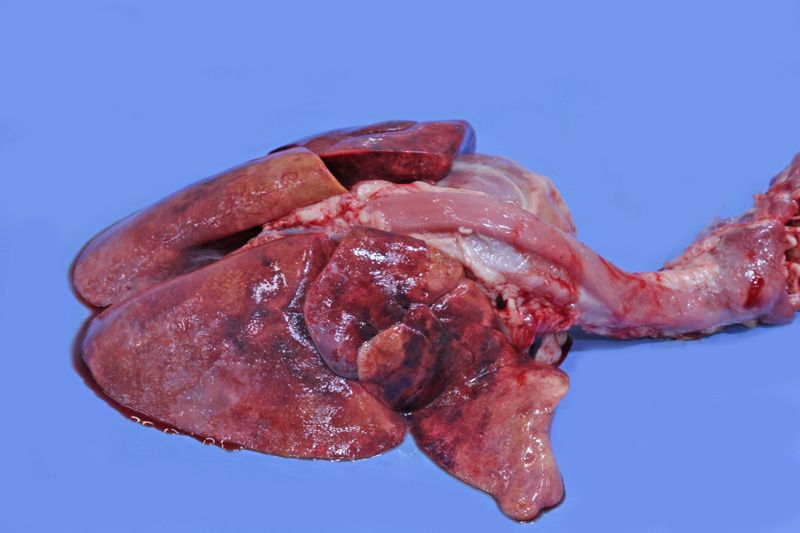
Gallagher et al. Irish Veterinary Journal 2012, 65:5                                                          Page 2 of 10
http://www.irishvetjournal.org/content/65/1/5

  A. vasorum was first reported in south west France,          buccal mucosal bleeding time (BMBT). Diagnostic
and has since been recognised in many other countries          images were retrospectively reviewed by a resident in
worldwide including south east England and Wales, Ire-         diagnostic imaging under the supervision of a European
land, Germany, Denmark, Canada, and south America              Diplomate in Diagnostic Imaging using a grade system
[1,3-11]. Recently there has been an increase in the           previously described [17].
number of reports of sporadic cases from countries and           Where faecal analysis was carried out, a modified
areas previously considered devoid of infection, particu-      Baermann technique was used by suspending a mini-
larly in the northern half of the United Kingdom               mum of 5 g of faeces wrapped in gauze in a funnel of
[12,13]. In data collated from referral centers, likely pre-   warm water. The solution was decanted 12 hours later,
sented with more complex and severe cases, there is an         and the sample was centrifuged and then the superna-
approximate mortality of 24% [7,8,10].                         tant was discarded and the sediment examined. Zinc
  Although A. vasorum is considered endemic in Ire-            sulphate floatation was performed by adding 330 g of
land, and anecdotally is confined to the East Coast,           zinc sulphate to 1000 ml of water followed by the addi-
there is no large case series published to date. The aim       tion and mixing of faeces. The less dense material then
of this study was to provide data on the geographic            floated to the top and the solution was strained and
location, variety of clinical signs, clinicopathological and   centrifuged. A coverslip was applied to the top of the
radiographic features, and the response to treatment           centrifuge tube allowing microscopic examination of the
and outcome in infected dogs from Ireland.                     material in contact with the coverslip [18]. If required,
                                                               faecal PCR was performed at Bristol University as pre-
Materials and methods                                          viously described [19].
The medical records of all dogs presenting to the Uni-           Bronchoalveolar lavage was carried out under general
versity College Dublin (UCD) Veterinary Hospital               anaesthesia by infusing a sterile 0.9% sodium chloride
between July 1999 and December 2010 were retrospec-            solution into the bronchi via a lavage catheter. The fluid
tively reviewed for a positive diagnosis of A. vasorum.        was immediately aspirated and samples submitted for
Reviewing the same time period, the computer records           cytological analysis and culture. Samples for cytology
of the parasitology and pathology departments were             were centrifuged and the sediment was examined micro-
interrogated using key words (Angiostrongylus vasorum,         scopically for the presence of L1 larvae with morpholo-
angiostrongylosis, A. vasorum and L1 larvae) for positive      gical characteristics indicative of A. vasorum [1].
faecal and post mortem diagnoses from external cases,            Follow-up telephone conversations were carried out
to provide additional information on geographic location       with all contactable clients that had a clinical case
and to limit the inherent geographic bias of the referred      referred to the UVH. Information was collected on the
clinical cases. The files from each clinical case were         type of parasite control administered prior to referral,
examined for data on geographical location, signalment,        possibility of ingestion of slugs/snails, known or pre-
presenting history and physical examination findings.          sumed contact with foxes, affected litter mates and any
Diagnostic tests, including clinicopathological analyses,      recurrence of clinical signs.
radiographic features, coagulation abnormalities and
other specific tests if performed were collated. Five cases    Results
have been reported previously, although in a small preli-      Angiostrongylosis was identified in 24 clinical cases and
minary case series or highlighting other specific aspects      25 external submissions (15 faecal samples and 10 post
not addressed in this paper [14-16].                           mortem examinations). This represented 0.17% of all
  Blood samples were obtained by jugular or cephalic           new case referrals (n = 13,998), 1.6% of faecal (n = 940)
venepucture and collected in ethylenediaminetetraacetic        and 2.6% of post mortem (n = 388) submissions over
acid (EDTA) for haematological analyses using either           the same time period.
the CELL-DYN 3500 (Abbott Laboratories) or ADVIA                 The majority of dogs (n = 44 (90%)) resided on the
2120 (Siemens Healthcare Diagnostics) systems. Samples         East Coast of Ireland. Of the remaining cases there were
were also placed in lithium heparin tubes for subsequent       two from county Kildare and a single case each from
biochemical analyses using the Randox Daytona (Ran-            counties Kilkenny, Galway and Roscommon (Figure 1).
dox) or Randox RX Imola (Randox) systems. If                     Of the 24 clinical cases there were nine (37.5%) males
requested sodium citrated samples collected for pro-           and 15 (62.5%) females. The median age was 20 (range
thrombin time (PT) and activated partial thromboplastin        4-144) months. The group comprised four cross breeds
time (APTT) were assessed using the KC4 Amelung                and 20 pedigree dogs including German Shepherd (n =
(Amelung). D-dimers were measured using the Minutex            3), Labrador Retriever (n = 2), Jack Russell Terrier (n =
D-dimer (Biopool), while a routine Simplate bleeding           2), Cocker Spaniel (n = 2), and one each of a variety of
time device (Organon Teknika) was used to quantify the         other breeds. From the telephone survey owners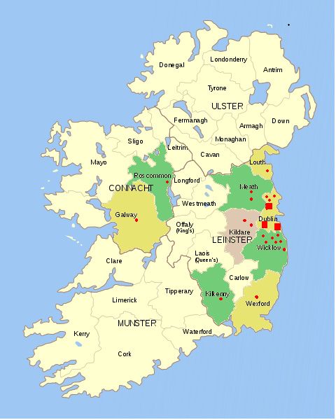
Gallagher et al. Irish Veterinary Journal 2012, 65:5                                                                             Page 3 of 10
http://www.irishvetjournal.org/content/65/1/5

                                                                             the animals with clinical evidence of a primary or sec-
                                                                             ondary coagulation defect, nine had either evidence of
                                                                             airway haemorrhage for which there was clinical evidence
                                                                             in five cases (haemoptysis) and diagnostic (radiographic
                                                                             (alveolar pattern) or cytological (ongoing or previous
                                                                             haemorrhage in BAL)) evidence in four. Neurological
                                                                             signs were noted in two animals and were considered to
                                                                             be secondary to intracranial haemorrhage which was
                                                                             confirmed in one case. The first animal presented with
                                                                             acute onset lethargy that progressed over 24 hours to stu-
                                                                             por, hyperexcitability, anisocoria, non-responsive mydria-
                                                                             tic pupils and eventually coma. The second animal
                                                                             presented with lethargy, acute onset collapse, unilateral
                                                                             hemiparesis and decreased menace bilaterally. Three ani-
                                                                             mals presenting with lameness had suspected haemar-
                                                                             throsis and one animal presenting primarily for
                                                                             abdominal pain had a retroperitoneal haematoma. Vague
                                                                             clinical signs including exercise intolerance, lethargy,
                                                                             weakness and failure to thrive were also identified and
                                                                             were the only clinical signs in two animals.
                                                                               Neutrophilia, anaemia and thrombocytopenia were the
                                                                             most common haematological abnormalities identified
                                                                             as presented in Table 2. Two cases were severely anae-
                                                                             mic (haematocrit < 0.20 L/L). Eosinophilia was present
 Figure 1 Geographical location of all cases of                              in only three cases. The median eosinophil count was
 angiostrongyloisis in clinical, faecal and post-mortem
                                                                             within the reference interval (0.3 × 10⁹/L (0-1.47 × 10⁹/
 submissions to UCD Veterinary Hospital between 1999 and
 2010. “red circle"- 1 case “red square"- 10 cases.                          L)) but individual values varied considerably (0-8.28 ×
                                                                             109/L) (Table 2). Biochemical abnormalities tended to
                                                                             be mild and included increases in creatine kinase (n =
reported that foxes were known to frequent the garden                        18 (75%)) concentration and amylase (n = 17 (71%)),
to which the animal had access in 16 of 21 (76%) cases.                      alkaline phosphatase (n = 16 (67%)) and alanine amino-
Eight of 23 (35%) dogs were previously observed eating                       transferase (n = 12 (50%)) activities. Hyperglobulinaemia
intermediate hosts. In four cases, another dog in the                        (n = 10 (42%)), hypoalbuminaemia (n = 10 (42%)) and
same household was affected. Two of these dogs died                          hyperphosphataemia (n = 8 (33%)) were also reported.
whilst undergoing investigation, as did their only respec-                   Mild hypercalcaemia (3.14 mmol/L and 3.16 mmol/L
tive housemate prior to referral.                                            (2.3-3.0 mmol/L)) was noted in two (8%) cases. Corre-
   Clinical signs included a variety of cardiorespiratory (n                 sponding phosphate values (0.8 mmol/L and 2.2 mmol/
= 15 (63%)), coagulopathic (n = 17 (71%)) and other (n =                     L) were within and above the reference interval (0.8-1.8
15 (63%)) less specific signs as presented in Table 1. Of                    mmol/L), respectively.

Table 1 Clinical signs reported in 24 animals diagnosed with angiostrongylosis
  Cardiorespiratory          n (%)                           Coagulopathic                      n (%)       Miscellaneous            n (%)
        Cough               10(42)              Clinical evidence of pulmonary haemorrhage      5 (21)    Exercise intolerance       6 (25)
      Dyspnoea               5 (21)       Diagnostic tests supporting pulmonary haemorrhage     4 (17)         Lethargy              6 (25)
     Tachypnoea              3 (12)                     Haemorrhage from wound                  4 (17)         Weakness               2 (8)
   Nasal discharge           1 (4)                             Haematoma                        4 (17)      Abdominal pain            1 (4)
    Heart murmur             1 (4)                               Epistaxis                      3 (13)      Failure to thrive         1 (4)
       Syncope               1 (4)                       Suspected haemarthrosis                3 (13)         Lameness              3 (13)
       Sneezing              1 (4)          CNS signs suspected secondary to haemorrhage         2 (8)
   Pneumothorax*             1 (4)                         Bruising/ecchymoses                   2 (8)
                                                               Haematuria                        1 (4)
*Developed five days post treatment, n number
Gallagher et al. Irish Veterinary Journal 2012, 65:5 Page 4 of 10
http://www.irishvetjournal.org/content/65/1/5
Table 2 Haematological abnormalities from 24 cases of angiostrongyslosis
Parameter Median Minimum Maximum % above reference interval % below reference interval
(Reference interval)
Haematocrit 0.38 0.13 0.63 4 42
(0.37 - 0.55 L/L)
Thrombocyte count 164.5 2 619 8 42
(150 - 500 × 109/L)
Neutrophil count 12.25 4.76 27.24 54 0
3 - 11.5 × 10⁹/L
Eosinophil count 0.3 0 8.28 13 0
(0 - 1.47 × 10⁹/L)
The PT and APTT were assessed in 18 animals, which days after a bulla was observed in one case, which was
included 15 of the 17 cases with evidence of coagulopa- being treated with fenbendazole, prednisolone and doxy-
thy (Table 3). Of the four (27%) cases with prolonged cycline. Repeat radiographs were available in six cases
values, two had reference interval BMBT and thrombo- that completed a full course of anthelminthic treatment.
cyte count, while the remaining animals each had either All continued to have radiographic abnormalities despite
severe thrombocytopenia or a prolonged BMBT. A a full clinical recovery and five of six that were tested
BMBT was performed in 8 of the coagulopathic animals demonstrating negative faecal Baermann flotations for a
and was prolonged in 5 (63%) cases. Three of these had median of 49 (range 1-203) days post treatment.
reference interval PT/APTT and thrombocyte count and Of the clinical cases, a diagnosis of angiostrongylosis
one each had either markedly prolonged PT and APTT was achieved by modified Baermann in 16 (72%) of 22
or moderate thrombocytopenia (57 × 109/L). Thrombo- cases. In one of these animals a final diagnosis was
cytopenia was severe enough to induce coagulopathy (≤ made by modified Baermann 10 months after numerous
50 × 10 9 /L) in four of ten animals and only one had negative faecals and two negative BALs. Negative results
additionally a mild prolongation in PT and APTT. In were obtained in six (28%) cases and a subsequent diag-
total, a reasonable explanation for the coagulopathic nosis was returned by post mortem (n = 4), BAL (n = 1)
signs was evident in 11 animals. Two of the remaining and faecal PCR (n = 1). Five (83%) of six cases in which
six, despite presenting with significant haemorrhage had a BAL was performed had negative results, in four of
reference interval PT, APTT and BMBT, with only one these modified Baermann was also negative. Whilst BAL
having a thrombocyte count below the reference interval was only positive in one case, three others had cytologi-
(98 × 10 9 /L) but above the value typically associated cal evidence supporting previous haemorrhage. Faecal
with overt haemorrhage. The remaining four had ade- PCR was performed in one case, confirming angiostron-
quate thrombocyte counts but a BMBT and/or PT and gylosis, after the modified Baermann suggested a diag-
APTT were not performed. In the seven cases without a nosis of Filaroides. Of the animals with one or more
documented clinical coagulopathy the thrombocyte false negative test results, 50% (n = 3) had recently
count was within reference interval in all but one that received an anthelminthic which had the potential to
was mildly decreased (148 × 10 9 /L). In four animals decrease their parasitic burden, making a subsequent
BMBT, PT and APTT were not performed. In the diagnosis challenging. Modified Baermann was not per-
remaining animals, BMBT (n = 2) PT and APTT (n = formed in two animals including one that died before
3) were within reference interval. the test was performed and one other which was posi-
Thoracic radiography was performed in 22 cases, nine- tive on zinc sulphate flotation. Details of those animals
teen of which were available for retrospective review. with one or more negative results and the anthelmintic
The results are presented in Table 4. Twelve animals they had received are presented in Table 5.
had mixed patterns, including bronchoalveolar and Four (16.7%) animals died before or within 24 hours
interstitial (n = 5) (Figure 2), bronchoalveolar alone (n = of instituting treatment. The median age of these ani-
6) and alveolar-interstitial (n = 1) were also reported. mals was six months. All presented with or developed
While most patterns were described as diffuse, a bron- severe dyspnoea with generalised alveolar or alveolar-
chial pattern was localised to the left hemi-thorax in interstitial patterns on thoracic radiography. Three cases
one case and alveolar patterns were localised in one had clinical and clinicopathological evidence of a bleed-
case each to the cranial thorax, the left hemi-thorax and ing diathesis.
the periphery. Five (of 8) cases with pleural fissures had A course of fenbendazole (50 mg/kg PO q 24 hours
a concurrent moderate to severe diffuse reticular or for 5-35 days, median (SIR) 10 (7.25-10) days) was
hazy interstitial pattern. A pneumothorax developed five administered in 19 cases. There was no significantGallagher et al. Irish Veterinary Journal 2012, 65:5 Page 5 of 10
http://www.irishvetjournal.org/content/65/1/5
Table 3 Coagulopathic test results in dogs with haemorrhagic diathesis associated with angiostrongylosis. Values in
red are abnormal
Case BMBT Thrombocyte PT APTT Clinical signs
(< 5 m) (150 - 500 × 109/L) (7 - 14 s) (15 - 25 s)
1 ND 116 9.8 14.2 Haematuria, Retroperitoneal haematoma
2 ND 7 18 26 Haemoptysis
3 >5 57 8 14 Haemorrhage post surgery & from a broken nail
4 >5 205 9.7 14.8 Suspected haemarthrosis (severe anaemia and lameness)
5 >8 175 22.3 250 Haemorrhage from tongue
6Gallagher et al. Irish Veterinary Journal 2012, 65:5 Page 6 of 10
http://www.irishvetjournal.org/content/65/1/5
Table 5 Diagnostic parasitological test results from cases
negative by one or more methodologies
Case MB BAL PCR PM Recent Anthelmintics
9467 - ve* - - +ve none
33248 - ve* - - +ve none
veΩ
34974 + ve -veΩ - - -
39262 - ve -ve - +ve none
* Ω
45801 - ve + ve - - Ivermectin 5 days prior to referral
52562 - ve - - +ve Selamectin 2 and 6 weeks prior to
referral
36236 - ve* -ve * - - -
¥
61218 - ve# -ve +ve - Fenbendazole immediately prior to
referral
*: Serial negative on MB or BAL, #: Filaroides suspected on MB, Ω: Cytological
evidence of intrapulmonary haemmorhage following BAL, ¥: Tested positive
by MB after 10 months
-ve: Negative
+ve: Positive
MB: Modified Baermann
PCR: Polymerase chain reaction
PM: Post Mortem
BAL: Bronchoalveolar lavage
adults were identified in the right heart or lungs of all
six cases (Figure 4). One animal (in which an antemor-
tem diagnosis was not made) had large numbers of
nematodes only within pneumonic areas of pulmonary
tissue.
Discussion
The present study illustrates that infection with A.
vasorum remains endemic in Ireland, is not confined to
Figure 2 Mixed pulmonary patterns on a dorsoventral and the East Coast, can occur in any age of dog and has a
lateral view.
widely varying clinical picture.
In the present study, approximately 90% of affected
animals either directly referred or from external post
There was initially a limited response to a full course of mortem or faecal submissions were from the East Coast
fenbendazole (50 mg/kg PO q 24 h for 10 days), but 10
months later following a positive diagnosis by modified
Baermann a full response to a prolonged (35 day) course
was recorded. Two recovered animals had recurrence of
similar clinical signs at a later date, neither had a parasi-
tological diagnosis but there was a full response to
appropriate treatment.
Post mortem examination was performed in all cases
that died (n = 4) or were euthanased (n = 2). A bleeding
diathesis was identified in five animals including pul-
monary congestion (n = 4) (Figure 3), injection site sub-
cutaneous haemorrhage (n = 2) and haemorrhagic
pleural effusion (n = 1). Cardiopulmonary pathology was
frequently noted including gross dilation or hypertrophy
of the right ventricle (n = 3), granulomatous pneumonia
Figure 3 Diffuse haemorrhagic consolidation of infected lungs.
(n = 2) and pulmonary fibrosis (n = 1). Larvae and/orGallagher et al. Irish Veterinary Journal 2012, 65:5 Page 7 of 10
http://www.irishvetjournal.org/content/65/1/5
clinical signs, regardless of age. However, as expected
based on a pre-patent period of 49-60 days, it is an unli-
kely differential in dogs two to three months of age and
younger. The youngest referred clinical case in this case
series was four months old.
Foxes are a known reservoir for infection. In the UK,
the overall prevalence in the fox population is 7.3%,
with absence of infection in foxes up to 2006 in north-
ern England and Scotland [23], areas where canine
angiostrongylosis was previously absent. However, five
cases have by now been reported from these latter areas
and may suggest that foxes are currently infected there
[12,13]. The fact that unrelated housemates were known
Figure 4 Adult female (Barber’s pole) within the heart. to be infected in this series exemplifies shared environ-
mental risk factors. Many owners did report exposure to
foxes and ingestion of snails but some did not. Further
of Ireland. These results may imply an endemic focus in study of infection in Irish foxes and other potential
this area. However, the two major counties represented hosts is therefore warranted.
are within the referral catchment area for the UCD Of the reported clinical features seen in naturally
Veterinary Hospital, likely creating a degree of bias. occurring angiostrongylosis, respiratory compromise is
Only five animals were from beyond this area with two common. Cough and dyspnoea were reported in 65%
from the West of Ireland. The only other published and 43% of dogs, respectively, in one recent report of 23
report of dogs affected in the West of Ireland was docu- cases [7], and in over 50% of dogs in two other series of
mented decades ago [6]. This may truly reflect a low eight and seven cases [8,10]. Cardiorespiratory compro-
prevalence beyond the East Coast. However, it could mise was a common complaint in the current study,
also reflect limited referral of cases from this geographic with cough, dyspnoea and tachypnoea frequently
location or a failure to consider the differential because reported. Despite demonstrating marked pulmonary
of a perception that angiostrongylosis does not exist pathology in experimentally infected animals, clinical
there. Nevertheless, worldwide there is evidence of area evidence of respiratory compromise tended to be mild
expansion, as demonstrated by a recent surge in spora- [24]. Similarly in over one third of animals in the cur-
dic cases in non-endemic areas and countries previously rent series radiographic evidence of pulmonary pathol-
considered disease free [13,20,21]. The fact that a diag- ogy ranged from mild to moderate, despite presenting
nosis was made from various different areas of Ireland with no overt respiratory signs. The absence of respira-
may support such expansion and suggests that a diagno- tory signs therefore should not preclude consideration
sis cannot be excluded based on geographic location of angiostrongylosis as a differential diagnosis. Equally
alone. the value of thoracic radiography in investigating angios-
Angiostrongylosis is considered to be a disease of pri- trongylosis in the absence of cardiorespiratory signs
marily young animals. In three separate reports of eight, should not be underestimated.
23 and 160 dogs, reported ages were less than 24 Although poorly understood, an association between a
months (100%), a median of ten months and one year of bleeding diatheses and angiostrongylosis has been firmly
age or younger (50%)[7,10,22]. The reason why older established and was the second most common clinical
dogs are less frequently affected are unclear and the sign observed in 35% of cases [7]. Isolated case reports
possible role of age resistance and immunity requires have described a varied spectrum of haemostatic defects
further study. The median age of 20 months in the pre- including haemoabdomen [25,26], multifocal brain and
sent study largely concurs with published data, with 75% spinal cord haemorrhage [27,28], gastrointestinal hae-
of animals diagnosed at less than 2.5 years old. How- morrhage [7], pleural effusion [17,29] and ecchymotic
ever, while young age appears to be a predisposing fac- and ocular haemorrhage [7,13]. Coagulopathy was the
tor, angiostrongylosis was diagnosed in this study in most common clinical feature of the current series
dogs of all ages, with a quarter of animals aged between occurring in approximately twice as many cases as
5 and 12 years, in keeping with other studies [7]. Infec- reported previously [7]. This higher proportion may
tion in older dogs could reflect a degree of immuno- reflect a greater likelihood of practitioners referring coa-
compromise predisposing to infection. Whatever the gulopathic dogs, rather than those with less severely cri-
reason, this emphasises that a diagnosis of angiostrongy- tical signs. Clinical signs of the coagulopathy tended to
losis should be considered in all dogs with appropriate vary widely in both location and severity. As aGallagher et al. Irish Veterinary Journal 2012, 65:5 Page 8 of 10 http://www.irishvetjournal.org/content/65/1/5 consequence, specific investigation for angiostrongylosis unlikely to be involved in one case with a concurrent is prudent in all animals presenting with unexplained hypophosphatemia. In conclusion there were no patho- haemorrhage. gonomic haematological or biochemical changes which Neurological signs secondary to A. vasorum are fre- were suggestive of angiostrongylosis in either this case quently reported but until recently infrequently investi- series or in other reports of naturally infected animals gated. The aetiology was presumed secondary to [7]. Certainly the absence of eosinophilia should not ischaemic (thrombosis or arterial emboli) or haemorrha- preclude consideration of angiostrongylosis. This finding gic insult to the central nervous system, with the latter is supported by experimental studies which despite ful- reported more commonly [27,28,30]. One of two neuro- minant disease demonstrate only minor alterations logically affected animals presenting in the current case [32,33]. series had multifocal brain haemorrhage demonstrated The stimulus for coagulopathic abnormalities and sub- both on computerisedtomography and post mortem sequent bleeding diathesis is unclear. Transient altera- examination [16]. The other recovered with sympto- tions in thrombocyte counts, PT, APTT, and Factor V matic therapy (fenbendazole) leaving the mechanism of and Factor VII concentrations are correlated in experi- neurological signs undetermined. As investigation of mental studies to stages of parasite development or host neurological disease can be both invasive and expensive inflammatory response. Consumptive coagulopathy or and considering the breath of neurological derange- immune-mediated platelet destruction have been pro- ments described secondary to angiostrongylosis, investi- posed in both experimental and naturally occurring gation of and treatment for A. vasorum should be infections [14,32,34-36] Severe thrombocytopenia was encouraged as it involves minimal expense and has the present in four cases in the current case series, of which potential to achieve a favourable outcome. three had no alterations in PT or APTT, supporting the A serendipitous diagnosis is possible in dogs with theory of immune-mediated thrombocyte destruction. A patent infection but in apparent good health [8,10]. In prolonged BMBT was observed in five animals with the present series angiostrongylosis was considered an clinical evidence of coagulopathy with reference interval incidental finding in two cases. One was being investi- thrombocyte counts in all cases, and normal PT and gated for exercise induced collapse already described in APTT in four, implying thrombocytopathy as a likely its familial breeding line, while another case presented mechanism. Clinical evidence of haemorrhage (haemop- for cough and exercise intolerance. In the latter case tysis) was seen in three cases despite having no PT, clinical, clinicopathological and post mortem findings APTT, thrombocyte count and BMBT abnormalities. identified a variety of other pathologies (hypothyroidism, Unfortunately, d-dimers were rarely assessed and fibri- arthritis, laryngeal paralysis, pulmonary fibrosis and nogen degradation product, fibrinogen concentrations angiostrongylosis) likely also contributing to the present- and thromboelastography were not performed, which ing history. Another animal displayed clinical and radio- limits the conclusions that can be drawn from these graphic evidence of severe pulmonary pathology, but data. Future studies are required to ascertain the exact never progressed to haemorrhagic diathesis, severe neu- cause of bleeding in dogs with angiostrongylosis. How- rological signs or death despite the diagnosis remaining ever, the current study suggests thrombocytopathy or undetermined and largely untreated for almost one year. thrombocytopenia may have a significant role in many These cases may imply a degree of immunotolerance in of the animals presenting with haemorrhage and that some animals or in the former cases represent recent with the tests routinely available for many veterinary infection not yet fully established. practitioners (PT, APTT, thrombocyte count and A perception exists that parasitism commonly induces BMBT), reference interval values cannot exclude the eosinophilia and this was the most common (39%) hae- risk of significant haemorrhage in infected dogs. matological alteration reported elsewhere [7]. By con- Interstitial, bronchial and alveolar radiographic pat- trast it was less frequently reported in the current cases terns are all commonly described while vascular patterns series, although when observed tended to be marked. are not in both naturally occurring and experimental There were no notably significant serum biochemical infections [17,37]. In the current study over 50% of changes. Dysregulated macrophage production of 1,25 cases were described as having an alveolar infiltrate. dihydroxycholecalciferol is the proposed mechanism for This is lower than previously described [17], differs in hypercalcaemia and hyperphosphataemia in humans being diffuse rather than multifocal or peripheral and with granulomatous disease and has been suspected in may be related to the duration of infection. Experimen- three confirmed cases of angiostrongylosis [31]. Only tal studies have demonstrated an association between two animals were hypercalcaemic in the current study the duration of infection and the pulmonary pattern but both were considered mild. Measurement of vitamin observed with alveolar patterns most common at D or its metabolites were not carried out but were patency (7-9 weeks post infection) which are
Gallagher et al. Irish Veterinary Journal 2012, 65:5                                                                         Page 9 of 10
http://www.irishvetjournal.org/content/65/1/5

histologically attributed to haemorrhage and granuloma-         A mortality rate of 25% was reported in this study,
tous inflammation [37]. In the current study, interstitial    this is similar to the combined mortality of three sepa-
infiltration was the most commonly observed radio-            rate UK case series [7,8,10], but does appear to be unu-
graphic feature. Pleural fissures were evident in approxi-    sually high when compared with two large case series in
mately half of the cases with many having a concurrent        isolation of 2 and 13% [7,44]. Four animals which died
interstitial pattern, possibly reflecting chronicity. The     within 24 hours had severe dyspnoea similar to another
progression of a bulla to pneumothorax seen in one            report [7]. Two animals were euthanized because of
case has been described previously [10]. The reasons for      either failure to confirm a diagnosis or to adequately
the differences in pattern and distribution between this      treat. The difference in mortality reported in various
and other studies are unclear. It is potentially related to   studies is likely to be associated with numerous factors
differences in radiological interpretation but may be         including, the duration of clinical signs prior to referral,
suggestive of more prolonged infection in many of the         the severity of the population affected, the presence or
cases seen. Despite radiographic abnormalities being          absence of typical clinical signs and the ability to make
detected, approximately one third had no evidence of          a prompt diagnosis or provide adequate treatment. It is
cough or dyspnoea, exemplifying that significant lung         unlikely to be associated with the treatment course
pathology can be present in the absence of obvious clin-      selected as it was similar in all three reports [7,44]. If A.
ical signs.                                                   vasorum is highly suspected an appropriate therapeutic
  Modified Baermann is considered the most sensitive,         trial is advised, as negative diagnostic tests do not pre-
currently available method for diagnosis of L1 larvae in      clude infection.
dogs infected with angiostrongylosis [5]. Accuracy for          In conclusion, angiostrongylosis is a recognised parasi-
detection of angiostrongylosis by modified Baermann           tic condition in Irish dogs. A diagnosis should be con-
was 100% in one review [7] although negative results          sidered in dogs from all areas and of all ages. The
occur prior to patency or in animals which intermit-          clinical presentation can be variable but should be con-
tently shed. Repeat faecal testing is therefore recom-        sidered particularly in dogs with clinical or radiographic
mended [8]. BAL also demonstrated good sensitivity in         respiratory and/or coagulopathic signs. A bleeding dia-
naturally occurring and experimental infections with A.       thesis was the most common feature and may be pre-
vasorum larvae identifying 70% and 100% of cases              sent despite normal coagulation. The routinely used
respectively [7,38]. By contrast in the current series, L1    parasitological tests can be repeatedly negative. While
larvae were only detected by modified Baermann and            the outcome is favourable in the majority of animals, a
BAL in 73% and 17% of cases respectively, despite             more guarded prognosis is advised for those animals
repeat sampling in some cases. Recently both serological      which present with severe dyspnoea, or in which a diag-
and PCR tests have been developed but are either not          nosis is challenging or the response to therapy is incom-
commercially available or not yet fully validated [39-43].    plete. Further research on diagnostic tests is required.
Thus while the modified Baermann remains the quickest
and most sensitive non-invasive diagnostic test available     Endnotes
ante-mortem, a negative result does not preclude dis-         Photographs courtesy of Brian Cloak in the Veterinary
ease, nor should it discourage a treatment trial.             Pathobiology Section in the UCD Veterinary Hospital
  Fenbendazole was the only anthelmintic used in this
retrospective study, with success reported in 95% of
                                                              Acknowledgements
cases as reported previously. The dose and duration is        The authors would specifically like to acknowledge Stephen Cahalan and
largely empirical ranging from 20 mg/kg to 50 mg/kg           Sean Hogan in the Veterinary Pathobiology Section for their help in
PO q 24 h for 5-21 days [7,10,44]. Authorised therapies       interrogating the pathology and parasitology databases respectively. In
                                                              addition, the staff of the UCD Veterinary Hospital and the Diagnostic
include imidacloprid/moxidectin (Advocate®, Bayer) for        Laboratory who were involved in the investigation and care of the animals.
treatment and prevention and milbemycin oxime (Mil-
bemax ® , Novartis Animal Healthy) for reducing the           Authors’ contributions
                                                              BG: Interrogated the case records, completed follow-ups and drafted the
level of infection [10,45,46]. Milbemycin oxime (Milbe-       manuscript. SB: Interrogated the case records. MZ: Individually assessed the
max®, Novartis Animal Healthy) requires increased fre-        radiographic features of each case. CTM: Supervised the study and drafted
quency (q 7 d for four weeks) of the standard dose (0.5       the manuscript. All authors read and approved the final manuscript.
mg/kg) but has a comparable efficacy to fenbendazole          Competing interests
[45]. Fenbendazole and imidacloprid/moxidectin have           The authors declare that they have no competing interests.
no significant difference in efficacy, radiographic fea-
                                                              Received: 28 September 2011 Accepted: 20 March 2012
tures or side effects when administered to mild to mod-       Published: 20 March 2012
erately naturally infected animals [44].
Gallagher et al. Irish Veterinary Journal 2012, 65:5 Page 10 of 10
http://www.irishvetjournal.org/content/65/1/5
References 31. Boag A, Murphy K, Connolly D: Hypercalcaemia associated with
1. Rosen L, Ash L, Wallace G: Life history of the canine lungworm Angiostrongylus vasorum in three dogs. J Small Anim Pract 2005,
Angiostrongylus vasorum (Baillet). Am J Vet Res 1970, 31(1):131. 46(2):79-84.
2. Bolt G, et al: The common frog (Rana temporaria) as a potential 32. Cury M, et al: Hematological and coagulation profiles in dogs
paratenic and intermediate host forAngiostrongylus vasorum. Parasitol experimentally infected with Angiostrongylus vasorum (Baillet, 1866). Vet
Res 1993, 79(5):428-430. Parasitol 2002, 104(2):139-149.
3. Taubert A, et al: Lungworm infections (Angiostrongylus vasorum, 33. Cury M, et al: Biochemical serum profiles in dogs experimentally infected
Crenosoma vulpis, Aelurostrongylus abstrusus) in dogs and cats in with Angiostrongylus vasorum (Baillet, 1866). Vet Parasitol 2005, 128(1-
Germany and Denmark in 2003-2007. Vet Parasitol 2009, 159(2):175-180. 2):121-127.
4. Barutzki D, Schaper R: Natural infections of Angiostrongylus vasorum and 34. Schelling C, et al: Coagulation abnormalities associated with acute
Crenosoma vulpis in dogs in Germany (2007-2009). Parasitol Res 2009, Angiostrongylus vasorum infection in dogs. Am J Vet Res 1986,
105(Suppl 1):39-48. 47(12):2669.
5. Morgan ER, et al: Angiostrongylus vasorum infection in dogs: 35. Gould S, McInnes E: Immune-mediated thrombocytopenia associated
presentation and risk factors. Vet Parasitol 2010, 173(3-4):255-261. with Angiostrongylus vasorum infection in a dog. J Small Anim Pract
6. Dodd K: Angiostrongylus vasorum (Baillet, 1866) infestation in a 1999, 40(5):227-232.
greyhound kennels. Vet Rec 1973, 92(8):195. 36. Ramsey IK, et al: Role of chronic disseminated intravascular coagulation
7. Chapman P, et al: Angiostrongylus vasorum infection in 23 dogs (1999- in a case of canine angiostrongylosis. Vet Rec 1996, 138(15):360-363.
2002). J Small Anim Pract 2004, 45(9):435-440. 37. Mahaffey M, et al: Experimental canine angiostrongylosis. II. Radiographic
8. Patteson M, et al: Angiostrongylus vasorum infection in seven dogs. Vet manifestations. The J Am Anim Hospital Association 1981, 17:499-502.
Rec 1993, 133(23):565. 38. Barçante J, et al: Cytological and parasitological analysis of
9. Bourque A, et al: Angiostrongylus vasorum infection in 2 dogs from bronchoalveolar lavage fluid for the diagnosis of Angiostrongylus
Newfoundland. Can Vet J 2002, 43(11):876. vasorum infection in dogs. Vet Parasitol 2008, 158(1-2):93-102.
10. Martin M, et al: Angiostroneylosis in Cornwall: clinical presentations of 39. Schnyder M, et al: An ELISA for sensitive and specific detection of
eight cases. J Small Anim Pract 1993, 34(1):20-25. circulating antigen of Angiostrongylus vasorum in serum samples of
11. Jones G, Neal C, Turner G: Angiostrongylus vasorum infection in dogs in naturally and experimentally infected dogs. Veterinary Parasitology ,
Cornwall. Vet Rec 1980, 106(4):83. Corrected Proof.
12. Yamakawa Y, et al: Emerging canine angiostrongylosis in northern 40. Al-Sabi M, et al: PCR detection of Angiostrongylus vasorum in faecal
England: five fatal cases. Vet Rec 2009, 164(5):149. samples of dogs and foxes. Parasitol Res 2010, 107(1):135-140.
13. Helm J, et al: A case of canine Angiostrongylus vasorum in Scotland 41. Verzberger-Epshtein I, et al: Serologic detection of Angiostrongylus
confirmed by PCR and sequence analysis. J Small Anim Pract 2009, vasorum infection in dogs. Vet Parasitol 2008, 151(1):53-60.
50(5):255-259. 42. Jefferies R, Morgan E, Shaw S: A SYBR green real-time PCR assay for the
14. O’Neill E, et al: Immune-mediated thrombocytopenia associated with detection of the nematode Angiostrongylus vasorum in definitive and
angiostrongylus vasorum infection in a Jack Russell terrier. Ir Vet J 2010, intermediate hosts. Vet Parasitol 2009, 166(1-2):112-118.
63(6):434-440. 43. Jefferies R, et al: Improved detection of canine Angiostrongylus vasorum
15. Brennan S, et al: Clinical signs, diagnosis and treatment of three dogs infection using real-time PCR and indirect ELISA. Parasitology Research
with angiostrongylosis in Ireland. Ir Vet J 2004, 57(2):103-109. 2011, 109:1577-1583.
16. Zarelli M, et al: Computer Tomography (CT) findings in a dog with 44. Willesen JL, et al: Efficacy and safety of imidacloprid/moxidectin spot-on
intracranial haemorrhage secondary to angiostrongylosis. Veterinary solution and fenbendazole in the treatment of dogs naturally infected
Radiology and Ultrasound . with Angiostrongylus vasorum (Baillet, 1866). Vet Parasitol 2007, 147(3-
17. Boag AK, et al: Radiographic findings in 16 dogs infected with 4):258-264.
Angiostrongylus vasorum. Vet Rec 2004, 154(14):426-430. 45. Conboy G: Natural infections of Crenosoma vulpis and Angiostrongylus
18. Zajac A, Conboy GA: Veterinary clinical parasitology. Wiley-Blackwell, vasorum in dogs in Atlantic Canada and their treatment with
Oxford; 2006. milbemycin oxime. Vet Rec 2004, 155(1):16.
19. Jefferies R, et al: Angiostrongylus vasorum from South America and 46. Schnyder M, et al: Larvicidal effect of imidacloprid/moxidectin spot-on
Europe represent distinct lineages. Parasitology 2009, 136(01):107-115. solution in dogs experimentally inoculated with Angiostrongylus
20. Tieri E, et al: Angiostrongylus vasorum in 20 dogs in the province of vasorum. Vet Parasitol 2009, 166(3-4):326-332.
Chieti, Italy. Vet Ital 2011, 47(1):77-88.
21. van Doorn D, et al: Autochthonous Angiostrongylus vasorum infection in doi:10.1186/2046-0481-65-5
dogs in The Netherlands. Vet Parasitol 2009, 162(1-2):163-166. Cite this article as: Gallagher et al.: Geographical, clinical,
22. Koch J, Willesen JL: Canine pulmonary angiostrongylosis: a update. The clinicopathological and radiographic features of canine
Vet Jour 2009, 179(3):348-359. angiostrongylosis in Irish dogs: a retrospective study. Irish Veterinary
23. Morgan E, et al: Angiostrongylus vasorum and Eucoleus aerophilus in Journal 2012 65:5.
foxes (Vulpes vulpes) in Great Britain. Vet Parasitol 2008, 154(1-2):48-57.
24. Prestwood A, et al: Experimental canine angiostrongylosis. I. Pathologic
manifestations. The J Am Anim Hospital Association 1981, 17:491-497.
25. Willesen J, Bjornvad C, Koch J: Acute haemoabdomen associated with
Angiostrongylus vasorum infection in a dog: a case report. Ir Vet J 2008,
61:591-593.
26. Humm K, Boag A: Unusual presentation of Angiostrongylus vasorum in Submit your next manuscript to BioMed Central
dogs. Vet Rec 2008, 162:632. and take full advantage of:
27. Wessmann A, et al: Brain and spinal cord haemorrhages associated with
Angiostrongylus vasorum infection in four dogs. Vet Rec 2006,
• Convenient online submission
158(25):858.
28. Garosi L, et al: Intracranial haemorrhage associated with Angiostrongylus • Thorough peer review
vasorum infection in three dogs. J Small Anim Pract 2005, 46(2):93-99. • No space constraints or color figure charges
29. Tebb A, Johnson V, Irwin P: Angiostrongylus vasorum (French heartworm)
in a dog imported into Australia. Aust Vet J 2007, 85(1-2):23-28. • Immediate publication on acceptance
30. Gredal H, et al: Acute neurological signs as the predominant clinical • Inclusion in PubMed, CAS, Scopus and Google Scholar
manifestation in four dogs with Angiostrongylus vasorum infections in
• Research which is freely available for redistribution
Denmark. Acta Vet Scand 2011, 53(1):43.
Submit your manuscript at
www.biomedcentral.com/submitYou can also read