Granulomatosis and Polyangiitis Presenting as Superficial Granulomatous Pyoderma
←
→
Page content transcription
If your browser does not render page correctly, please read the page content below
Fortune J Health Sci 2019; 2 (1): 009-013 DOI: 10.26502/fjhs006
Case Report
Granulomatosis and Polyangiitis Presenting as Superficial
Granulomatous Pyoderma
Sophia Zhang1*, Ellen Roh2, Timothy Patton3
1
University of Pittsburgh School of Medicine, 3550 Terrace St, Pittsburgh, PA 15213, USA
2
Massachusetts General Hospital, Department of Dermatology, 55 Fruit Street, Boston, Massachusetts, USA
3
Department of Dermatology, University of Pittsburgh School of Medicine, 3550 Terrace St, Pittsburgh, PA 15213,
USA
*
Corresponding Author: Sophia Zhang, University of Pittsburgh School of Medicine, 3550 Terrace St,
Pittsburgh, PA 15213, USA, E-mail: shz49@pitt.edu
Received: 27 November 2018; Revised: 12 January 2019; Accepted: 16 January 2019; Published: 21 January
2019
Abstract
Superficial granulomatous pyoderma (SGP) is one of four subtypes of pyoderma gangrenosum (PG), an uncommon
neutrophilic dermatosis, that presents as superficial, indolent ulcerative nodules or plaques that arise independently
from an associated underlying disease [1]. Granulomatosis and polyangiitis (GPA) is a systemic, cytoplasmic
ANCA (cANCA) positive vasculitis of small and medium vessels classically presenting with a triad of vascular,
pulmonary, and renal complications [2]. We report a case of a limited variant of GPA presenting as SGP in a 57-
year-old male with multiple nonhealing, painful ulcers on the upper extremities, torso, and neck with histologic
features of superficial ulcers with predominantly plasma cell infiltrate consistent with atypical SGP. Over the
following year, violaceous ulcers appeared, and a larger wedge biopsy demonstrated sinus tract formation with
underlying plasmocytosis with histiocytes and giant cells, neutrophilic inflammation, and focal granuloma formation
without vasculitis. The patient developed pulmonary symptoms and fever in the setting of markedly elevated
cANCA. A chest CT revealed a lung nodule with fine needle aspiration (FNA) demonstrating acute and
granulomatous inflammation but no organisms. Based on compatible clinical and histological findings, the diagnosis
of GPA presenting as SGP was established. This case of a variant of GPA initially limited to the integument and
mistaken for SGP characterizes the clinical and histopathological presentations of two rare diseases and supports the
notion that GPA should be included in the differential diagnosis for SGP even before systemic symptoms and
elevated cANCA arise to ensure early diagnosis and treatment.
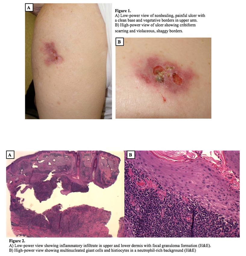
Fortune Journal of Health Sciences - Vol. 2 No. 1 - Mar 2019 9Fortune J Health Sci 2019; 2 (1): 009-013 DOI: 10.26502/fjhs006 Keywords: Dermatology; Granulomatosis and Polyangiitis; Superficial Granulomatous Pyoderma; Autoimmune; cANCA 1. Introduction Superficial granulomatous pyoderma (SGP), also referred to as vegatitive pyoderma gangrenosum in some sources, is a subtype of pyoderma gangrenosum (PG), a rare neutrophilic dermatosis that often occurs with autoinflammatory disorders such as inflammatory bowel disease, hematologic disorders, and arthritis [1]. Unlike classic PG, SGP usually manifests as a more superficial, indolent ulcerative nodule or plaque that lacks an association with an underlying systemic or autoimmune disease [1,3]. Granulomatosis and polyangiitis (GPA) is a vasculitis of small and medium vessels classically presenting with a triad of vascular, pulmonary, and renal complications [12]. Some cases of protracted superficial variants of GPA presenting as PG have been reported, but cases of GPA presenting as SGP are very rare [4-6]. We present a case of a limited variant of GPA presenting as SGP in a 57-year-old male. 2. Case Report A 57-year-old white male presented with multiple nonhealing, painful ulcers on the upper extremities, torso, and neck over a period of almost 1 year. Past medical history was unremarkable. The patient’s initial lesion occurred on the leg following a car accident. A punch biopsy performed at that time demonstrated a superficial ulcer with a predominately plasma cell infiltrate, and a presumptive diagnosis of atypical pyoderma gangrenosum (PG) was made. SPEP/UPEP were within normal limits. The lesion was treated with intralesional (IL) steroids and resolved. Over the following year, the patient developed subsequent 3-5cm violaceous ulcers with shaggy borders and cribiform scarring on the upper extremities, torso, and neck (Figure 1). No systemic symptoms were present. Work- up for the etiology of the ulcers included CBC, CMP, UA, and SPEP/UPEP, all of which were within normal limits. HIV, RPR, and FTA-ABS testing were negative, and tissue cultures for bacteria and fungi were negative. A larger wedge biopsy was taken that demonstrated sinus tract formation with underlying plasmocytosis with histiocytes and giant cells, neutrophilic inflammation, and focal granuloma formation without the presence of vasculitis (Figure 2). When treated with intralesional steroids, there was mild, transient improvement but new lesions would develop. Systemic steroids led to moderate improvement, but this was discontinued due to peptic ulcer disease. Cyclosporine (3 mg/kg) also led to moderate improvement. While on cyclosporine, the patient developed pulmonary symptoms and fever, and a chest CT was performed, revealing a lung nodule. Fine needle aspiration (FNA) of the lung nodule demonstrated acute and granulomatous inflammation but no organisms. Cytoplasmic ANCA (cANCA) was markedly elevated. Repeat BUN/creatinine and UA were within normal limits. The patient was diagnosed with GPA and treated with systemic corticosteroids Fortune Journal of Health Sciences - Vol. 2 No. 1 - Mar 2019 10
Fortune J Health Sci 2019; 2 (1): 009-013                                                  DOI: 10.26502/fjhs006
in combination with oral cyclophosphamide. Both the skin and lung lesions completely resolved over the next few
months following therapy.

3. Discussion
GPA and SGP are rare diseases of unknown etiologies that can be difficult to diagnose [3]. Skin lesions are present
in half of GPA cases and can have various clinical presentations, including palpable purpura, necrotic ulcerations
resembling PG, verrucous nodules (usually on elbows), subcutaneous nodules, and gingival hyperplasia [7]. Our
patient presented with a rare case of a limited, protracted variant of GPA manifesting as SGP.

The classic histopathologic description of SGP is that of a 3-layered granuloma with an innermost zone of
neutrophils, a surrounding layer of granulomatous inflammation with histiocytes and giant cells, and an outermost

Fortune Journal of Health Sciences - Vol. 2 No. 1 - Mar 2019                                                       11
Fortune J Health Sci 2019; 2 (1): 009-013                                                   DOI: 10.26502/fjhs006
layer of plasma cells and eosinophils [1,3,7]. SGP, like PG in general, is a diagnosis of exclusion, and other causes
of chronic ulceration such as infection, malignancy, or autoimmune disease need to be excluded [3,6].

In addition to compatible biopsy findings, definitive diagnosis of SGP relies on clinical presentation and course of
disease [1,8]. Clinical appearance of SGP is usually a sterile, well-defined superficial ulcer with a clean base and no
undermining borders most commonly occurring on the trunk and rarely on the face [1,3,6-8]. In contrast to classic
PG, SGP typically presents as slowly progressing, more superficial granulomatous lesions and as such has a more
favorable prognosis and is not associated with underlying systemic or autoimmune disease [1,3,6-11]. SGP usually
resolves with local, topical or intralesional anti-inflammatory or anti-microbial agents and does not require more
aggressive systemic immunosuppressive agents that may be required in the treatment PG lesions or GPA, although a
handful of cases of more aggressive SGP variants on the face, head, and neck have been reported [9-11].

A protracted superficial variant of GPA has been described that is characterized by ulcerating, necrotizing lesions of
mucosa and skin confined to cutaneous and upper respiratory tract [4]. Limited variants of GPA have also been
described in the absence of renal disease [4]. If untreated, progression to more fatal multi-organ involvement seen in
classic GPA may develop, justifying early treatment with aggressive systemic immunosuppressive agents.

Some cases of atypical GPA presenting as PG have placed GPA on the differential for chronic, painful ulcers, but
only a few cases of GPA presenting as SGP have been reported [4-6]. Recognizing rare variants of GPA limited to
the integument can ensure appropriate treatment to prevent further systemic involvement, permanent wound
scarring, progression to renal failure, or death.

We present this rare case of GPA presenting as SGP to help characterize the clinical and histopathological
presentations of 2 rare diseases and to support the notion that limited variants of GPA should be included in the
differential diagnosis for SGP for proper long-term monitoring and treatment of potential systemic complications
that may arise in the future.

References
    1.   Ruocco E, Sangiuliano S, Gravina AG, Miranda A, Nicoletti G. Pyoderma gangrenosum: an updated
         review. J Eur Acad Dermatol Venereol 23 (2009): 1008-1017.
    2.   Vanoli J, Riva M, Vergnano B, et al. Granulomatosis with polyangiitis presenting with diffuse alveolar
         hemorrhage requiring extracorporeal membrane oxygenation with rapid multiorgan relapse: A case report.
         Medicine (Baltimore) 96 (2007): e6024.
    3.   Tollefson MM, Cook-Norris RH, Theos A, Davis DM. Superficial granulomatous pyoderma: a case in an
         11-year-old girl and review of the literature. Pediatr Dermatol 27 (2007): 496-499.
    4.   Sinovich V, Snow J. Protracted superficial Wegener's granulomatosis. Australas J Dermatol 44 (2003):
         207-214.

Fortune Journal of Health Sciences - Vol. 2 No. 1 - Mar 2019                                                        12
Fortune J Health Sci 2019; 2 (1): 009-013 DOI: 10.26502/fjhs006
5. Kuchel J, Lee S. Cutaneous Wegener's granulomatosis: a variant or atypical localized form? Australas J
Dermatol 44 (2003): 129-135.
6. Rosina P, Papagrigoraki A, Colato C. A case of superficial granulomatous pyoderma mimicking a basal
cell carcinoma. Acta Dermatovenerol Croat 22 (2014): 48-51.
7. Persing SM, Laub D, Jr. Superficial granulomatous pyoderma of the face: a case report and review of the
literature. Eplasty 12 (2012): e56.
8. Panes-Rodriguez A, Arregui-Murua MA, Gutierrez-Tamara P, Borja-Consigliere HA, Rodriguez-Perez I,
Tuneu-Valls A. Vulvar superficial granulomatous pyoderma successfully treated with dapsone. Clin Exp
Dermatol 42 (2017): 230-232.
9. D'Epiro S, Salvi M, Mattozzi C, et al. Facial superficial granulomatous pyoderma. Int Wound J 12 (2015):
737-738.
10. Contreras-Verduzco FA, Espinosa-Padilla SE, Orozco-Covarrubias L, Alva-Chaire A, Rojas-Maruri CM,
Saez-de-Ocariz M. Pulmonary nodules and nodular scleritis in a teenager with superficial granulomatous
pyoderma gangrenosum. Pediatr Dermatol 35 (2018): e35-e38.
11. Ibrahim O, Bunick CG, Srivastava B, Lazova R, Ko CJ, Watsky KL. The role of infliximab in the treatment
of superficial granulomatous pyoderma of the head and neck. J Am Acad Dermatol 71 (2014): e222-e225.
Citation: Sophia Zhang, Ellen Roh, Timothy Patton. Granulomatosis and Polyangiitis Presenting as Superficial
Granulomatous Pyoderma. Fortune Journal of Health Sciences 2 (2019): 009-013.
This article is an open access article distributed under the terms and conditions of the
Creative Commons Attribution (CC-BY) license 4.0
Fortune Journal of Health Sciences - Vol. 2 No. 1 - Mar 2019 13You can also read