Unexplained ascites, a sign for neuroendocrine carcinoma
←
→
Page content transcription
If your browser does not render page correctly, please read the page content below
CASE REPORT

Unexplained ascites, a sign for neuroendocrine carcinoma

Mohd Khairul Mohd Kamil, MD1, Ngiu Chai Soon, FRCP1, Nurismah Md Isa, MBBS2, Yazmin Yaacob, MBBS3,
Deborah Chew Chia Hsin, MBBS1, Wong Zhiqin, FRCP1, Raja Affendi Raja Ali, FRCP1

1
 Gastroenterology Unit, Faculty of Medicine, UKM Medical Centre, Kuala Lumpur, Malaysia, 2 Department of Pathology, Faculty
of Medicine, UKM Medical Centre, Kuala Lumpur, Malaysia, 3Department of Radiology, Faculty of Medicine, UKM Medical
Centre, Kuala Lumpur, Malaysia

SUMMARY
Neuroendocrine neoplasm is an epithelial neoplasm with
                                                                C and HIV serology were negative. Peritoneal fluid analysis

predominant neuroendocrine differentiation that can arise
                                                                revealed serum ascites albumin gradient was 5g/L with no

from many organs in the body. We reported a rare case of
                                                                evidence of malignant cells and the cultures for bacteria and

gastric neuroendocrine carcinoma which accounts for less
                                                                mycobacterium were negative. Subsequently, abdominal

than 1% of all gastric tumours that is associated with poor
                                                                ultrasound demonstrated a ‘hidden’ epigastric mass.

prognosis. The recognition of this rare tumour in early stage
                                                                Computed tomography of the abdomen (figure 1A) revealed

is challenging and high suspicious into it might bring to
                                                                a ‘hidden’ mass in the posterior wall of the gastric antrum

early detection and so forth might improve the
                                                                measuring 2.1 x 4.5cm with numerous intra-abdominal

prognostication.
                                                                lymph nodes involving perigastric area, greater omentum,
                                                                peritoneum, mesenteric and rectovesical pouch. PET scan was
                                                                not performed in view of extensive disease. Endoscopically,

INTRODUCTION
                                                                gastric mucosa and subsmucosa were normal and endoscopic
                                                                ultrasound (figure 1B) was performed with fine needle
The first two cases of gastric neuroendocrine tumour (NET)      aspiration cytology and biopsy of gastric mass was taken.
were described by Askanazy in 1923 and in 1961.                 Histologically, the biopsy revealed a poorly differentiated
Subsequently, Christodoulopoulos and Klotz (1961) published     NEC with high index of proliferation of malignant cells of
79 cases in the literature, noting that their diagnosis was     grade 3 (figure 2A-D). Chromogranin A level was not raised,
usually delayed due to the late presentation and diagnosis is   at less than 36.7 ng/ml. Surgical treatment was not indicated
often made during autopsy. This late presentation is due to     for this patient as he had distant metastasis and poorest
slow growing tumour and asymptomatic except if it is            prognosis (poorly differentiated carcinoma). Chemotherapy
hormone secreting tumor or compressive effect of the tumor.     was initiated which comprised of cisplastin and etoposide as
NETs of the stomach comprise less than 1% of gastric            the chemotherapy is standard management for patients with
neoplasms. In the pre-endoscopy era, they comprised 1.9% of     advanced disease. However, the patient developed life-
all carcinoids, but in more recent studies, 10% to 30% of all   threatening post-chemotherapy neutropenic sepsis and with
carcinoids are reported in the stomach.1 According to the       the background of this aggressive disease, the oncology team
most recent World Health Organization (WHO) classification,     had decided for palliation.
NETs that involve the stomach are generally divided into

                                                                DISCUSSION
well-differentiated NETs, well differentiated neuroendocrine
carcinomas (NECs), and poorly differentiated NECs.2
                                                                Gastric neuroendocrine carcinoma (NEC) is an uncommon

CASE REPORT
                                                                tumor, usually associated with highly malignant biological
                                                                behavior with extremely poor prognosis. NEC is composed of
We report a case of a 47-year-old man, an active smoker         uniform small round cells with salt and pepper chromatin
presented with 3 months history of abdominal distension,        along with specific immunohistochemical staining that
loss of appetite and weight loss. He has no significant         required to confirm the diagnosis.
personal and family history of malignancy and chronic liver
disease. Physical examination revealed a gross ascites,         Gastric NET is classified into pathological grading and
bilateral pitting oedema and bilateral pleural effusion. He     clinical staging. In the 2010, based on the WHO pathological
has no stigmata of chronic liver disease and                    grading criteria, NETs of the stomach are defined as
lymphadenopathy.                                                neoplasms with neuroendocrine differentiation, including
                                                                neuroendocrine tumors (NETs) and NECs either well or poorly
Initial   laboratory    investigations    showed    normal      differentiated carcinoma arising from the stomach.2
haemoglobin level, total white blood cells count, platelet      American Joint Committee on Cancer (AJCC) endorsed
count, renal and liver profiles with serum albumin of 34g/L.    staging neuroendocrine neoplasm using specified TNM-based
Serum amylase, lactate dehydrogenase, immunoglobulins,          system. Recent study has demonstrated that malignant
tumour markers; alpha fetoprotein, carcinoembryonic             disease, defined by direct invasion of adjacent organs by
antigen and CA19.9 were within normal range. Hepatitis B,       tumor, lymph node metastases, or distant organ spread, may
This article was accepted: 15 September 2017
Corresponding Author: Chai Soon Ngiu
Email: csngiu@ppukm.ukm.edu.my

    60                                                                                    Med J Malaysia Vol 73 No 1 February 2018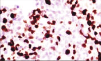
Unexplained ascites, a sign for neuroendocrine carcinoma
Fig. 1: A) Contrasted computed tomography of the abdomen showed a mass in the posterior wall of the stomach measuring 2.1 x 4.5
cm (Arrow). B) Endoscopic ultrasound showed a heterogenous mass measuring 69 mm x 71mm near antrum (Arrow).
a b
c d
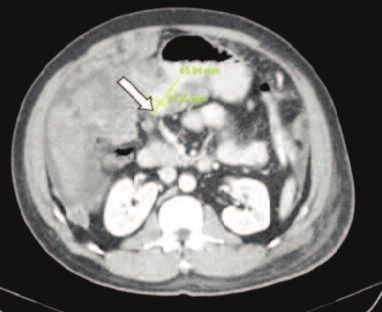
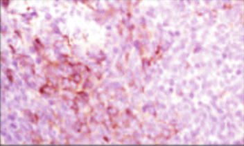
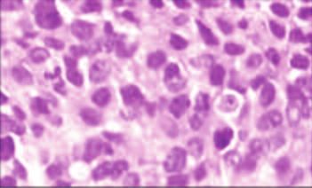
Fig. 2: A) Histopathological examination of the tissue from the stomach (HE stain with magnification 400x) showed high grade of
mitotic cells; B) Ki-67 preparation showed high index of proliferation of the malignant cells; C) CK AE1/AE3 preparations and
D) NSE preparation showed positive results.
have 5-year survival rates as high as 77% to 95% when In our case, histologically the tumour was infiltrated by
treated aggressively with resection of primary tumor and malignant cells and had high mitotic activity with Ki-67
adjunctive therapy.3 proliferation index of 80%. These malignant cells were
positive to NSE and CD56 that confirmed the diagnosis of
NETs are commonly divided according to the primary site; gastric NEC. Combination of debulking surgery and
foregut (lung, bronchus, stomach or duodenum), midgut chemotherapy is recommended. However, the role of surgery
(jejunum, ileum, appendix or proximal colon) and hindgut in not clear in patient with high-grade poorly differentiated
(distal colon or rectum). Recently, the WHO proposed a new NEC. In view of rapid progressive nature of disease, palliative
diagnostic criteria mainly depend on the rate of cellular chemotherapy is generally favoured as in our patient.
proliferation and the proliferative rate of the tumor is Cisplatin-based regimens are preferred chemotherapy in
assessed based on number of mitoses/10 high power filed or advanced NEC4 due to a superior response that can lead to a
the percentage of neoplastic cells immunolabelling for Ki-67, decrease in tumour size and our patient is currently receiving
a proliferative marker.2 a combination of cisplatinum and etoposide chemotherapy.
Med J Malaysia Vol 73 No 1 February 2018 61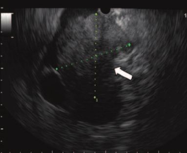
Case Report

Unlike our case, somatostatin analogues therapy is beneficial    REFERENCES
in patients with functioning NEC and G1/G2 small intestine.5     1.   Svorcan P, Maksimovic B, Djordjevic J. A Rare Gastric Carcinoma-
                                                                      Neuroendocrine Tumors. In: Ismaili N, editor, Management of Gastric
                                                                      Cancer. InTech 2011.

CONCLUSION
                                                                 2.   Bosman FT, Carneiro F, Hruban RH, Theise ND. WHO classification of
                                                                      tumours of the digestive system. World Health Organization; 2010.
This case illustrates that the diagnosis of NEC is challenging   3.   Norton JA, Kivlen M, Li M, Schneider D, Chuter T, Jensen RT. Morbidity
                                                                      and mortality of aggressive resection in patients with advanced
and can be missed in early stage of the disease due to a slow         neuroendocrine tumors. Arch Surg 2003; 138(8): 859-66.
growing tumour and not typical carcinoid syndrome                4.   Kang SH, Kim KH, Seo SH, An MS, Ha TK, Park HK, et al. Neuroendocrine
manifestation. We hope that highly suspicious of this rare            carcinoma of the stomach: A case report. World J Gastrointest Surg. 2014
                                                                      Apr 27; 6(4): 77.
tumour will lead to early recognition and might improve the
                                                                 5.   Rinke A, Müller HH, Schade-Brittinger C, Klose KJ, Barth P, Wied M, et al.
prognostication of the patient.                                       Placebo-controlled, double-blind, prospective, randomized study on the
                                                                      effect of octreotide LAR in the control of tumor growth in patients with
                                                                      metastatic neuroendocrine midgut tumors: a report from the PROMID
                                                                      Study Group. J Clin Oncol 2009; 27: 4656-63.

62                                                                                                  Med J Malaysia Vol 73 No 1 February 2018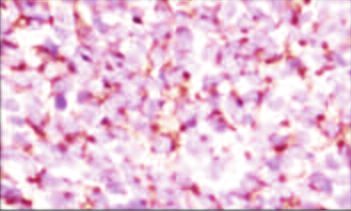
You can also read