Case Report Two Cases of Ectopic Pregnancy Mimicking Gestational Trophoblastic Disease
←
→
Page content transcription
If your browser does not render page correctly, please read the page content below
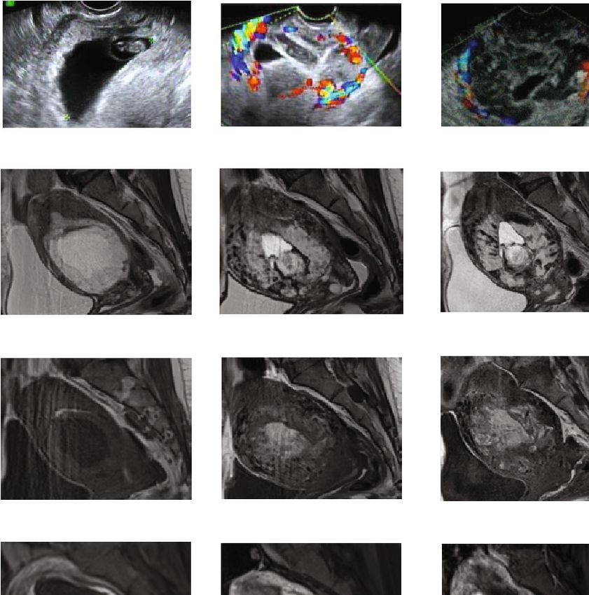
Hindawi
Case Reports in Obstetrics and Gynecology
Volume 2020, Article ID 2417428, 4 pages
https://doi.org/10.1155/2020/2417428

Case Report
Two Cases of Ectopic Pregnancy Mimicking Gestational
Trophoblastic Disease

 Haruka Suzuki, Yoshitsugu Chigusa, Junzo Hamanishi, Masaki Mandai, and Eiji Kondoh
 Department of Gynecology and Obstetrics, Kyoto University, 54 Shogoin Kawahara-cho, Sakyo, Kyoto 606-8507, Japan

 Correspondence should be addressed to Eiji Kondoh; kondo@kuhp.kyoto-u.ac.jp

 Received 4 March 2020; Accepted 8 June 2020; Published 16 June 2020

 Academic Editor: Kyousuke Takeuchi

 Copyright © 2020 Haruka Suzuki et al. This is an open access article distributed under the Creative Commons Attribution License,
 which permits unrestricted use, distribution, and reproduction in any medium, provided the original work is properly cited.

 A well-known typical feature of ectopic pregnancy is an evident gestational sac structure outside of the uterus. However, some cases
 show atypical appearance that is described as a heterogeneous hypervascular mass. We report two cases of ectopic pregnancy that
 presented heterogeneous findings mimicking gestational trophoblastic diseases but were correctly diagnosed as ectopic pregnancies
 on MRI. The first case was an interstitial pregnancy in which the patient underwent surgical treatment. The second case was a
 cesarean scar pregnancy that was treated conservatively but showed spurious enlargement of pregnancy-related lesions after the
 treatment. Both cases lacked myometrial invasion on MRI, and the patients were diagnosed with ectopic pregnancies. Invasive
 findings on MRI may discriminate ectopic pregnancy from trophoblastic tumors and avoid unnecessary hysterectomy.

1. Introduction hospital with a complaint of amenorrhea over the previous
 7 weeks and 3 days. She had mild lower abdominal pain,
Ectopic implantation occurs in approximately 1−2% of all and her vital signs were stable. The serum human chorionic
pregnancies. Fallopian tube pregnancy accounts for 90% gonadotropin (hCG) β level was 63,557 mIU/ml, which indi-
of ectopic pregnancies. Other unusual implantation sites cated pregnancy. Transvaginal ultrasonography (TV-USG)
include the uterine cervix, interstitium, ovary, abdominal did not show a GS, but an eccentric, 38-mm heterogeneous
cavity, and cesarean scar tissue [1]. Magnetic resonance mass was detected in the uterine interstitium. MRI was per-
imaging (MRI) is a useful diagnostic modality for detec- formed at 7 weeks and 5 days of pregnancy, and a heteroge-
tion of an ectopic pregnancy. Typically, an extrauterine neously enhanced hypervascular mass was identified on the
gestational sac (GS) is exhibited as a high-intensity cystic right side of the uterine fundus (Figures 1(a)–1(c)). The mass
structure surrounded by a thick wall on T2-weighted measured 52 mm, which was larger than a GS at the corre-
images and is enhanced in the early phase [2]. However, sponding estimated gestational age. An ectopic pregnancy
not all cases of ectopic pregnancy show these typical image was primarily suspected, but the possibility of GTD was also
findings. considered because of the atypical images and the size of the
 We report two cases of ectopic pregnancy that presented mass. She did not desire fertility preservation, and an abdom-
heterogeneous findings mimicking gestational trophoblastic inal hysterectomy was performed on the same day. The mac-
disease (GTD) on MRI. roscopic contents of the mass included a villous component,
 an embryo, and a blood clot (Figures 1(d) and 1(e)). The
2. Case 1 crown-rump length (CRL) of the embryo was 10 mm,
 which corresponded to 7 weeks of pregnancy. Histopatho-
A 40-year-old, gravida 5, para 2 patient with a history of two logic examination showed normal villi. The final diagnosis
spontaneous miscarriages and one right tubal pregnancy was interstitial pregnancy. The postoperative course was
treated with laparoscopic right salpingectomy consulted our uneventful, and she was discharged on postoperative day 7.
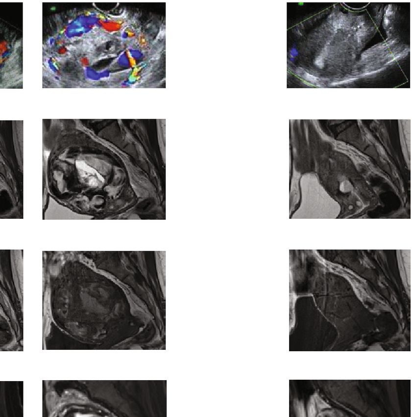
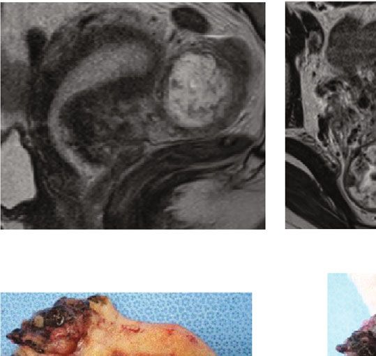
2 Case Reports in Obstetrics and Gynecology

 (a) (b) (c)

 (e)

 (d)

Figure 1: MR image performed at 7 weeks and 5 days in case 1 shows an interstitial pregnancy. A heterogeneously-enhanced hypervascular
mass is identified on the right side of the uterine fundus. The mass measures 52 mm, which is larger than a GS at the corresponding estimated
gestational age. Arrows indicate the pregnancy-associated lesion. (a) A sagittal T2-weighted MR image. (b) An axial T2-weighted MR image.
(c) An enhanced axial MR image. (d) Gross pathology of the uterus and the pregnancy-related tissue. (e) An enlarged view of the
implantation site.

3. Case 2 4. Discussion
A 38-year-old, gravida 3, para 2 patient with a history of 2 The differential diagnosis of ectopic pregnancy and GTD by
cesarean sections was referred to our department and hos- imaging features is sometimes difficult. GTD is a group of
pitalized at 8 weeks and 3 days of pregnancy due to a sus- rare tumors that can originate in any product of conception.
pected cesarean scar pregnancy. The hCG β level was Although GTD in ectopic pregnancy is quite rare, Tasha
228,454 mIU/ml. TV-USG showed the GS with a live fetus reported that GTD was found in approximately 18 of 100
on the cesarean scar of the uterus. The CRL was 19.3 mm. ectopic pregnancies [3]. GTD is visualized as a heteroge-
MRI findings also confirmed a cesarean scar pregnancy. neously hyperintense tumor on MRI [4]. In some cases of
The patient requested termination, but she wanted fertility ectopic pregnancy, particularly interstitial, cervical, or cesar-
preservation and decided to undergo medical treatment. ean scar pregnancy, the pregnancy-related tissue shows an
Methotrexate and potassium chloride were locally injected atypical appearance that is described as a heterogeneous
under ultrasonic guidance at 9 weeks and 2 days of hypervascular mass on USG and MRI [5, 6]. When a hema-
pregnancy. toma exists around the mass, the size of the mass appears
 One month after treatment, the serum hCG β level had to be larger than that of the GS at the same gestational age.
decreased to 1,506 mIU/ml. However, TV-USG revealed a The embryo is obscured by the hematoma, which makes
hypervascular lesion in the uterus. Enhanced MRI showed a diagnosis difficult. The mass even shows spurious enlarge-
strongly enhanced hypervascular mass that was larger than ment after medical treatment due to the formation of the
that before the treatment (Figure 2). Sequential GTD was hematoma [7]. This is probably because the muscle layer of
suspected because of the heterogeneous change and growth the uterus stretches in an interstitial or cesarean scar preg-
of the mass, but an invasive finding such as a fluffy appear- nancy, which allows the formation of growing hematoma,
ance of tumor burden was absent on MRI; thus, the patient whereas the fallopian tube tears easily in a tubal pregnancy.
underwent careful follow-up. The lesion continued to grow One of the differences between an ectopic pregnancy and
gradually as shown on serial MRI on days 67 and 95; how- GTD may be an invasive finding in the myometrium on
ever, the size of the enhanced lesion did not change, and MRI [4]. Actually, in our cases, the pregnancy-associated
the enlarged part was considered to be a hematoma. The mass was heterogeneous and deceptively looked similar to
serum hCG β level continued to decrease. Menstruation ectopic GTD; however, the invasive findings were absent,
restarted on day 120, necrotic tissue was extruded from the and the radiologist correctly diagnosed them as ectopic preg-
uterus on day 133, and the serum HCG β level became neg- nancies. In case 2, the ectopic lesion enlarged and showed
ative (

Case Reports in Obstetrics and Gynecology 3

 (a)

 (b)

 (c)

 (d)
 Before the treatment Day 32 Day 67 Day 95 day 165
 100000
 MTX+KCL

 1000
 1506
 300
 100
 10
4 Case Reports in Obstetrics and Gynecology

References
 [1] D. M. Panelli, C. H. Phillips, and P. C. Brady, “Incidence, diag-
 nosis and management of tubal and nontubal ectopic pregnan-
 cies: a review,” Fertility Research and Practice, vol. 1, no. 1,
 p. 15, 2015.
 [2] K. Tamai, T. Koyama, and K. Togashi, “MR features of ectopic
 pregnancy,” European Radiology, vol. 17, no. 12, pp. 3236–
 3246, 2007.
 [3] I. Tasha, E. Kroi, A. Karameta, R. Shahinaj, and N. Manoku,
 “Prevalence of gestational trophoblastic disease in ectopic
 pregnancy,” Journal of prenatal medicine, vol. 4, no. 2,
 pp. 26–29, 2010.
 [4] S. Dhanda, S. Ramani, and M. Thakur, “Gestational tropho-
 blastic disease: a multimodality imaging approach with impact
 on diagnosis and management,” Radiology Research and Prac-
 tice, vol. 2014, 842712 pages, 2014.
 [5] M. Rheinboldt and S. Ibrahim, “Atypical presentation of a
 large interstitial pregnancy,” Emergency Radiology, vol. 20,
 no. 3, pp. 251–254, 2013.
 [6] T. E. Ackerman, C. S. Levi, S. M. Dashefsky, S. C. Holt, and
 D. J. Lindsay, “Interstitial line: sonographic finding in intersti-
 tial (cornual) ectopic pregnancy,” Radiology, vol. 189, no. 1,
 pp. 83–87, 1993.
 [7] G. Guzowski and P. Sieroszewski, “Invasive ultrasound in the
 management of cervical ectopic pregnancy,” European Journal
 of Obstetrics, Gynecology, and Reproductive Biology, vol. 172,
 pp. 7–9, 2014.
 [8] A. M. Gillespie, E. A. Lidbury, J. A. Tidy, and B. W. Hancock,
 “The clinical presentation, treatment, and outcome of patients
 diagnosed with possible ectopic molar gestation,” Interna-
 tional Journal of Gynecological Cancer, vol. 14, no. 2,
 pp. 366–369, 2004.
 [9] A. Monteagudo, V. K. Minior, C. Stephenson, S. Monda, and
 I. E. Timor-Tritsch, “Non-surgical management of live ectopic
 pregnancy with ultrasound-guided local injection: a case
 series,” Ultrasound in Obstetrics & Gynecology, vol. 25, no. 3,
 pp. 282–288, 2005.
[10] R. Pirjani, L. Bayani, and M. Shirazi, “Successful local and sys-
 temic medical treatment of cesarean scar pregnancy and a sub-
 sequent term pregnancy after treatment: a case series,” Iranian
 Journal of Reproductive Medicine, vol. 13, no. 7, pp. 445–450,
 2015.
[11] V. Dadhwal, D. Deka, B. Ghosh, and S. Mittal, “Successful
 management of live ectopic pregnancy with high beta-hCG
 titres by ultrasound-guided potassium chloride injection and
 systemic methotrexate,” Archives of Gynecology and Obstetrics,
 vol. 280, no. 5, pp. 799–801, 2009.
[12] S. Petousis, C. Margioula-Siarkou, I. Kalogiannidis et al.,
 “Conservative management of cervical pregnancy with intra-
 muscular administration of methotrexate and KCl injection:
 case report and review of the literature,” World Journal of Clin-
 ical Cases, vol. 3, no. 1, pp. 81–84, 2015.
You can also read