Integrating The prefrontal cortex - Introduction to Neuroscience: Systems neuroscience - Weizmann Institute of ...
←
→
Page content transcription
If your browser does not render page correctly, please read the page content below
Introduction to Neuroscience:
    Systems neuroscience

    Integrating
    The prefrontal cortex

           Ofer Yizhar
      Dept. of Neurobiology
What does the prefrontal cortex do?

Executive functions:   Cognitive, or executive, control refers to the ability to coordinate thought and action and direct it
                       toward obtaining goals.

                       It is needed to overcome local considerations, plan and orchestrate complex sequences of behavior,
                       and prioritize goals and subgoals. Simply stated, you do not need executive control to grab a beer,
                       but you will need it to finish college.

                                                                                                                        Miller and Wallis, 2010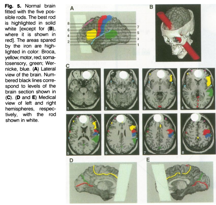
The cortical stages of the perception-action cycle

                                                     Fuster, 2000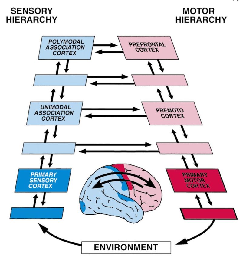
The lobotomists
              •   London 1935, Second International Congress of Neurology: John
                  Fulton and Carlyle Jacobsen present work on prefrontal lesions in
                  chimps, which reduced “tantrums” and made them “happier”.

John Fulton

                    Fulton                      Muniz            ?

              •   Egas Muniz asks Fulton if such surgery can be performed on
                  psychiatric patients. Fulton rejects the idea.

Egas Moniz
                               Boettcher, L. B., & Menacho, S. T. (2017).   https://doi.org/10.3171/2017.6.FOCUS17249
The lobotomists
             •   November 1935: Moniz performs “frontal leucotomy” on several
                 psychiatric patients in Lisbon.

             •   The goal of the operation was “to remove some of the long fibres
                 that connected the frontal lobes to other major brain centres”
Egas Moniz

                           •   The mode of action: ethanol injection into the white
                               matter tracts under the prefrontal cortex

                           •   Later patients were treated with a “leucotome”,
                               making circular lesions of white matter fibers.

                           •   1949 - Moniz receives the Nobel Prize for Physiology
                               and Medicine for the development of leucotomy.
                                 Boettcher, L. B., & Menacho, S. T. (2017).   https://doi.org/10.3171/2017.6.FOCUS17249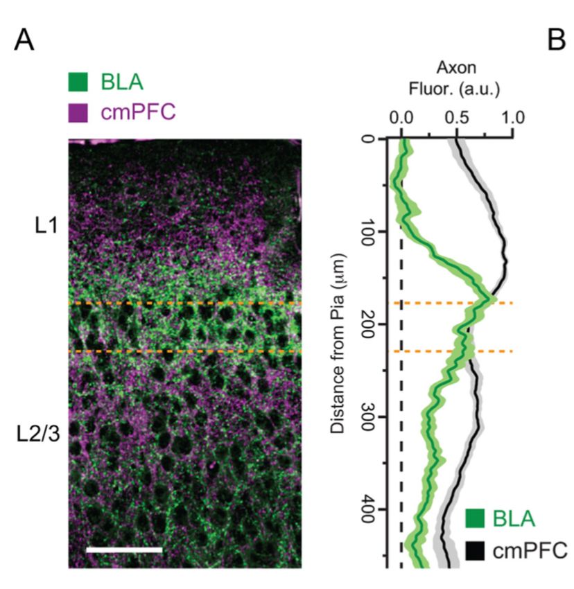
The lobotomists
                    •   Walter Freeman, an American neurologist, adopts Moniz’s procedure and
                        develops a more rapid method for lobotomy

                    •   The major advance: can be performed as an “office procedure” without
                        complex surgical equipment.

  Egas Moniz

Walter Freeman II           Transorbital lobotomy
The lobotomists
                    •   Approx. 40,000 psychiatric patients were lobotomized by Freeman and his
                        colleagues in the US from 1946 to the 1970s.

                    •   Criticism of the procedure mounted, mainly regarding the efficacy and the lack of
                        consistent long-term follow-up on patients.
  Egas Moniz
                    •   A majority of lobotomies was performed on women and minorities (up to 70% in
                        some cases). In Japan - mostly done on children with behavior “problems”.

                    •   Very minimal follow-up on patients after their discharge -> poor scientific evidence
                        for the efficacy of the procedure and its side effects.

                    •   Calls to revoke Moniz’s Nobel Prize were rejected by the Nobel committee.

                                                               Further reading: “The Lobotomist” by Jack El-Hai
Walter Freeman II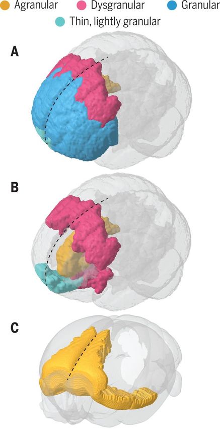
Experiments of chance: what have we learned from
                accidental lesions?

                              “The equilibrium or balance, so to speak,
                              between his intellectual faculties and
                              animal propensities, seems to have been
                              destroyed. He is fitful, irreverent, indulging
                              at times in the grossest profanity (which
                              was not previously his custom),
                              manifesting but little deference for his
                              fellows, impatient of restraint or advice
                              when it conflicts with his desires….”

                              John Harlow, "Recovery from the Passage
                              of an Iron Bar through the Head”, 1868

Phineas P. Gage (1823–1860)
Experiments of chance: what have we learned from
               accidental lesions?
                    Mapping the connec-vity damage to Gage’s brain:

                    •Only 4% of the cortex was directly injured by the metal rod.

                    •~11% of leE frontal lobe WM was damaged.

                    •Broca’s area was spared, along with motor, somatosensory and Wernicke’s
                     area.

                    •The rod went through the ventromedial area of both hemispheres and
                     exited dorsomedially.

                    •PaLern consistent with several modern cases, which showed similar
                     behavioral changes (“Their ability to make ra-onal decisions in personal and
                     social maLers is invariably compromised and so is their processing of
                     emo-on. On the contrary, their ability to tackle the logic of an abstract
                     problem, to perform calcula-ons, and to call up appropriate knowledge and
                     aLend to it remains intact.”)

                                                                            Damasio et al., Science 1994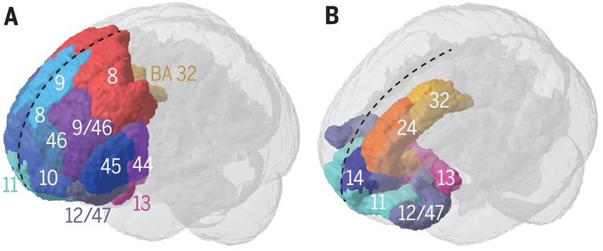
Experiments of chance: what have we learned from
accidental lesions?
PFC damage leads to very little overt impairment, but can be devastating.
• Executive functions: alertness, set (Wisconsin card sorting
task), task switching, rule learning, working memory Cognitive
• Decision making, value and rule learning deficits
• Depression
Emotional
• Euphoria
• Hyper / Hypokinesia, perseverance in old behavioral patterns Motor / arousal
• Social deficits - social anxiety, theory of mind, social Social
motivation
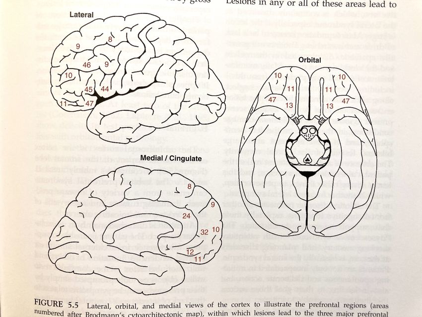
“The Prefrontal Cortex”, Joaquim Fuster (4th ed.) Ch. 5Anatomy of the prefrontal cortex
Brodmann (1909): the prefrontal cortex in primates is defined as the frontal granular region (anatomical/
cytoarchitectonic definition).
Dorsolateral granular PFC - unique to primates;
Dorsomedial, ventromedial and orbital (dysgranular and agranular) exist in all mammals to some extent.
Dorsomedial
Ventromedial
Dorsolateral
Ventrolateral
Orbital
Carlén, Science 358, 478–482 (2017)Problem: the “PFC” in many animals is mostly agranular
Does this mean that rodents and carnivores have no PFC?
Rose and Woolsey (1948): the prefrontal cortex should be defined as the termination field of the mediodorsal
thalamus (hodological definition)
Human prefrontal cortex Mouse prefrontal cortex
Carlén, Science 358, 478–482 (2017)Cytoarchitectonics of the PFC
Human prefrontal cortex
Granular cortex: contains layer 4 with large “granule cells”
that receive direct thalamic input; agranular cortex has no
layer 4; dysgranular - less pronounced L4.
Granular Dysgranular Agranular
Carlén, Science 358, 478–482 (2017) Beul & Hilgetag, Front. Neuroanat., 2015Thalamic input to agranular cortex arrives at L1, L3
VPM-S1 POM-S1 MD - mPFC projections Other inputs
Sermet et al. eLife 2019 Delevich et al., JN 2015 Little & Carter 2012A canonical microcircuit for agranular cortex?
The canonical microcircuit in
(granular) S1:
Strong recurrence in L4, ascending
inputs L4->L2/3, descending from
L2/3->L5A
Lefort et al., Neuron 2009
Organization of connectivity
in (agranular) M1:
Strong recurrence in L2/3,
ascending from L5A->L2 (might
replace L4-L2/3)
Weiler et al., Nat Neuro 2008The medial prefrontal cortex is conserved across
mammalian species
“The orbital and medial prefrontal areas are especially well connected with the medial and anterior
nuclei of the thalamus, the prepiriform cortex, the hippocampus, the amygdala, and the
hypothalamus.
The cortex of the dorsolateral prefrontal convexity is profusely connected with other frontal areas
homolaterally and-through the corpus callosum-contralaterally, with the hippocampus, and with the
temporal and parietal cortex.” (Fuster, 2000)
Fuster, Psychobiology 2000 Burgos-Robles et al., 2019Long-range connectivity of the PFC
Monkey PFC connectivity Rodent PFC connectivity
Connectivity with MD is conserved across mammals;
Some prefrontal connections are unique to granular DLPFC/VLPFC in primates, but are replaced by other PFC
regions in rodents;
Medial PFC regions connect heavily with the hippocampus and amygdala in all mammals.
Tsutui et al., Front. Sys. Neurosci 2016Subregion specificity of prefrontal syndromes
• Lateral prefrontal lesions (areas 8, 9, 10,
46):
- Attention disorder (selective/inclusive,
exclusive);
- Apathy (“frontal neglect”);
- Perseveration;
- Working memory and planning deficits
- Spoken language disorders (mainly left
hemisphere).
- Depression
“The Prefrontal Cortex”, Joaquim Fuster (4th ed.) Ch. 5Subregion specificity of prefrontal syndromes
• Orbital lesions (areas 11,13):
- Attention deficit (mainly exclusive)
- Hypermotility
- Impulsivity and compulsivity
- Perseveration, bad decision making
- Disinhibition, disrupted moral judgement
- Sociopathy
- Disinhibition of instinctual behaviors
“The Prefrontal Cortex”, Joaquim Fuster (4th ed.) Ch. 5Subregion specificity of prefrontal syndromes
• Medial lesions (areas 8-10, 12, 24, 32):
- Action initiation difficulties
- Cataplexy (loss of whole-body muscle
tone during intense emotion)
- Apathy - most prevalent disorder
- Social deficits: theory of mind
impairments, aggression
“The Prefrontal Cortex”, Joaquim Fuster (4th ed.) Ch. 5Identifying prefrontal dysfunction
• Executive function tasks are particularly difficult for people with
lateral prefrontal damage
WCST experiment: https://www.psytoolkit.org/experiment-library/experiment_wcst.html
Grant and Berg, Journal of Experimental Psychology, 38, 404-411 (1948) Brenda Milner, JAMA 1963Identifying prefrontal dysfunction
• Executive function tasks are particularly difficult for people with
prefrontal damage
Brenda Milner, JAMA 1963Identifying prefrontal dysfunction
• Executive function tasks are particularly difficult for people with
prefrontal damage
“The impairment shown by patients after frontal lobectomy reveals itself
as a strong perseverative tendency. In extreme cases, a patient may sort
all 128 cards to one preferred category (for example, form), despite the
experimenter repeatedly telling him that his responses are wrong.”
Brenda Milner, JAMA 1963Behavioral/cognitive functions of the PFC
Working memory: the ability to remember and manipulate information over a brief period (in the
order of seconds).
Definitions:
Limited capacity: originally proposed as 7 items; recent studies suggest a limit of 4 items.
Associated with increased PFC activity: PFC activity increases with increased working
memory load (fMRI and ephys evidence).
Individual differences in WM capacity are associated with variation in several important
abilities, including control of attention, non-verbal reasoning ability and academic performance.
Working memory = Reference memory
But what distinguishes working memory from short-term memory?
Is it more about the rate of forgetting than a unique acquisition mechanism?
WM experiment: http://try.cognitionlab.com/demo/demo_types/sternberg/shell.htmlReaction time and gamma oscillations during WM
Subjects: two hospitalized epilepsy patients implanted with intracranial electrode
arrays for monitoring seizure activity.
High WM load Low WM load
Howard et al., 2003Definition and putative mechanism of WM in humans:
Working memory as a basis for long-term memory
(WM —> LTM)
Language habits as a template for WM performance
(“contramponist” vs “loddenapish”)
Baddeley A. Working memory. Science 1992;255:556–9.Prefrontal cortex is required for working memory
Butters and Pandya, Science 1969Neural correlates of working memory
Fuster and Alexander, Science 1971Neural correlates of working memory
The oculomotor delayed-response (ODR) task:
~30% of DLPFC neurons around the principal sulcus (PS) show delay-period persistent firing activity after
presentation of a cue and before making a motor response.
Cooling the PFC impairs WM performance:
Funahashi et al., 1989 Chafee and Goldman-Rakic, 2000Neural correlates of working memory
The oculomotor delayed-response (ODR) task:
~30% of DLPFC neurons around the principal sulcus (PS) show delay-period persistent firing activity after
presentation of a cue and before making a motor response.
Where else in the brain can you find delay-period activity?
Funahashi et al., 1989Neural correlates of working memory
The oculomotor delayed-response (ODR) task:
~30% of DLPFC neurons around the principal sulcus (PS) show delay-period persistent firing activity after
presentation of a cue and before making a motor response.
Where else in the brain can you find delay-period activity?
Gamma Oscillations (in PFC, parietal and temporal cortex) during
performance of the Sternberg task in two human subjects:
Howard et al., 2003Neural correlates of working memory
The oculomotor delayed-response (ODR) task:
~30% of DLPFC neurons around the principal sulcus (PS) show delay-period persistent firing activity after
presentation of a cue and before making a motor response.
Where else in the brain can you find delay-period activity?
Leavitt et al., Trends Neurosci 2017Neural correlates of working memory
The vibrotactile comparison task (Ranulfo Romo):
Monkey has to compare two vibrational stimuli applied to the finger, and make a choice (press right / press
left) based on which one was higher frequency.
Romo et al., Nature 1999What are the mechanisms of WM-related persistent activity?
• Cell-intrinsic mechanism: unique properties of prefrontal neurons that allow sustained firing
• Network mechanisms:
• Local circuit dynamics
• Cross-regional dynamics (cortico-cortical, thalamocortical)
• Synaptic mechanisms
** Persistent activity is *not* unique to WM. It is found in a variety of brain regions during behaviors
(e.g. motor control) and is a general feature of neural circuit function. (see Major and Tank 2004 for review)
Kandel, Principles of Neural Science Ch. 67What are the mechanisms of WM-related persistent activity?
• Cell-intrinsic mechanism: unique properties of prefrontal
neurons that allow sustained firing
• Persistent firing in the absence of synaptic input - can be
observed in some neuron types in the cortex.
• Depends on the activation of calcium-activated non-specific
cation (CAN) channels.
• Sustained activity is enhanced by cholinergic agonists (e.g.
carbachol) acting through muscarinic acetylcholine receptors
(entorhinal cortex postsubicular neurons)
Yoshida and Hasselmo, J Neurosci 2009What are the mechanisms of WM-related persistent activity?
• Cell-intrinsic mechanism: unique properties of prefrontal
neurons that allow sustained firing
• Persistent firing in the absence of synaptic input - can be
observed in some neuron types in the cortex.
• Depends on the activation of calcium-activated non-specific
cation (CAN) channels.
• Sustained activity is enhanced by cholinergic agonists (e.g.
carbachol) acting through muscarinic acetylcholine receptors
• Dopamine receptor inhibition impairs persistent firing in vivo.
Sawaguchi, Matsumura, Kubota , Neurosci Res 1988What are the mechanisms of WM-related persistent activity?
• Cell-intrinsic mechanism: unique properties of prefrontal
neurons that allow sustained firing
• Persistent firing in the absence of synaptic input - can be
observed in some neuron types in the cortex.
• Depends on the activation of calcium-activated non-specific
cation (CAN) channels.
• Sustained activity is enhanced by cholinergic agonists (e.g.
carbachol) acting through muscarinic acetylcholine receptors
• Dopamine receptor inhibition impairs persistent firing in vivo.
DA: Dopamine administration
Fluphenazine: D2 antagonist
Sawaguchi, Matsumura, Kubota , Neurosci Res 1988What are the mechanisms of WM-related persistent activity?
• Cell-intrinsic mechanism: unique properties of prefrontal
neurons that allow sustained firing
• Persistent firing in the absence of synaptic input - can be
observed in some neuron types in the cortex.
• Depends on the activation of calcium-activated non-specific
cation (CAN) channels.
• Sustained activity is enhanced by cholinergic agonists (e.g.
carbachol) acting through muscarinic acetylcholine receptors
• Dopamine receptor inhibition impairs persistent firing in vivo.
SCH23390: D1 antagonist
Sawaguchi and Goldman-Rakic, Science 1991What are the mechanisms of WM-related persistent activity?
• Cell-intrinsic mechanism: unique properties of prefrontal
neurons that allow sustained firing
• Network mechanisms:
• Local circuit dynamics
• Cross-regional dynamics (cortico-cortical,
thalamocortical)
• Synaptic mechanisms
Kandel, Principles of Neural Science Ch. 67What are the mechanisms of WM-related persistent activity?
• Cell-intrinsic mechanism: unique properties of prefrontal Ferret prefrontal cortical slices show intrinsic UP/DOWN state dynamics
neurons that allow sustained firing
• Network mechanisms:
• Local circuit dynamics
• Cross-regional dynamics (cortico-cortical,
thalamocortical) Voltage-clamp recordings show that UP states involve balanced
excitation/inhibition
• Synaptic mechanisms
Shu et al., Nature 2003What are the mechanisms of WM-related persistent activity?
• Cell-intrinsic mechanism: unique properties of prefrontal
neurons that allow sustained firing
• Network mechanisms:
• Local circuit dynamics
• Cross-regional dynamics (cortico-cortical,
thalamocortical)
• Synaptic mechanisms
Kandel, Principles of Neural Science Ch. 67What are the mechanisms of WM-related persistent activity?
• Cell-intrinsic mechanism: unique properties of prefrontal
neurons that allow sustained firing
• Network mechanisms:
• Local circuit dynamics
• Cross-regional dynamics (cortico-cortical,
thalamocortical)
• Synaptic mechanisms
Mongillo, Barak, Tsodyks, Science 2008Does the PFC have unique temporal properties
that support WM?
• Recordings pooled from several labs across different anatomical
locations along the sensory -> prefrontal hierarchy
• Measure autocorrelation decay in single-neuron spike trains
• Prefrontal regions show slower decay timecourse than sensory
regions.
Murray et al., Nat Neuro 2014Does the PFC have unique temporal properties
that support WM?
• Recordings pooled from several labs across different anatomical
locations along the sensory -> prefrontal hierarchy
• Measure autocorrelation decay in single-neuron spike trains
• Prefrontal regions show slower decay timecourse than sensory
regions.In search of circuit mechanisms: can rodents do working memory?
The 8-arm maze for rats (Olton et al., 1970): Delayed alternation on a T-maze (spontaneous/cued)
Working memory: remove after 4 arm visits, check if Spontaneous alternation: animal is removed after receiving
animal returns to previously-visited arms reward on one side, has to alternate to receive the second
reward.
Cued version: a light/sound/odor indicates correct side;
animal is released after a delay.
Potential problem with spatial tasks: animals can “cheat” by “postural cueing”.In search of circuit mechanisms: can rodents do working memory?
The odor span task:
Rats can remember up to 24
odors in this task
Operant delayed non-match to sample:
Automated, many trials, but might also
suffer from “postural cueing”
Overall: delayed comparison tasks >>> delayed reaction tasks.PFC neuronal activity during WM in rodents
Fujisawa et al., Nat Neurosci 2008WM in rodents is associated with long-range synchrony
• Mice performing a T-maze task with a sample (“forced-choice”)
phase followed by a choice phase.
• Simultaneous mPFC single-unit + hippocampal LFP recording
• mPFC neurons show phase-locking to hippocampal theta
oscillations
• Phase-locking is stronger during the “choice” phase of the task
compared with the “sample” phase
• Also: mice with a schizophrenia associated mutation (Df16) show
impaired phase-locking and impaired WM performance.
Sigurdsson et al., Nature 2011Prefrontal cortex and working memory: summary • Working memory performance is impaired by prefrontal lesions; particularly by lesions to the middle region of the principal sulcus • Working memory tasks in primates: the delayed alternation task, the oculomotor delayed- response task (different flavors); the vibrotactile comparison task. • Persistent activity in the PFC (and other regions) during WM tasks in monkeys • Models of WM-related persistent activity including cellular, local-circuit and long-range interactions. • A synaptic theory of WM permits retention of information in the circuit with minimal spiking (energy-efficient) • Rodent WM tasks are mainly based on spatial alternation • Ephys correlates: “tiling” of the delay period, hippocampal-prefrontal phase-locking, impairment in a mouse model of schizophrenia.
Prefrontal regulation of fear learning

               Pavlovian fear conditioning: A previously safe stimulus (CS+) is paired
               with an aversive outcome (US).
Prefrontal regulation of fear learning
Pavlovian fear conditioning: A previously safe stimulus (CS+) is paired
with an aversive outcome (US).
The CS+ stimulus triggers a conditioned response
Extinction training: repeated presentation of the CS+ (cupcakes)
without the US (electrical shock) triggers new learning that “overrides”
the conditioned response.Prefrontal regulation of fear learning
Aversive conditioning: Stimuli are paired with an electrical shock (in
humans!) over several trials. Partial reinforcement. SCR measurements.
Reversal learning: The previously “safe” stimulus is now associated
with electrical shock.
Skin conductance response (SCR) in response to
CS (Face A) increases during conditioning,
decreases during reversal.
vmPFC (Brodmann’s Area 32/10): differential
activity to CS decreases during conditioning,
increases during extinction.
Schiller et al., 2008Prefrontal regulation of fear learning
Infralimbic cortex (IL) neurons fire to the CS
during extinction:
Milad & Quirk, Nature 2002Prefrontal regulation of fear learning
Stimulation of the infralimbic cortex (IL)
decreases freezing on Day 2:
unstimulated (open triangles), unpaired IL stimulated
(open squares) and paired IL stimulated (filled squares)
Milad & Quirk, Nature 2002Prefrontal regulation of fear learning
Optogenetic excitation of the infralimbic Optogenetic inhibition of the infralimbic
cortex (IL) decreases freezing: cortex (IL) impairs extinction learning:
Do-Monte et al., J Neurosci 2015Prefrontal regulation of fear learning Circuit mechanism of extinction learning: Auditory stimuli reach the BLA from cortex and thalamus; Excitatory BLA neurons project to PL and IL; PL projection to BLA is mostly excitatory; IL projection has strong feed-forward inhibitory component through the intercalated cells. Johanssen and Herry., Nat Neurosci 2015
Representation of reward value and prediction error coding
Prediction error coding in the ACC: Monkeys trained on a simple reward association task show activity in
ACC that tracks the type of error performed.

                                                                                           Matsumoto et al., Nat Neuro 2007
The role of PFC in social behavior:
Prefrontal cortex volume in macaques is associated with social network size, and
with social rank within the group.
Sallet et al., 2011The role of PFC in social behavior:
Prefrontal cortex volume in macaques is associated with social network size, and
with social rank within the group.
Social network size correlated with increased BOLD signal correlations between
rPFC and STS.
Sallet et al., 2011The role of PFC in social behavior:
In mice, excitatory synaptic strength in the mPFC correlates with social rank
(Wang et al., 2011)
Wang et al., 2011The role of PFC in social behavior:
Excitation or inhibition of dmPFC led to winning/losing in the tube test. Following
repeated encounters, mice that consistently “won” showed increased synaptic strength
in MD-PFC connections (Zhou et al., 2017)
Chemogenetic inhibition: Optogenetic excitation: MD-PFC plasticity is sufficient:
Zhou et al., Science 2017The role of PFC in social behavior:
In human neurosurgical patients with ventromedial prefrontal lesions: deficits in
emotion recognition (Jenkins et al., 2014; Rudebeck et al., 2007)
No social deficit in dmPFC or OFC lesioned patients.
Jenkins et al, 2014The role of PFC in social behavior:
Mice show preference toward “emotionally-altered” conspecifics (mice exposed to electrical
shock, other acute stressors, liquid reward or social enrichment)
Scheggia et al., Nat Neuro 2019The role of PFC in social behavior:
Mice show preference toward “emotionally-altered” conspecifics (mice exposed to electrical
shock, other acute stressors, liquid reward or social enrichment)
Silencing of prefrontal somatostatin-expressing interneurons eliminates the preference
toward an “emotionally-altered” conspecific.
Silencing PV neurons had no effect on preference
score, but changed overall sociability
Scheggia et al., Nat Neuro 2019Autism-associated changes of GABAergic inhibition in the PFC
Decreased GABA receptor binding in the frontal cortex of humans with autism (Oblak et
al., 2009)
Reduced interneuron density in prefrontal cortex of autism patients (Zikopoulous and
Barbas, 2013, Hashemi et al., 2016)
Figure 4. Examples of pseudocolored images of [3H]-muscimol binding (15 nM) in autistic (a) and control (b) cases. Solid black arrows
demonstrate the location of sampling in the supragranular layers; arrows with dashed lines indicate sampling in the infragranular layers.
In the [3H]muscimol binding experiments, the number of GABAA receptors in the supragranular (P 5 5.16 ! 10"6) and infragranular layers
(P 5 0.04) was significantly (!!) decreased (c). [Color figure can be viewed online at www.interscience.wiley.com]
Oblak et al., 2009 Hashemi et al., 2016Dysfunction of PFC in psychiatric disorders
Reduced glucose metabolism was observed in the subgenual anterior cingulate
area (BA 24/25) in patients suffering from major depression or bipolar disorder
(during depressive stages)
Reduced glucose metabolism was observed in the subgenual anterior cingulate
area (BA 24/25) in patients suffering from major depression or bipolar disorder
(during both manic and depressive stages)
Drevets et al., Nature 1997Deep-brain stimulation for major depression
Summary: the PFC is involved in multiple high-level cognitive/behavioral
processes. Circuit mechanisms (outside of fear learning) are only partially
elucidated.

                            Working Memory

                              Emotional regulation

                                  Decision making

                                   Value representation, prediction error

                            Social cognition

                      Attention

               Language
You can also read