JOURNAL OF CLINICAL CHIROPRACTIC PEDIATRICS
←
→
Page content transcription
If your browser does not render page correctly, please read the page content below

JOURNAL OF CLINICAL
                              CHIROPRACTIC PEDIATRICS

                                            VOLUME 17   •   NO. 2   •   JULY 2018

                               PUBLICATION OF THE COUNCIL ON CHIROPRACTIC PEDIATRICS
                                     INTERNATIONAL CHIROPRACTORS ASSOCIATION

Volume 17, No. 2, July 2018             JOURNAL OF CLINICAL CHIROPRACTIC PEDIATRICS
EDITORS
Sharon Vallone, DC, DICCP, FICCP                                        BOARD OF REVIEWERS
Cheryl Hawk, DC, PhD
                                                                        Cathrin Alvestad Slettebo, DC, MSc
                                                                        Sola, Norway
EDITORIAL BOARD
                                                                        Tracy Barnes, DC, DICCP, CKTI
Clinton Daniels, DC, MS, DAAPM                                          Louisville, KY, USA
VA Puget Sound Health Care System,
Tacoma, WA, USA                                                         Faraneh Carnegie-Hargreaves, DC
                                                                        South Windsor, CT, USA
Peter N. Fysh, DC, FICCP
Professor Emeritus, Palmer College of                                   Marion Willard Evan, Jr., DC, PhD, MCHES
Chiropractic West, San Jose, CA, USA                                    Texas Chiropractic College, Pasadena, TX, USA

Aupama Kizhakkeveettil, BAMS                                            Jean Elizabeth Grabowski
(Ayurveda), MAOM, LAC                                                   Kentuckiana Children’s Center, Louisville, KY, USA
Southern California Unversity of                                        Alison K. Hazelbaker, MA, PhD, IBCLC
Health Sciences, Whittier, CA, USA                                      Columbus, OH, USA
Dana J. Lawrence, DC, MMedEd, MA                                        Valerie Lavigne, DC, FICP, MScApp, IBCLC
Palmer College of Chiropractic,                                         Kirkland, QC, Canada
Davenport, IA, USA
                                                                        Robert A. Leach, DC, MS, CHES
Joyce Miller, DC, PhD                                                   Starkville, MS, USA
AECC University, Bournemouth, UK
                                                                        Amy Sarah Miller, DC, MSc
Molly Rangnath, MA                                                      Bournemouth University, Bournemouth, UK
Falls Church, VA, USA
                                                                        Stephanie O’Neill-Bhogal, DC, DICCP
Lora Tanis, DC, DICCP                                                   Life Chiropractic College West, Hayward, CA, USA
W. Milford, NJ, USA
                                                                        Mark T. Pfefer, RN, MS, DC
                                                                        Cleveland University, Overland Park, KS, USA
                                                                        Katherine A. Pohlman, DC, MS, DICCP
                                                                        Parker University, Dallas, TX, USA
              The Journal of Clinical Chiropractic Pediatrics (JCCP)    Carol Prevost, DC, DICCP
              is the official peer-reviewed journal of the Council on
                                                                        Palmer College of Chiropractic, Port Orange, FL, USA
              Chiropractic Pediatrics, 6400 Arlington Boulevard,
              Suite 800, Falls Church, Virginia 22042, USA              Veronica Pryme, MSc(Chiro), MSc(Paeds)
              Copyright by the Council on Chiropractic Pediatrics.      Bergan, Norway
              All rights reserved.
                                                                        Richard Strunk, DC, MS
Editorial Correspondence: Correspondence should be sent to:             Hamden, CT, USA
Editor, JCCP
                                                                        Meghan Van Loon, PT, DC, DICCP
ICA Council on Chiropractic Pediatrics
6400 Arlington Boulevard, Suite 800
                                                                        New York Chiropractic College, Seneca Falls, NY, USA
Falls Church, Virginia 22042, U.S.A.
                                                                        Sue A. Weber, DC, MSc(Paeds), FEAC, FRCC
Email: pediatricscouncil@chiropractic.org                               Stockholm, Sweden
or svallonedc@aol.com

                                JOURNAL OF CLINICAL CHIROPRACTIC PEDIATRICS                                  Volume 17, No. 2, July 2018
TABLE OF CONTENTS

  VOLUME 17, NUMBER 2                                                                   					                                                                JULY 2018

  Editorial
  Remembering Maxine McMullen, RN, DC, FICCP . . . . . . . . . . . . . . . . . . . . . . . . . . . . . . . . . . . . . . . . . . . . . . . 1430
  By Sharon Vallone, DC, DICCP, FICCP

  Benign joint hypermobility – developing clinical significance . . . . . . . . . . . . . . . . . . . . . . . . . . . . . . . . . . . 1431
  By Peter N. Fysh, DC, FICCP

  Portable pad or pen and paper: Preference of mothers completing an outcomes instrument: a cross- 1441
  sectional survey . . . . . . . . . . . . . . . . . . . . . . . . . . . . . . . . . . . . . . . . . . . . . . . . . . . . . . . . . . . . . . . . . . . . . . . . . .
  By Mandy Hiew, MSc, Derek Shawn Lo Tiap Kwong, MSc, Zicheng Mok, MSc, Yun Han Tee, MSc and Joyce
  Miller, BS, DC, PhD

  Early intervention: Improvement in motor developmental speech delay in a 2-year-old male fol-
  lowing chiropractic care: a case report . . . . . . . . . . . . . . . . . . . . . . . . . . . . . . . . . . . . . . . . . . . . . . . . . . . . . . 1444
  By Andrew Dorough, DC

  Chiropractic management of musculoskeletal disorders associated with a neonatal clavicle frac-
  ture: a case report . . . . . . . . . . . . . . . . . . . . . . . . . . . . . . . . . . . . . . . . . . . . . . . . . . . . . . . . . . . . . . . . . . . . . . . . 1449
  By Amelie Bourque, DC

  The chiropractor’s role in the interdisciplinary care of the infant with faltering growth: two case
  reports . . . . . . . . . . . . . . . . . . . . . . . . . . . . . . . . . . . . . . . . . . . . . . . . . . . . . . . . . . . . . . . . . . . . . . . . . . . . . . . . . . . 1456
  By Linda Alida Bernadette Anna Overschie, BSc, MSc Chiropractic, DC

  JOURNAL ABSTRACTS . . . . . . . . . . . . . . . . . . . . . . . . . . . . . . . . . . . . . . . . . . . . . . . . . . . . . . . . . . . . . . . . . . . . . 1462

              Publishing Offices:
               ICA Council on Chiropractic Pediatrics
               6400 Arlington Boulevard, Suite 800, Falls Church, Virginia 22042 U.S.A.
Volume 17, No. 2, July 2018                                 JOURNAL OF CLINICAL CHIROPRACTIC PEDIATRICS
GUIDELINES FOR AUTHORS

The Journal of Clinical Chiropractic Pediatrics welcomes origi-   The paper must include an abstract or summary. This ab-
nal and scholarly manuscripts for peer‑review and con-            stract/summary should state the purpose of the paper (ob-
sideration for publication. Topics must pertain to the field      jective), procedures, methods, main findings (results) and
of pediatrics which includes pregnancy and adolescence.           principal conclusions. Also, any key words or phrases that
Manuscripts should not have been published before or sub-         will assist indexers should be provided.
mitted to another publication.
                                                                  References must be cited for all materials derived from the
The following will be considered:                                 works of other people and previously published works.
                                                                  Reference numbers in superscript must be assigned in the
Case Reports and Case Series — presentations of individual        order of citation in the paper.
or groups of cases deemed to be of interest to the profes-
sional and scholarly community.                                   Tables — Each table or figure should be on a separate page
                                                                  and not imbedded in the manuscript. If the table is from
Pilot Studies or Hypothesis — papers which, while very            another publication, permission to publish must be granted
broad, present with a clear hypotheses and suggest a foun-        and the publication acknowledged.
dation for future, in‑depth studies.
                                                                  Photographs — Photographs may be in color or in grayscale
Literature Reviews — studies of existing papers and books         and scanned at 300 dpi with sharp contrast. Patient photo-
presented with the intention of supporting and encourag-          graphs must have consent form signed by the individual or
ing new and continuing study.                                     parent or guardian in the case of a minor.

Technical Descriptions — reports of new analytical/diag-          Informed Consent — If the research/study involves experi-
nostic tools for assessment and delivery of care. Controlled,     mental investigations performed on humans the manu-
Large Scale Studies — usually, but not necessarily, performed     script must include a statement that informed consent was
at a college or research facility. May be double-blinded.         obtained from the individuals involved in the investigation.

Commentaries — presentations of opinion on trends within          Patient Anonymity — Patient names or any information that
the profession or current events, pertaining to pediatric and     could identify a specific patient should be avoided. All case
adolescent chiropractic care.                                     reports, with or without identifying photographs accompa-
                                                                  nying a manuscript must have a consent form signed by
Guidelines for submission                                         the individual or parent or guardian in the case of a minor.
                                                                  These are to include any requests for blocking faces, etc.
All manuscripts are accepted purely for consideration.
They must be original works and should not be under con-          Acknowledgements — Any illustrations from other publi-
sideration by any other journal or publisher at the time of       cations must be acknowledged. It is the author’s responsi-
submission. They must be accompanied by a TRANSFER                bility to obtain written permission from the publisher and/
OF COPYRIGHT form, signed by all authors and by the               or author for their use.
employer if the paper is the result of a “work for hire.” It
is understood that while the manuscript is under consider-        All manuscripts deemed appropriate for publication by the
ation it will not be sent to any other publication. In the case   editor will be sent blind to at least two reviewers. If the man-
of multiple authors, a transmittal letter should designate        uscript is accepted, the author will be notified. If substantive
one author as correspondent.                                      changes are required, the paper will be returned to the au-
                                                                  thor and the author must re-submit a clean copy of the re-
Manuscripts may be sent to editor at svallonedc@aol.com.          vised manuscript. Author will be given a tentative date for
Manuscript should be in document style MS Word (or com-           publication if accepted. Manuscripts not accepted for publi-
patible) and unformatted. PDFs will not be accepted.              cation will be returned to the author without comment.

1424                                JOURNAL OF CLINICAL CHIROPRACTIC PEDIATRICS                            Volume 17, No. 2, July 2018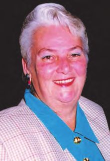
Instructions to Authors — Summary
See Uniform Requirements for Manuscripts Submitted to          page. Use page break function to separate page, not repeat-
Biomedical Journals for detailed information                   ed line breaks to get to a new page.
http://www.icmje.org/.                                         • Title page
                                                               • Abstract
General formatting guidelines                                  • Manuscript
• All submission components must be submitted                  • Acknowledgements
  electronically.                                              • References
• Only manuscripts in English are accepted.                    • Tables
• Submit manuscripts as Microsoft Word documents.              • Figures
• Use 1” margins on all sides
• Use Arial 12 point black font                                Title page
• Capitalize only the first letter in the title, and any       • Title of article–ONLY CAPITALIZE FIRST LETTER OF
  proper nouns.                                                  FIRST WORD
• Do not justify text.                                         • Running head (limited to 40 characters)
• Do not use column function                                   • Word count (excluding references, tables and figures)
• Number all pages at bottom right.                            • Number of tables
• Double-space manuscript. Single-space references,            • Number of figures
  tables or figure legends.                                    • Authors
• Do not abbreviate words or terms the first time they are       o Name, with all degrees (do not include Bachelor’s
  introduced; at that time, provide the abbreviation in            level degrees)
  parentheses and use it from that point forward.                o Current title/position and affiliation, including city,
• Number citations consecutively using superscripted               state and country
  Arabic numerals and place all references in a Reference      • Corresponding author
  section immediately at the end of your section.                o Name
• Run spell check and grammar check after completing the         o Mailing address, phone, fax
  manuscript. Use American English spelling and units            o E-mail address; provide alternative e-mail address
  of measurement.                                                  if possible

Submission Components                                          Abstract–not to exceed 250 words. It may be structured or
• JCCP authorship form–submit separately from manu-            unstructured. Structured abstracts usually include the fol-
script. All authorship forms may be combined in a single       lowing sections: Purpose, Methods (include study design
PDF. Each author must complete this form, scan and return      in this section), Results, Conclusion. For case reports and
it electronically to the editor before the manuscript can be   case series, see document, “Instructions for Case Reports
processed.                                                     and Case Series.”
• JCCP Patient (or Parent/Guardian) Permission to Pub-
lish Form–one form for each case (1 for case report; mul-      Manuscript Components
tiple individual forms for case series) – all forms may be     Manuscript length will vary with the type of article; in gen-
combined as a single PDF.                                      eral, manuscripts are expected to be 1,500-3,000 words in
• Permission to acknowledge forms: All individuals named       length, excluding references, tables and figures. These may
in the Acknowledgements section of the manuscript must         vary with the type of article. For case reports and case se-
sign a permission form. The corresponding author may use       ries, see, “Instructions for Case Reports and Case Series.”
his or her own form, or use the one JCCP provides—submit       In general, for manuscripts reporting research studies, the
separately from manuscript. All permission forms may be        order of components is:
combined as a single PDF.                                      • Introduction: succinctly describe the relevant literature
• Cover letter–submit as separate document, either Word        supporting the need for the study.
or PDF.                                                        • Methods: describe the methods used to accomplish the
                                                               study, in detail sufficient to allow the informed reader to
The following items MUST be submitted as a Word                evaluate their appropriateness.
document.                                                      • Results: present the results of the study, without interpre-
                                                               tation.
Cover letter–Explain why your manuscript is appropriate        • Discussion: describe limitations of the study; interpret
for JCCP.                                                      results; compare results to those of other relevant studies;
                                                               discuss value and implications of the study.
Document– Each of the following should be on a separate        • Inclusion of appendices is discouraged.

Volume 17, No. 2, July 2018              JOURNAL OF CLINICAL CHIROPRACTIC PEDIATRICS                                    1425
Instructions to Authors — Summary
Tables                                                                Acknowledgements
• Number tables consecutively in text, using Arabic                   Include a statement disclosing any funding support for the
  numerals (1, 2, 3 etc.)                                             project or project personnel, or any other potential conflicts
• Place each table on a separate page at the end of the               of interest. Acknowledge only individuals or organizations
  section, immediately following the References section.              who provided input or resources to the project that were
• Use “table” function in Word to construct tables; do NOT            above and beyond their usual responsibilities. All individu-
use tab or space keys to form columns and rows. Use table             als acknowledged must provide written permission to use
“normal” style to construct table. Do not insert vertical lines       their name; these permissions must accompany the manu-
between columns; do not use grids. Place horizontal line              script at the time of submission (scan documents and sub-
under table title and at end of table, separating the table           mit electronically).
from any footnotes. You may place horizontal lines under
headings in the table for clarity.                                    Reference format–examples
• Use footnotes to explain details at bottom of the table (be-        • Journal article: Jefferies LJ, Milanese SF, Grimmer-Somers
low a horizontal line). Identify using either superscripted           KA. Epidemiology of adolescent spinal pain: A systematic
lower-case letters or standard footnote symbols (sequence:            overview. Spine 2007;32:2630-2637.
*,†, ‡, §, ||, ¶, **, ††). Sequence the footnotes in the order text   • Book: Task Force on Community Preventive Services.
is read—from left to right and then down.                             Guide to Community Preventive Services. New York: Ox-
• Use left-justification to align numbers in columns.                 ford University Press; 2005.
                                                                      • Website/webpages: Author. Title. Name of website. URL.
Figures                                                               Date of publication. Updated date (if applicable). Date ac-
• Place figure title and legend on page with the figure.              cessed. Example: Fox F. Promoting and sustaining collabor-
• Figures must be submitted electronically. Acceptable file           ative networks in pediatrics. Pew Research Center. http://
formats: DOC, JPG, PDF. Figures may be embedded at the                www.pewinternet.org/2013/06/14/promoting-and-sus-
end of the manuscript text file or loaded as separate files for       taining-collaborative-networks-in-pediatrics/. Published
submission purposes. Should not be imbedded within the                June 14, 2013. Accessed September 3, 2017.
manuscript text
• Hand-drawn illustrations are not acceptable.                        Permission to acknowledge forms
• Provide documentation of permission for any figures that            All individuals named in the Acknowledgements section
are not original.                                                     of the manuscript must sign a permission form. The cor-
                                                                      responding author may use his or her own form, or use the
                                                                      one JCCP provides.

                                                     Title Page Format
Running Head:                                                         Corresponding Author
Word count (excluding references, tables and figures):                Name
Number of tables:                                                     Address
Number of figures:                                                    Phone Number:
                                                                      Fax:
Authors (in correct order)                                            Email:
Name, degrees
Current title/position and institution (if applicable)
City, State, Country

1426                                  JOURNAL OF CLINICAL CHIROPRACTIC PEDIATRICS                             Volume 17, No. 2, July 2018
Journal of Clinical Chiropractic Pediatrics Authorship Form
   Materials published in Journal of Clinical Chiropractic Pediatrics online are covered by copyright. All rights are reserved under United
   States and international copyright and other laws and conventions.
   Each author must read and sign the statements on 1) authorship responsibility and contribution, 2) financial disclosure and conflict
   of interest, 3) copyright transfer. The corresponding author must sign the Acknowledgement Statement and email the completed
   form to Svallonedc@aol.com to initiate manuscript processing.
   Manuscript title: ___________________________________________________________________________________________________
   1. Authorship Responsibility and Contribution
   • I certify that this submission represents original work, and that neither this submission nor a substantially similar one has been
   published or is under consideration for publication elsewhere in any medium (paper or electronic). I also affirm that this submission
   is not subject to copyright or any other rights except those of the current authors.
   • I certify that if so requested by the editor, I will provide the data or cooperate in obtaining the data on which this submission is
   based, for review by the journal’s designated representative(s).
   • I agree that the corresponding author may represent me to review proofs and make other decisions regarding the submission.
   I have approved the submission.
   • I certify that I meet the criteria for authorship, having made substantive contribution to the manuscript as indicated below (check
   all that apply).
             ___ Development of project concept or hypothesis
             ___ Study design and development of methodology
             ___ Project implementation
             ___ Data collection and management
             ___ Data analysis and interpretation of results
             ___ Literature search and review
             ___ Manuscript writing
             ___ Other (specify contribution)______________________________________________________________________________
   2. Financial Disclosure and Conflict of Interest
   I certify that all sources of extramural support of this submission, and the role of any funding agencies in the conduct of the study
   have been clearly described in the Acknowledgements section of the submission.
   Check one of the following two statements:
   q I certify that I have no financial interests, relationships or affiliations related to the project or materials addressed in the submission.
   OR
   q I certify that any potential conflicts of interest, including financial interests, relationships or affiliations related to this submission
   are disclosed in the Acknowledgements section of the manuscript.
   3. Copyright Transfer
   In consideration of the action of the Journal of Clinical Chiropractic Pediatrics in reviewing and editing this submission (including
   manuscripts, tables, figures and any supplemental documents), I hereby transfer, assign, or otherwise convey all copyright owner-
   ship including all rights and incidental thereto, exclusively to the Journal of Clinical Chiropractic Pediatrics.
   I also understand that if the manuscript is not accepted for publication by the Journal of Clinical Chiropractic Pediatrics I will be noti-
   fied and the transfer of copyright will be null and void.

   Signature					                                             e-mail address					                                        date signed

   Acknowledgement statement to be signed by corresponding author
   All individuals named in Acknowledgements section should provide written permission. I certify that:
   • All individuals who have made substantive contributions to the submission but who do not qualify as authors have been named,
   along with their specific contribution in the Acknowledgements.
   • All individuals so named have provided me with their written permission to be named.
   • If no Acknowledgement section is included in the submission, there are no other contributors to the manuscript.

   Corresponding Author Signature			                          e-mail address					                                        date signed

Volume 17, No. 2, July 2018                      JOURNAL OF CLINICAL CHIROPRACTIC PEDIATRICS                                                   1427
Journal of Clinical Chiropractic Pediatrics
Patient Consent Form for Case Report
Print name:__________________________________________________________________________________________
If patient is a minor, print parent/guardian name: ________________________________________________________
I have read the information about me/minor and/or seen the photograph to be published.
I give my consent for this material to appear in a scientific journal.
I understand the following:
(1) My name/minor’s name will not be attached to the material. The authors of the article will make every attempt
to keep my identity/minor’s identity anonymous. I understand, however, that they cannot guarantee complete
anonymity. It is possible that someone, such as someone who works in this clinic or one of my relatives, might be
able to identify me/minor.
(2) The material will only be published in a scientific journal.
(3) The material will not be used for advertising.
Signed:_________________________________________________ Today’s date: ______________________________
(if patient is a minor, parent or guardian signs.)
Journal of Clinical Chiropractic Pediatrics
Permission to Acknowledge
I give my permission to be acknowledged in the manuscript,
____________________________________________________________________________________________________
which is to be submitted to the Journal of Clinical Chiropractic Pediatrics.
____________________________________________________ ___________________________________________
Signature Date Signed
____________________________________________________
Print Name
1428 JOURNAL OF CLINICAL CHIROPRACTIC PEDIATRICS Volume 17, No. 2, July 2018Instructions for Case Reports and Case Series
Abstract tient’s presenting demographics, other relevant character-
The abstract should be 250 words or fewer. It may be either istics, complaint(s) and related symptomatology.
structured or unstructured. If structured, use the same sec-
tions as described below for the components of the report • Intervention and outcomes: Describe the course of treat-
(Introduction, Case Presentation, Intervention and Out- ment, including frequency and duration, and summarize
comes, Discussion). the patient’s clinical outcomes, using recognized outcome
measures if possible. Include whether informed consent
Case Report Components was obtained and if there were any adverse events reported.
• Introduction: State why this case is unusual or important. • Discussion: Succinctly state the important aspects of the
case, in terms of its implications for patient care in general,
•Methods: describe the search engine and key words used or for specific patient populations or conditions. You may
to review previously published literature on the subject also compare/contrast the case to other cases in the pub-
lished literature. Be cautious about overstating the impor-
• Case presentation: Provide a brief summary of the pa- tance/implications of your case.
Evidence-based Case Report Instructions
An Evidence-based Case Report (EBCR) is NOT the same as a traditional case report. The EBCR focuses on an answerable
clinical question, how it was explored in the search, appraising the results and how it applies to the case, along with the
integration of this information with the patient interaction. The final stage in this process is to audit the results.
These are the steps to include:1,2
• Brief summary of the chief complaint: 50-100 words
• Briefly describe the clinical case: 250-400 words
• Explain how you developed the clinical question: 200-300 words
• Explain your search for evidence (key words, databases used, number of articles retrieved): 50-100 words
• Evaluate the articles retrieved: critically appraise the evidence for validitiy and relevance: 200-300 words
• Describe how you made your clinical decision by applying these findings to the case, including how you considered and
integrated the patient’s preferences and values: 250-400 words
• Evaluate your performance: 50-100 words
1. Heneghan C, Badenoch D. Evidence-based Medicine Toolkit, 2nd ed. Oxford, UK: Blackwell Publishing, 2006.
http://onlinelibrary.wiley.com/doi/10.1002/9780470750605.index/summary (download pdf of “all chapters” for free copy
of the publication)
2. Jones-Harris AR. The evidence-based case report: a resource pack for chiropractors. Clin Chiropr 2003;6 73-84. (download
for free from www.chiro.org/cases/FULL/Evidence-based_Case_Report.pdf)
Additional interesting articles to read about EBM and writing and EBCR:
Review an example of an EBCR at:
https://www-ncbi-nlm-nih-gov.uws.idm.oclc.org/pmc/articles/PMC1126937/pdf/302.pdf
Iran J Pediatr. 2010 Sep; 20(3): 261–268. Evidence Based Medicine in Pediatric Practice: Brief Review
https://www.ncbi.nlm.nih.gov/pmc/articles/PMC3446038/
J Can Chiropr Assoc. 2014 March; 58(1): 6–7. Evidence-based case reports
http://pubmedcentralcanada.ca/pmcc/articles/PMC3924510/
3 BMJ. Vol 7, Issue 3, 2002, Evidence-Based Medicine in Practice: EBM Notebook
http://ebm.bmj.com/content/7/3/68
Volume 17, No. 2, July 2018 JOURNAL OF CLINICAL CHIROPRACTIC PEDIATRICS 1429Editorial
Remembering Maxine McMullen, RN, DC, FICCP
By Sharon Vallone, DC, DICCP, FICCP the education of graduate doc-
tors of chiropractic who wanted
There are people who come into our lives who gently nudge to broaden their knowledge and
(or in some cases, firmly bump!) us off the path we thought gain new skills to serve this pop-
we were following with keen focus. They broaden our ho- ulation that was so special to her.
rizon and challenge us to excel by raising the bar. Whether
inspired by awe (or “fear,”) beguiled by humor, instilled Dr. McMullen’s dedication to
with confidence, embraced with love or a kaleidoscope of the supporting the chiroprac-
all those experiences, we follow their lead which soon be- tic profession and the growing
comes the way of our own hearts as we begin to embrace body of chiropractors who were
the gifts they confidently declare that we have and must pursuing further education in
own so that we can be of the greatest service to the children chiropractic pediatrics was inex-
who need our care and advocacy. One such very special haustible. Next, she teamed up
and awesome woman crossed my path in 1992 and irrevo- with the ICA again and created
cably changed the course of the years to follow. a venue to publish research, case reports and commentar-
ies to support the field clinician and encourage research in
It was with deep sorrow that the International Chiroprac- the field. Her hope was that chiropractic pediatrics would
tors Association and chiropractors around the world heard have a seat at the table when long term planning of pediat-
of the passing of Dr. Maxine McMullen, one of the icons ric healthcare was conducted.
of chiropractic and a pioneer of chiropractic pediatrics. Dr.
McMullen passed away January 16, 2018 leaving a legacy With the long time support of Molly Rangnath, the Jour-
of the love, leadership and encouragement for her family, nal of Clinical Chiropractic was born and as its editor, Dr.
friends and students. Dr. McMullen believed life was to be McMullen tirelessly encouraged authors to contribute and
led to the fullest and she dedicated hers to her family of ori- build a foundation of pediatric chiropractic literature. The
gin and her family of chiropractic encompassing thousands journal carries on today with the continued dedication of
of patients, students and colleagues, many who claimed her Molly Rangnath and carrying her mission and vision fur-
friendship over the years. ther, we hope, as an open access journal. Interested chiro-
practors, healthcare professionals and web surfing families
Her history, as shared so lovingly by her sister, encompassed alike, will hopefully not only find an avenue of publishing
caring for babies and students from her years in New Zea- their work, but also as a resource that will support their
land as a surgical nurse, to her years of professorship and healthcare choices and management.
leadership at Palmer College as the first female academic
dean of a chiropractic college. While also running a private Dr Maxine, I do believe you will be remembered everyday
practice, she continued to teach and served on the Nation- as each of your students lay their hands on a child with the
al Board of Examiners and as vice president in 1999 and knowledge that they have the skill to make a difference in
2001 of the International Chiropractic Association. Serving their health and well being. You also constantly cajoled us
in many other capacities within the ICA, she was then the to attend to our stress levels and mental health as well as
founder and first chair of the ICA’s Council on Chiroprac- you attended to you’re young patients. Your words ring out
tic Pediatrics, developing the professions first postgradu- in my memory as a reminder to take life as it comes, slowly
ate diplomate in chiropractic pediatrics, the Diplomate in and with gratitude….”Don’t Sweat the Small Stuff!”. Thank
Clinical Chiropractic Pediatrics (DICCP) which fostered you, dear Friend. Until we meet again.
1430 JOURNAL OF CLINICAL CHIROPRACTIC PEDIATRICS Volume 17, No. 2, July 2018Benign joint hypermobility — developing clinical significance
By Peter N. Fysh, DC, FICCP1
1. Emeritus Professor, Palmer College of Chiropractic West, San Jose, California;
Chairman, Board of Examiners, ICA Council on Chiropractic Pediatrics
Email: drfysh@gmail.com
ABSTRACT
The aim of this paper is to examine the clinical significance of joint hypermobility, and to suggest some diagnostic and
management protocols which might be used in a chiropractic practice. Joint hypermobility is a largely unrecognized
condition that is little understood, little talked about and often misdiagnosed. Clinicians may encounter patients
with joint hypermobility but fail to appreciate the significance in terms of overall morbidity. The clinical significance
of joint hypermobility is examined from many aspects. Considerations include the effect of joint hypermobility on
different body structures as well as during pregnancy, on newborn, school-aged and adolescent conditions and the
effect of different sports on the hypermobile child. Finally, the effects of joint hypermobility on spinal adjusting, and
the modifications thereof, are discussed.
Key words: joint hypermobility, newborn, infant, child, pregnancy, sport, chiropractic, spinal adjusting, motor
development, disc degeneration, scoliosis, attention deficit hyperactivity disorder.
History of Joint Hypermobility Hypermobility may occur in a few joints (pauciarticular) or
An early clinical description of hypermobility was attribut- in multiple joints throughout the body (polyarticular). All
ed to Hippocrates in the fourth century B.C., wherein he de- joints have mobility; it is when joints demonstrate the abil-
scribed the Scythians, a race of nomadic equestrian warriors ity to move excessively that issues occur. Joint mobility can
who inhabited a region which is now the Ukraine.1 One of be considered as a sliding scale, with some patients falling
the Scythians’ main problems noted was the hyperlaxity of at the stiff jointed end of the scale, while others fall at the
their elbow and shoulder joints which made it difficult for other end, i.e. the hypermobile end of the scale. The remain-
them to draw their bows or launch their javelins effectively. ing patients who fall somewhere in the middle of the range,
The clinical significance of hypermobility was not further with joint hypermobility but without demonstrable clinical
reported until the late nineteenth century, when physicians symptoms may go unnoticed clinically, leading to a frus-
were energetically describing and naming medical syn- trating life of undiagnosed pain and disability. Hypermo-
dromes. During this period, the hypermobile character of bility (provided it is looked for) is seen commonly in clinical
joints became an important feature of conditions, notably in practice. Measurement scales for joint hypermobility have
the Ehlers-Danlos and Marfan syndromes. been devised, which allow an individual to be assigned a
hypermobility rating. More will be discussed on rating sys-
Definition and Characteristics of Joint Hypermobility tems in a later section of this paper.
Hypermobile joints are defined as those that typically move
beyond the normally accepted ranges of motion, taking into Most of the research publications in the literature describe
consideration age, sex, and ethnic background. The maxi- syndrome manifestations and associated management pro-
mal range of movement that a joint is capable of is deter- tocols. For many years, discussions in the literature have
mined by the degree of tightness of the restraining liga- emphasized and reported on patients with increased levels
ments. Thus, it has been determined that the primary cause of joint hypermobility, such as in Ehlers-Danlos or Marfan
of hypermobility is ligament laxity. Epidemiological studies syndrome. Fewer studies have discussed the type of hyper-
have determined that hypermobility is seen in up to 10% mobility that has no associated syndrome, but which affects
of individuals in Western populations and as high as 25% a larger percentage of the population. It is the author’s con-
in other populations.2 The incidence of joint hypermobil- tention that chiropractors more frequently encounter this
ity within individual families suggest genetic inheritance, latter type of patient, and it is on this group that greater
while the incidence difference between genders would im- emphasis is placed in this paper.
ply a hormonal contribution. Joint hypermobility seems to
be transmitted by an autosomal pattern, and first-degree Benign Joint Hypermobility Syndrome
relatives with the disorder can be identified in many cases. Joint hypermobility is a term used to describe excess joint
movement. However, when joint hypermobility leads to
Volume 17, No. 2, July 2018 JOURNAL OF CLINICAL CHIROPRACTIC PEDIATRICS 1431Benign joint hypermobility - developing clinical significance
symptoms in joints or other areas of the body, it is called The collagen that exists within the ligaments and joints of
Benign Joint Hypermobility Syndrome (BJHS). The char- the skeleton is mainly composed of collagen type I and type
acteristics of BJHS involve proprioception impairment, in- III. Types I and III are the major constituents of ligament tis-
creased frequency of pain within joints and the tendency sue, with type I collagen accounting for approximately 90%
to injure soft tissues while performing physical activities. and type III for the remainder.6
Most papers in the literature relating to joint hypermobility
discuss this form known as BJHS. Genetic Inheritance
A study of joint hypermobility by Bridges, demonstrated
Hypermobility of the joints is a common clinical finding in that up to 65% of patients with joint hypermobility had
children, although not symptomatic in the majority. In gen- first-degree family members with a history of joint hyper-
eral, girls have greater joint mobility than boys of the same mobility.7
age, with ranges usually being greater on the non-dominant
side of the body. In studies which included race, Asians In a large chiropractic practice, it is not uncommon to find 3
have been found to be more mobile than Caucasians.3 to 4 generations of family members demonstrating various
symptoms associated with joint hypermobility.
Causes of Joint Hypermobility
Joint strength is dependent upon the supporting ligament Assessment of Joint Hypermobility
structure that crosses the joint space. Ligaments are com- The most widely used method of joint hypermobility assess-
posed mainly of collagen, so it is important that we discuss ment is to test whether a patient can perform a standard set
the collagen factors that contribute to joint hypermobility. of maneuvers, providing a numerical score, known as the
There are 28 known types of collagen in the body, identified Beighton score. Unfortunately, many clinicians omit these
as types I through XXVIII. tests from their examination; as a result, joint hypermobility
is often overlooked and its importance passes undetected.
Collagen is the main component of connective tissue and is
the most abundant protein in the body, making up between Beighton Score
25% and 35% of the whole-body protein content. It is most- The Beighton score for assessing joint hypermobility is con-
ly found in fibrous tissues such as tendons, ligaments and sidered the gold standard for diagnosis, because it is quick,
skin. Collagen tissues may be rigid, as in bones, compliant it is easy to use, and it has high intra-rater reliability.8
as in tendons, or have a gradient from stiff to flexible as in
cartilage. Collagen is also abundant in the tissues of blood The 9-point scale is based on the following assessments:
vessels, the digestive tract, intervertebral discs and viscera.
In muscle tissue, collagen makes up about 6 percent of the 1. passive apposition of the thumbs to touch the flexor
tissue serving as a major component of the endomysium, aspect of the forearm,
the tissue that sheaths each individual muscle fiber. The fi- 2. passive dorsiflexion of the 5th fingers beyond 90°,
broblast is the most common cell that creates collagen and 3. hyperextension of the elbows beyond 10°,
plays a critical role in tissue repair and wound healing.4 4. hyperextension of the knees beyond 10°, and
5. ability to place the palms of both hands flat on the floor,
Collagen Variants with knees in extension.
Different types of collagen serve different purposes in the
body. By this method a score can be assigned, with a maximum
of nine points, one point for each thumb, one point for each
Type I collagen is the most abundant type of collagen in the 5th finger, one point for each elbow, one point for each knee,
human body. It is present in scar tissue, tendons, ligaments, and one point for the ability to place the hands flat on the
muscles, bone, skin and viscera. floor (spinal hypermobility). The maximum score is 9. A
score of four or greater, on the 9-point scale, confirms the
Type II collagen forms articular cartilage and hyaline car- classification of hypermobility.
tilage. It makes up 85 to 90% of collagen found in articular
cartilage. The Beighton score is a useful starting point, but it has a
few shortcomings. For instance, it gives no indication of the
Type III collagen is an essential component of ligaments, severity of the hypermobility throughout the body. It mere-
vascular structures, arterial walls and veins, skin and the ly indicates how widely that hypermobility is distributed
digestive tract. Some studies have suggested the possibility throughout the musculoskeletal system. Because collagen is
that type III collagen deficiency may be implicated in con- ubiquitous throughout the body, it became increasingly ap-
genital heart disease.5 parent that organ systems may also become involved and
1432 JOURNAL OF CLINICAL CHIROPRACTIC PEDIATRICS Volume 17, No. 2, July 2018Peter N. Fysh, DC, FICCP
should be considered as part of any evaluation. Further, and gender. In this regard, we will further examine specific
certain individuals in different ethnic groups can demon- problems of each age group and gender with appropriate
strate striking hypermobility, without any apparent symp- recommendations for care.
tomatology.
Joint Hypermobility in Infants
Brighton Criteria The onset of hypermobility can be recognized at birth and,
The British Society of Rheumatology addressed the issue in because it can significantly affect the newborn, diagnosis
1999 and developed an updated evaluation system which becomes important for the pediatric population. The recog-
became known as the Brighton Criteria.9 nition of potential problems is important when examining
the infant as is the importance of the recently recognized
The advantage seen with the Brighton Criteria is that it in- effects linked with slowed motor development. Infants who
corporates symptomatology, thereby increasing the speci- test positive for joint hypermobility typically can bend fur-
ficity for the diagnosis of benign joint hypermobility syn- ther than typical. As a result, the trunk and extremity joints
drome (BJHS). can appear weak and floppy. This increased flexibility also
affects the muscles causing them to appear similarly floppy
The Brighton Criteria requirements are classified into major and weak.
and minor criteria. According to the Brighton Criteria, be-
nign joint hypermobility syndrome (BJHS) is diagnosed in Delayed Motor Development
the presence of 2 major and 2 minor criteria or 4 minor cri- Joint hypermobility is associated with an increased inci-
teria. Two minor criteria will suffice if there are first degree dence of delayed motor development in infants.10 Muscle
relatives with a diagnosis of BJHS. weakness leads to difficulty sitting upright. Normal de-
velopmental milestones suggest that an infant should be
Major Criteria able to sit unsupported in the upright, seated position by
• A Beighton score of 4 or greater (either currently or 6 months of age. Infants with joint hypermobility may not
historically) only be late sitting, but when they eventually sit, the spine
• Arthralgia for longer than 3 months in 4 or more joints characteristically flexes forward into kyphosis.
Minor Criteria Three joints have been found to be significantly associated
• Beighton score of less than 4 with motor delay. Joint hypermobility associated with mo-
• Arthralgia in 1 to 3 joints for more than 3 months tor delay is seen significantly with hip abduction, elbow
• Back pain for more than 3 months hyperextension and foot dorsiflexion.10 It is recommended
• Spondylosis, spondylolysis or spondylolisthesis that particular attention should be given to evaluation of
• Dislocation/subluxation in more than 1 joint, or in 1 joint these three joints when examining infants. Flexibility of
on more than one occasion the hip joints can affect an infant’s ability to get up into a
• Soft tissue inflammation (epicondylitis, tenosynovitis, kneeling position or on to all fours. A hypermobile infant,
bursitis) in more than 3 locations on their tummy, will typically position their legs wider
• Marfanoid habitus (tall, slim, arm span/body height ratio apart than usual, making it more difficult for them to flex
>1.03, upper/lower segment ratioBenign joint hypermobility - developing clinical significance
possibly genetically determined.13 Carter and Wilkinson increase in the time taken post-partum for the maternal pel-
showed that children who have congenital hip dysplasia, vis to regain stability?
and their first-degree relatives, tend toward generalized
joint hypermobility.14 Wynne-Davies reported on the as- Concerning low back and pelvic pain, Mogren identified
sociation between joint hypermobility and congenital hip that women with joint hypermobility had more persistent
dislocation as far back as the early 1970s.15 A study by Carr low back and pelvic pain after pregnancy and had signifi-
demonstrated that children with congenitally dislocated cantly earlier onset of pain during pregnancy.18
hips had significantly more joint laxity than did controls.16
Knoepp identified that benign joint hypermobility syn-
Developmental Dysplasia of the Hip drome may facilitate spontaneous vaginal birth but does
The term developmental dysplasia of the hip (DDH) which not appear to be a risk factor for pelvic floor disorders in
is now more commonly used, describes the whole range of the first decade after childbirth.19
deformities involving the growing hip, including frank dis-
location, subluxation and instability, and dysplasia of the Calguneri conducted a study of changes in peripheral joint
femoral head and acetabulum. Early diagnosis and treat- laxity occurring during pregnancy in 68 females. A signifi-
ment for DDH are critical. Screening for this condition is cant increase in joint laxity measured during the last trimes-
of utmost importance and traditionally has involved ortho- ter of pregnancy was greater than measurements from the
pedic testing after delivery (Ortolani test, Barlow test, and same individuals after parturition. When primigravida and
others). An awareness of a family history of generalized multigravida were compared, a highly significant increase
joint hypermobility can be of importance in early identifica- in laxity was found in women having their second baby
tion and management. over those having their first though no further increase in
laxity occurred in subsequent pregnancies.20
Joint Hypermobility in Childhood
In a British study by Adib, a group of 125 children (64 fe- A French study of obstetric outcomes in women with joint
males) with joint pain were evaluated to help determine the hypermobility indicated no significant increase in the inci-
etiology of their symptoms. Examination for hypermobility dence of deliveries by cesarean section or premature births:
revealed that 94% scored more than 4/9 on the Beighton the incidence of both multiple and singular spontaneous
scale for generalized hypermobility. The joints most fre- abortion however, was significantly higher.21
quently involved were knees (92%), elbows (87%), wrists
(82%), hand metacarpophalangeal joints (79%), and ankles Sandoz conducted a radiographic study of pubic symphy-
(75%). The major presenting complaint was arthralgia in sis stability post-partum.22 With subjects standing, weight-
74%, abnormal gait in 10%, apparent joint deformity in 10% bearing on one leg, it was determined that significant uni-
and back pain in 6%. The mean age at first walking was lateral height deviation on the weight-bearing side of the
15 months; 48% were considered ‘clumsy’ and 36% as hav- pubic symphysis could be measured, indicating hypermo-
ing poor coordination in early childhood. Twelve percent bility of the joint supporting structure. Serial radiography
had ‘clicky’ hips at birth and 4% had congenitally dislocat- determined that this hypermobility persisted for up to 6
able hips. Urinary tract infections were present in 13 and months post-partum. No information was provided how-
6% of the female and male cases, respectively. Thirteen and ever relating to the subjects’ joint hypermobility status or
14%, respectively, had speech and learning difficulties diag- family history of joint hypermobility.
nosed. A history of recurrent joint sprains was seen in 20%
and actual subluxation/dislocation of joints in 10%. Forty Joint Hypermobility and Musculoskeletal Pain
per cent had experienced problems with handwriting tasks, Children and adolescents with increased joint laxity have
48% had major limitations of school-based physical educa- been found to frequently suffer from chronic musculoskel-
tion activities, 67% other physical activities and 41% had etal pain complaints.23, 24
missed significant periods of schooling because of symp-
toms. Forty-three per cent described a history of easy bruis- One study found that 81% of Israeli school-children with
ing.17 fibromyalgia had joint hypermobility,23 and another study
based in the United States reported that 40% of adolescents
Effect of Joint Hypermobility During Pregnancy with fibromyalgia also had joint hypermobility.26
Questions one might consider, relating to joint hypermobil-
ity during pregnancy, include 1) does benign joint hyper- An increasing number of studies have demonstrated a sig-
mobility during pregnancy cause an increase in low back or nificantly increased incidence of back pain in subjects with
pelvic pain; 2) does benign joint hypermobility change birth BJHS. Morris (2017) conducted an extensive literature re-
outcomes; and 3) does benign joint hypermobility cause an view on the topic of hypermobility and musculoskeletal
1434 JOURNAL OF CLINICAL CHIROPRACTIC PEDIATRICS Volume 17, No. 2, July 2018Peter N. Fysh, DC, FICCP
pain in adolescents. 27 the Cobb’s angle, degree of apical vertebral rotation, the
number of vertebrae within the curve or the age of the sub-
Joint Hypermobility and Intervertebral Disc Degeneration jects. Joint hypermobility prevalence however, was found
A controlled study was carried out on male subjects aged to be higher in children with single curve scoliosis than
between 20 – 30 years with lumbar disc herniation diag- in children with double curve scoliosis. The prevalence
nosed by MRI. Joint hypermobility scores were evaluated of generalized joint hypermobility in girls with idiopathic
based on the Beighton scale. The prevalence of joint hyper- scoliosis varied by age. The younger the subject, the more
mobility equal to or greater than 4/9 was significantly great- likely she was to have joint hypermobility. The prevalence
er in the study group (13.2%) than in controls (5.1%).26 An for each group was determined to be 9–12 years (34.2%),
increasing number of studies have been conducted to de- 13–15 years (25.6%), and 16–18 years (5.6%).37 These find-
termine the incidence of disc herniation in adolescents.29-33 ings suggest that children with a higher risk of developing
A controlled MRI study of 39 students at 15 years-of-age adolescent scoliosis can be identified earlier by performing
identified disc degeneration was present in 15 (38%) of the assessments for joint hypermobility. The prevalence and se-
children with LBP and in 10 (26%) of the control subjects.29 verity of scoliosis is higher in girls than in boys. For mild
Although an increasing number of studies have identified curves (10° to 20°) the ratio has been reported to be 1.4 to
disc degeneration as a cause of low back pain in children, 1, whereas for more severe curves the ratio is reported as
no studies could be located that considered collagen gene 7.2 to 1.
mutations to be a potential cause of early deterioration of
the intervertebral disc. This is an area of research that may A study of 2600 female junior high school students dem-
help refine the etiology of intervertebral disc degeneration onstrated that classical ballet training was most common
in the pediatric population. in adolescent girls with idiopathic scoliosis. The odds of
adolescent idiopathic scoliosis developing increased as the
Joint Hypermobility and Osteoarthritis frequency of training, the number of years of experience,
Joint hypermobility is common, familial and associated and duration of training in ballet increased.38
with joint pain and osteoarthritis. A U.S. study of 130 adult
patients demonstrated a statistically significant association Children with joint hypermobility have increased joint
between joint hypermobility and the premature develop- flexibility and are more likely to develop adolescent idio-
ment of osteoarthritis.7 There is increasing evidence that pathic scoliosis. Joint hypermobility is a physical character-
joint hypermobility is an important, yet largely unacknowl- istic that provides a distinct advantage for participation in
edged, risk factor in the pathogenesis of osteoarthritis (OA). classical ballet. Subjects with joint hypermobility may be
Hypermobility might be considered to place additional drawn to ballet as a sport because their body is better able
stress on the cartilage supporting and insulating the joint to perform the required movements. This fact may help to
capsule, resulting in premature degeneration. Remember, explain the predominance of ballet dancers with scoliosis.
we have already identified type II collagen as being respon- Dancers who are naturally predisposed to their sport may
sible for the formation of most of the articular cartilage in proceed to higher levels of performance, being reflected in
joints. Genes have been identified as the strongest risk fac- their frequency of training and duration and years of expe-
tor for OA in the general population and mutations in the rience. They perform at a higher level because their body
genes for Collagen I, II, IX and X1 have been implicated in affords them this ability.
osteoarthritis.34 A further study by Mustafa suggests that
female hip OA is linked to a defect in the type IX collagen Effect of Joint Hypermobility on Muscles
gene.35 A clinically consistent finding in patients with BJHS is
tight paraspinal muscles. Affected patients seek out exer-
Association between Joint Hypermobility and Adolescent cise routines and home care to help with this affliction but
Idiopathic Scoliosis the effects appear to be only short term. Involuntary mus-
The prevalence of BJHS in a group of 1584 adolescents, 14 cle contraction in these patients is likely associated with
years of age, was 60.6% in girls and 36.7% in boys, when us- nerve compression or irritation of the nerve supply to the
ing the standard Beighton cut-off score of ≥4.36 involved muscles. The increased motion of the spinal joint
capsules, being a cause of mechanical stimulation, could be
Controlled studies to assess the incidence of joint hypermo- a result of irritation of the free nerve endings (nociceptors)
bility in adolescents with idiopathic scoliosis demonstrated in the intervertebral facet joint capsules and associated liga-
that joint hypermobility occurred in 51% of the study group, ments. Peripheral nociceptive fibers transmit sensory im-
whereas in the control group, joint hypermobility was iden- pulses to the spinal cord, performing a “loop”, similar to
tified in only 19% of cases.37 No significant relationship was the neurology of deep tendon reflexes, before transmitting
found between the prevalence for joint hypermobility and a motor stimulus out to skeletal muscle fibers. The brain
Volume 17, No. 2, July 2018 JOURNAL OF CLINICAL CHIROPRACTIC PEDIATRICS 1435Benign joint hypermobility - developing clinical significance
is simultaneously informed of the painful stimuli but can III collagen, but who are not as far along the hypermobil-
significantly suppress the pain signals by secreting endog- ity scale as those with EDS, may well have some of these
enous opioids. The result of this muscle stimulation activ- vascular signs and symptoms as seemingly unrelated
ity likely produces the common finding of tight paraspinal findings. Patients with varicose veins are commonly seen
muscles in patients with FLL. to have joint hypermobility. Along with a high frequency
of occurrence of varicose veins, hemorrhoids and uterine
Patients with tight muscles associated with BJHS will fre- prolapse have also been identified.40 The development of
quently find their way into yoga classes where the exercise varicose veins during pregnancy is recognized as being due
routines stretch and loosen the tight spinal muscles. Because to hormonal mechanisms which create venous dilatation.
such patients naturally have increased joint flexibility they The contributing factors are mechanical obstruction of the
find yoga movements quite easy to perform. “Pilates” ex- venous outflow in the pelvis due to the increasing size of
ercise routines also seem to have similar effects on patients the baby, increase in the circulating blood volume and hor-
with BJHS. Massage also would appear to help, but the ef- monal effects causing smooth muscle dilatation with an in-
fect is only short term. Because these patients’ muscles are hibition of normal contractility.
chronically tight, possibly due to the build-up of lactate due
to anaerobic metabolism, patients with FLL will often com- Pulmonary Embolism
plain of intense muscle soreness when tissues are pressed A pulmonary embolism occurs when an embolus, usually a
upon as in a deep soft tissue massage. It is not uncommon blood clot, blocks the blood flowing through an artery that
for such patients to have been previously diagnosed with feeds the lung. The most common cause of these emboli is
fibromyalgia. An increasing number of studies, previously deep vein thrombosis (DVT) in an extremity. The cause of
discussed, have shown a significant link between fibromy- DVT can be an injury to the muscles, soft tissues or blood
algia and BJHS.26, 39 vessels causing clots to form which travel to the lungs. An
inherited weakness of the tissues forming the walls of the
Infants and toddlers with joint hypermobility tend to have blood vessels could be a precipitating factor in pulmonary
tight hip muscles affecting their ability to crawl, walk and embolism in which type III collagen rich systemic arteries
balance. This may contribute to a delay in their ability to may be predisposed to undergo dissection, aneurysm, or
sit independently, often sitting with a very rounded back rupture. A study to evaluate the hypermobility of patients
or sitting cross-legged in the “W” position (flexed, inter- diagnosed with pulmonary embolism may well add to the
nally rotated hips, with flexed knees). These infants often knowledge base of BJHS.
skip crawling completely, being inclined to bottom shuffle
instead. Children with joint hypermobility tend to develop Effect of Joint Hypermobility on Bones
co-ordination and attention problems as they get older.17 When treating a large cohort of patients, it may often be
This characteristic may be attributed to their having tight noticed that patients who present with joint hypermobility
muscles and, since movement helps to loosen and stretch also have a history of one or more fractures. The question
out tight muscles, it may be that their more frequent mobil- arises: is there a link between joint hypermobility and frac-
ity provides them with some level of comfort. The question tures?
arises: Could such children be labeled as “hyperactive” sim-
ply due to the difficulty they have in sitting still in class? Joint hypermobility syndrome is a characteristic feature of
osteogenic imperfecta (OI), a disorder caused by genetic
Effect of Joint Hypermobility on Vascular Tissues defects that affect the body’s ability to make strong bones.
Joint hypermobility and Ehlers-Danlos syndrome (EDS) has The affected individual has too little type I collagen or a
been well documented. The increased flexibility and fragil- poor quality of type I collagen due to a mutation in one of
ity of the soft connective tissues in such patients results in a the type I collagen genes. Collagen is the major protein of
wide range of changes in the skin, ligaments, joints, blood the body’s connective tissue and genetic mutations that in-
vessels and internal organs. Ehlers-Danlos syndrome has terfere with collagen production result in fragile bones that
been sub-classified into six types based on the associated break easily. Type I collagen defect is also associated with
clinical manifestations. The type IV classification of EDS ligament laxity, so it is likely that the possible link between
has increased vascular findings.50 These findings include JHS and OI is noticeable.
easy bruising, early onset of varicose veins, fragile arteries,
intestinal symptoms and uterine fragility or rupture. Ab- OI has various subtypes, some of which are lethal. OI Type
normalities with the expression of collagen type III have I is the most common and mildest form of OI and the two
been identified as being associated with EDS. conditions may therefore be linked clinically. The following
list describes the clinical features of the mildest form of OI.
Patients with a similar problem in the expression of type • Most common and mildest type of OI.
1436 JOURNAL OF CLINICAL CHIROPRACTIC PEDIATRICS Volume 17, No. 2, July 2018You can also read