ECR 2021 - European Medical Journal
←
→
Page content transcription
If your browser does not render page correctly, please read the page content below

RADIOLOGY
Vol. 2.1 April 2021                                         emjreviews.com

                             ECR 2021
                             EDITOR’S PICK
                             Giant Cell Arteritis: Navigating Beyond
                             the Headache

                             INTERVIEWS
                             The President of the European Society
                             of Radiology (ESR) and four influential
                             members discuss their positions within
                             the society and provide insight into
                             their areas of expertise.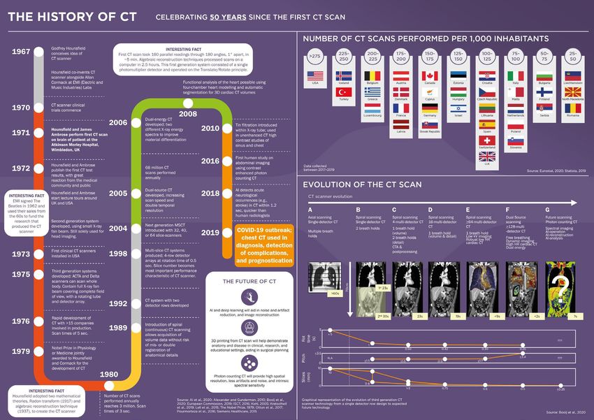
Contents

    +   EDITORIAL BOARD                                                     4

    +   WELCOME                                                             7

    +   FOREWORD                                                            9

    +   CONGRESS REVIEW
        Review of the European Congress of Radiology (ECR) 2021, 3rd–7th   12
        March 2021

    +   CONGRESS FEATURES
        Pairing CT and Laboratory Data to Predict Prognosis in COVID-19    22
        Katherine Colvin

    +   ABSTRACT REVIEWS                                                   24

    +   CONGRESS INTERVIEWS
        Prof Michael Fuchsjäger                                            40
        Dr Elizabeth Loney                                                 44
        Dr Philip Robinson                                                 46
        Prof Apostolos Karantanas                                          48
        Prof Olivera Nikolic                                               50

2       RADIOLOGY • April 2021                                                  EMJ
“Summaries of key abstracts presented at the
  congress are also included and are written by the
  presenters themselves, covering topics such as
  artificial intelligence analysis systems to support
  prostate cancer diagnostic imaging and a proposal
  for a new prognostic grading system in achalasia
  using dynamic barium swallow. ”

  Spencer Gore, CEO

   +    THE HISTORY OF CT                                                                   52

   +    FEATURES
        Artificial Intelligence in Radiology: An Exciting Future, but                       54
        Ethically Complex
        Adrian Brady
        An Ultrasound Phantom for Stenosing Flexor Tenosynovitis                            58
        Joseph Gartrell Willis et al.

   +    ARTICLES
        Editor’s Pick: Giant Cell Arteritis: Navigating Beyond the Headache                 66
        Harkins and Conway
        Breast Lesion Characterisation with Diffusion-Weighted Imaging Versus               75
        Dynamic Contrast-Enhanced-MRI: A Prospective Observational Study in
        a Tertiary Care Hospital
        Singh et al.
        Sonohysterography: A Formidable Diagnostic Tool in the Evaluation of                83
        the Caesarean Scar Defect in Comparison to MRI
        Amreen et al.
        Left Hemicolectomy for Intussusception of the Transverse Colon                      90
        Caused by a Giant Benign Lipoma
        Birmingham

Creative Commons Attribution-Non Commercial 4.0                         April 2021 • RADIOLOGY   3
Editorial Board
    Editor-in-Chief
    Dr Sophie Willis              City, University of London, UK

    Editorial Board
    Prof Dr Jean de la Rosette    Academic Medical Center (AMC), Netherlands
    Prof Eduard Ruiz-Castañé      Fundació Puigvert, Spain

    Prof Christian Jürgens        BG Trauma Hospital Hamburg, Germany

    Dr Olusola Michael Adeleke    NHS Clinical Entrepreneur Fellow, NHS England, UK

    Prof Roger Dmochowski         Vanderbilt University Medical Center, USA

    Prof Aad van der Lugt         Erasmus University Medical Center, Netherlands

    Dr Cetin Erol                 Ankara University, Turkey

    Dr Luke Dixon                 Imperial College Healthcare NHS Trust, UK

    Dr Sanjog Kalra               Einstein Medical Center, USA

    Dr Paul Bezzina               University of Malta, Malta

    Yasmeen Malik                 St George’s University of London, UK

    Dr Nicholas Kipshidze         New York Cardiovascular Research, USA

                                                                         VIEW IN FULL

4        RADIOLOGY • April 2021                                                         EMJ
Aims and Scope                                                    We are always keen to hear from healthcare professionals
                                                                    wishing to discuss potential submissions, please email:
  EMJ is an online only, peer-reviewed, open access general
                                                                    editorial.assistant@emjreviews.com
  journal, targeted towards readers in the medical sciences.
  We aim to make all our articles accessible to readers from
                                                                    To submit a paper, use our online submission site:
  any medical discipline.
                                                                    www.editorialmanager.com/e-m-j
  EMJ allows healthcare professionals to stay abreast of
                                                                    Submission details can be found through our website:
  key advances and opinions across Europe.
                                                                    www.emjreviews.com/contributors/authors
  EMJ aims to support healthcare professionals in
  continuously developing their knowledge, effectiveness,           Reprints
  and productivity. The editorial policy is designed to
  encourage discussion among this peer group.                       All articles included in EMJ are available as reprints
                                                                    (minimum order 1,000). Please contact
  EMJ is published quarterly and comprises review articles,         hello@emjreviews.com if you would like to order reprints.
  case reports, practice guides, theoretical discussions, and
  original research.
                                                                    Distribution and Readership
  EMJ also publishes 18 therapeutic area journals, which
  provide concise coverage of salient developments at               EMJ is distributed through controlled circulation to
  the leading European congresses. These are published              healthcare professionals in the relevant fields
  annually, approximately 6 weeks after the relevant                across Europe.
  congress. Further details can be found on our website:
  www.emjreviews.com                                                Indexing and Availability
                                                                    EMJ is indexed on DOAJ, the Royal Society of Medicine,
  Editorial Expertise
                                                                    and Google Scholar®; selected articles are indexed in
  EMJ is supported by various levels of expertise:                  PubMed Central®.
  • Guidance from an Editorial Board consisting of leading
                                                                    EMJ is available through the websites of our leading
    authorities from a wide variety of disciplines.
                                                                    partners and collaborating societies.
  • Invited contributors are recognised authorities from
    their respective fields.                                        EMJ journals are all available via our website:
                                                                    www.emjreviews.com
  • Peer review, which is conducted by EMJ’s Peer Review
    Panel as well as other experts appointed due to their
    knowledge of a specific topic.                                  Open Access
  • An experienced team of editors and technical editors.           This is an open-access journal in accordance with the
                                                                    Creative Commons Attribution-Non Commercial 4.0
  Peer Review                                                       (CC BY-NC 4.0) license.

  On submission, all articles are assessed by the editorial
  team to determine their suitability for the journal and           Congress Notice
  appropriateness for peer review.                                  Staff members attend medical congresses as reporters
                                                                    when required.
  Editorial staff, following consultation with either a
  member of the Editorial Board or the author(s) if
  necessary, identify three appropriate reviewers, who are          This Publication
  selected based on their specialist knowledge in the
  relevant area.                                                    ISSN 2633-9978

  All peer review is double blind.                                  EMJ Radiology is published once
                                                                    a year. For subscription details please visit:
  Following review, papers are either accepted without              www.emjreviews.com
  modification, returned to the author(s) to incorporate
  required changes, or rejected.                                    All information obtained by EMJ and each of the
                                                                    contributions from various sources is as current and
  Editorial staff have final discretion over any                    accurate as possible. However, due to human or
  proposed amendments.                                              mechanical errors, EMJ and the contributors cannot
                                                                    guarantee the accuracy, adequacy, or completeness of
  Submissions                                                       any information, and cannot be held responsible for
  We welcome contributions from professionals,                      any errors or omissions. EMJ is completely independent
  consultants, academics, and industry leaders on relevant          of the review event (ECR 2021) and the use of the
  and topical subjects.                                             organisations does not constitute endorsement or media
                                                                    partnership in any form whatsoever.
  We seek papers with the most current, interesting, and
  relevant information in each therapeutic area and accept          Front cover and contents photograph: Vienna, Austria,
  original research, review articles, case reports, and features.   home of the ECR 2021. © rh2010 / 123rf.com

Creative Commons Attribution-Non Commercial 4.0                                                  April 2021 • RADIOLOGY         5
EMJ Radiology
Chairman of Advisory Board                     Finance Manager
Prof Jonathan Sackier                          Antony Kindell
Chief Executive Officer                        Head of Recruitment
Spencer Gore                                   Karen Lee
Chief Commercial Officer                       Head of Operations
Daniel Healy                                   Keith Moule
Managing Director                              Operations Manager
Dan Scott                                      Nikki Curtis
Head of Marketing                              Operations Assistants
Marc Koskela                                   Satkartar Chaggar, Emma Knight,
                                               April McCaffrey
Head of Commercial
Michael McConaghy                              Editor
                                               Evgenia Koutsouki
Performance Manager
Darren Brace                                   Deputy Managing Editor
                                               Sam Davis
Senior Project Managers
Kelly Byrne, Hayley Cooper, Nabihah Durrani,   Content & Editorial Assistants
Millie McGowan, Max Roy                        Michaila Byrne, Isabel O’Brien
Client Services Senior Project Managers        Content Assistant
Vanessa Frimpong, Alexander Skedd,             Cheyenne Eugene
Caleb Wright
                                               Editorial Co-ordinators
Project Managers                               Katherine Colvin, Anaya Malik
Emilie De Meritens, Tilly Flack, Antonio
Grier, Robert Hancox, Rebecca Harrison,        Editorial Assistants
Andrew Hodding, Mark Kirwan, Lewis Mackie,     Janet Nzisa, Louise Rogers, Theo Wolf
Thomas Madden, Jack Moore, Billy Nicholson,    Design Managers
Aleksandar Popovic                             Tian Mullarkey, Stacey Rivers
Client Services Executive Project Manager      Graphic Designers
Mariana Napoleao                               Gennaro Draisci, Roy Ikoroha, Emma Rayner
Client Services Associate Project Managers     Junior Designer
Jessica Alcock, Andrew Le Baigue               Steven Paul
Sales Administrator
Simi Ige                                       Digital and Data Innovation Manager
                                               Louis Jonesco
Head of Client Services
Courtney Jones                                 Marketing Co-ordinator
                                               Noah Banienuba
Head of Special Projects
Jayne Logan                                    Business Analyst
                                               Rajdeep Bhangoo

6    RADIOLOGY • April 2021                                                                EMJ
Welcome
  Dear Readers,                                                 technology in medical imaging, with several ECR
                                                                sessions presenting and embracing this new era.
  Welcome to the second issue of EMJ Radiology
  following a successful launch last year! This                 We also spoke with the European Society
  open-access eJournal covers the most important                of Radiology (ERS) President, Prof Dr
  developments in radiology through interviews                  Michael Fuchsjäger, and four ESR Scientific
  and articles from experts within the field and a
                                                                Subcommittee Chairpeople: Dr Elizabeth Loney,
  congress review presenting the highlights from
                                                                Dr Philip Robinson, Prof Apostolos Karantanas,
  the European Congress of Radiology (ECR) 2021.
                                                                and Prof Olivera Nikolić. The interviewees spoke
  As the world adapts to virtual means of                       about their respective areas of expertise and
  communication, this year, the ECR took place                  what the future of radiology holds.
  both virtually and in Vienna, Austria. Hundreds of
  presentations, studies, and even an unexpected                Peer-reviewed articles included in this issue
  yoga session were delivered. Summaries of                     present the latest developments in the field.
  key abstracts presented at the congress are                   Singh et al. deliver a prospective observational
  also included and written by the presenters                   study on breast lesion characterisation with
  themselves, covering topics such as artificial                diffusion-weighted imaging versus dynamic
  intelligence analysis systems to support prostate             contrast-enhanced MRI; Birmingham discusses
  cancer diagnostic imaging and a proposal for
                                                                left hemicolectomy for intussusception of the
  a new prognostic grading system in achalasia
                                                                transverse colon caused by a giant benign
  using dynamic barium swallow.
                                                                lipoma; and, Conway and Harkins discuss
  With countries worldwide coping with the                      medical imaging in giant cell arteritis.
  coronavirus disease (COVID-19) pandemic, the
  effects of this novel coronavirus in radiology                I would like to take this moment to thank all the
  was a major topic this year. Another topic was                contributors, Editorial Board, and editorial team
  the advancement in artificial intelligence and 3D             for their help in creating this special eJournal.

                          Spencer Gore
                          Chief Executive Officer, EMG-Health

Creative Commons Attribution-Non Commercial 4.0                                        April 2021 • RADIOLOGY   7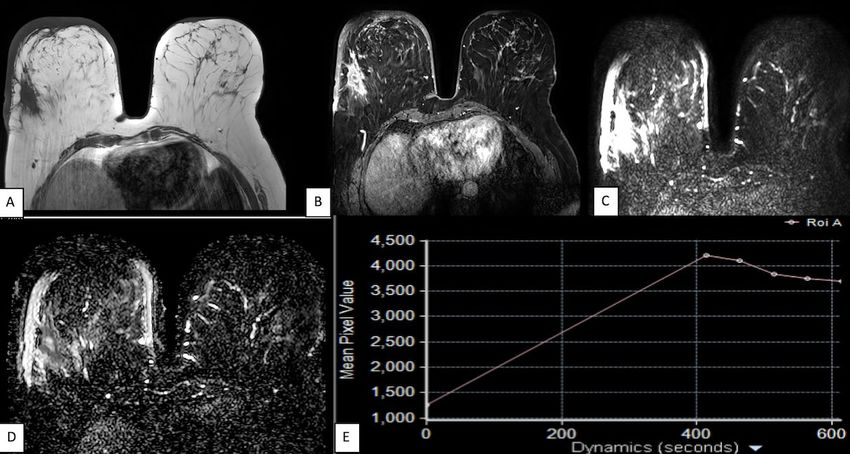
Want a daily dose
of healthcare?
Sign up for free to our daily newsletter
to feed your inbox with regular
healthcare updates.

                                                 EMJREVIEWS.COM   /SUBSCRIBE
‘The go to place for healthcare professionals’
Foreword
  Dear Readers,                                               improve image quality, can be found in the
                                                              following Congress Review.
  It is with great pleasure that I welcome you to
  the latest issue of EMJ Radiology following the             Key members of the European Society of
  success of the first launch last year, bringing             Radiology (ESR) were interviewed about
  you the latest developments in radiology. They              the impact of COVID-19 on radiology, facial
  say that ‘change is the only constant’, and                 trauma    imaging,    genitourinary   imaging,
  this has been displayed by the scientists and               and much more. Notable areas of clinical
  healthcare professionals (medical, nursing, as              interest provided in this journal include left
  well as allied health care professionals such               hemicolectomy for intussusception of the
  as radiographers) who have worked tirelessly                transverse colon caused by a giant benign
  to adapt with the changing times. This open-                lipoma and sonohysterography use in the
  access eJournal will deliver these developments             evaluation of the caesarean scar.
  brought about by leading experts following the
  European Congress of Radiology (ECR) 2021.                  The paper that I have selected as the Editor’s
                                                              Pick covers CT and MRI angiography, temporal
  The implications of the coronavirus disease                 artery biopsy, ultrasound, and PET scanning
  (COVID-19) meant that not everyone was able                 for giant cell arteritis and the challenges faced
  to attend the congress this year, which is why it           in the diagnosis pathway, a topic that would
  was delivered virtually and in Vienna, Austria. It          interest radiographers too.
  showcased the latest innovations in radiology
  from all over the world, covering interesting               I hope that you all enjoy reading EMJ Radiology,
  topics and challenges in the field by bringing              an issue that should be of great interest to
  together international authorities. Highlights              a wide range of healthcare professionals,
  from the event, including an in-depth review                including radiologists and diagnostic and
  of a session from ECR on deep learning to                   therapeutic radiographers.

                          Yasmeen Malik
                          Senior Lecturer, St George’s University of London, London, UK

Creative Commons Attribution-Non Commercial 4.0                                           April 2021 • RADIOLOGY   9
Available now.   EMJ 6.1 2021
                 Feature
                 Rare 2030 Final Policy Conference: Summary of the Recommendations of the
                 Rare 2030 Foresight Study

                 Interviews
                 Addressing the Unmet Need in Treatment of Nonmelanoma Skin Cancers:
                 Interviews with Two Key Opinion Leaders

                 The Treatment Landscape of Atopic Dermatitis: Interviews with Three
                 Consultant Dermatologists

                 Is CD37-Targeted Therapy a Viable Alternative in the Treatment of Diffuse Large
                 B-cell Lymphoma?

                 Articles
                 Editor’s Pick: The Correlation Between Stroke and Coronavirus Disease
                 (COVID-19): Where is the Evidence?

                 Emerging Treatments for Crohn’s Disease: Cells, Surgery, and Novel
                 Therapeutics

                 Pancreatic β Cell Senescence: Mechanisms and Association with Diabetes

                 And even more...

                 Subscribe for free.

                    EMJREVIEWS.COM     /SUBSCRIBE
Congress Review
Review of the European Congress of
Radiology (ECR) 2021
Location: ECR 2021
Date: 3rd–7th March 2021
Citation: EMJ Radiol. 2021;2[1]:12-21. Congress Review.
V
IENNA’S musical culture was a by Prof Fuchsjäger, embracing is an
central element throughout the all-encompassing word and radiology
opening ceremony of this year’s is the one thing that attendees
European Congress of Radiology (ECR), collectively embraced.
which opened to the music of Johan
In addressing the audience during the
Strauss’s 'Die Fledermaus', performed by
opening ceremony, Prof Fuchsjäger
the Philharmonic Five. The musical element
emphasised that “At the outbreak of
continued to manifest itself as the musicians
a global pandemic, the ESR chose
performed classical pieces throughout the
to embrace the innovative and wide-
ceremony ranging from 'Giuditta' to 'Don
reaching opportunities that only learning
Giovanni'. Although the coronavirus disease
can provide.”
(COVID-19) pandemic forced yet another
congress to switch to a virtual setting, The ESR online platform offered five
Vienna’s character and style was diffused parallel streams with a plethora of sessions
throughout the congress. and themes to choose from, which made
navigation an engaging and stimulating
In what now seems to be an ironic twist of experience for attendees. Channel 1
fate due to the COVID-19 pandemic, last contained live broadcasts from a dedicated
year the European Society of Radiology studio in Vienna acting as a hub and
(ESR) President Prof Michael Fuchsjäger hosting pop-up events from academic
chose ‘embracing’ as the main theme of this and industry partners in 20 different
year’s ECR. Underpinning this theme cities around the world. This channel also
was Gustav Klimt's painting 'The Kiss’, broadcast classes of yoga, mindfulness,
which served as the signature image dance, and Aikido, in line with the special
of the congress. As elegantly put theme of this year’s congress: wellbeing.Fuchsjäger characterised as “a
“At the outbreak of a global pandemic, luminary of exceptional style
the ESR chose to embrace the innovative and wisdom.”
and wide-reaching opportunities that The three recipients for this
only learning can provide.” year’s Gold Medal of the
ESR were Prof Fiona Gilbert,
Cambridge, UK; Prof Paul M.
Prof Fuchsjäger expressed his belief that “if
Parizel, Perth, Australia; and Prof Francesco
we can remain resilient and focussed in the face
Sardanelli, Milan, Italy. The award is presented
of adversity, we can serve our patients even
to outstanding scientists who work in radiology
more effectively.”
or life sciences and have been active in the
Ranging from plenary lectures and workshops organisation and establishment of radiology in
to new horizons sessions and industry symposia, national or international organisations. Each of
this congress had something for everyone. With the recipients chose a favourite piece of music to
presentations on artificial intelligence, radiomics, be played at the ceremony, which ranged from
thera(g)nostics, and Star Wars-themed image cool jazz to Tchaikovsky. Combining an element
interpretation quizzes, there was a wide range of of surprise with a touch of Viennese culture, the
topics and sessions to choose from. Of course, opening ceremony closed with Prof Fuchsjäger
there was an abundance of sessions on COVID-19, joining the soprano Valentina Nafornita to sing a
ranging from its radiological manifestations, duet from 'Don Giovanni'.
diagnosis, and the use of artificial intelligence. This was a truly memorable congress, hosted on
a highly innovative platform paving the way for
This year’s recipients of honorary membership,
more virtual events organised by the ESR. In the
which is awarded to individuals for their
closing ceremony, Prof Fuchsjäger emphasised
scientific excellence, international reputation,
that “we adapt, we innovate, and we embrace
and achievements in national or international
opportunities, no matter what form our annual
radiology, were Prof Sanjiv Sam Gambhir,
meeting might take.”
Stanford, California, USA; Prof James P.
Borgstede, Colorado, Denver, USA; and Prof Pek- In this issue of EMJ Radiology our team has
Lan Khong, Hong Kong, SAR, China. A particularly covered a selection of the topics from ECR,
touching tribute was paid to Prof Gambhir aiming to impart some of the knowledge shared
who sadly passed away in July 2020, who Prof at the congress. ■
ECR 2021 REVIEWED
14 RADIOLOGY • April 2021 EMJCardiovascular
Magnetic Resonance in
Sudden Cardiac Death
S
UDDEN cardiac death is unexpected that quantification of grey zone on LGE images
mortality occurring as a result of cardiac accurately identified a group at high risk of
causes and within a short time period. future arrhythmia. Dr Cochet emphasised:
The majority of these events are attributable to “This strategy clearly outperformed the one
ventricular arrhythmias. Underlying structural based on LVEF measurements, which was
cardiac abnormalities, including postinfarction poorly discriminate.”
scar tissue, are found in most cases of
sudden cardiac death. During the ECR On the topic of nonischaemic dilated
2021 Master Class titled ‘Cardiac cardiomyopathy, Dr Cochet stated:
Imaging in Arrhythmia And “As opposed to ischaemic heart
Sudden Cardiac Death’, Dr “This disease, in which LGE may
Hubert Cochet, University of strategy clearly recruit additional participants
Bordeaux, Bordeaux, France, for implantable cardioverter
outperformed the
discussed the role of late defibrillator implantation, LGE
one based on LVEF in dilated cardiomyopathy
gadolinium enhanced (LGE)
cardiac magnetic resonance measurements, may be more useful to prevent
(CMR) in scar characterisation. which was poorly unnecessary implantations
discriminate.” in a subset of patients who
The current approach for the are currently considered for
primary prevention of sudden implantation but whose risk
death relies on the measurement of profile is rather low.” A further
left ventricular ejection fraction (LVEF). series of studies have illustrated that LGE
Dr Cochet noted that this methodology cannot qualifications could also be used in hypertrophic
be considered satisfactory because a large cardiomyopathy to improve primary prevention.
proportion of patients dying suddenly remain
undetected, with >80% of them having LVEF Lastly, the importance of CMR in the aetiological
>35%. To overcome LVEF limitations, analysis diagnosis of patients presenting with ventricular
of LGE CMR images has been proposed. arrhythmia was discussed. Dr Cochet stressed
Dr Cochet highlighted a recent study that that CMR should be performed even when
monitored approximately 1,000 patients with first-line echocardiography and angiography
coronary artery disease over 6 years and found are negative. ■
Creative Commons Attribution-Non Commercial 4.0 April 2021 • RADIOLOGY 15Functional MRI for Assessment of Cervical Cancer
T
HE USE of functional MRI (fMRI) in the
evaluation of cervical cancer is increasing.
While standard MRI and conventional “In
imaging techniques, such as ultrasound helping
and CT, are effective at detecting delineate small
tumours that show anatomical distortional tumours, such as
and changes in tissue appearance, they
often fail to identify the small-volume
adenocarcinoma of
active tumours, either when they first the cervix, dynamic
present or in early disease relapse; contrast-enhanced
the anatomic and functional imaging (DCE) MRI is
ability of fMRI makes this possible. Prof favourable”
Evis Sala, Honorary Consultant Radiologist,
Addenbrooke’s Hospital, Cambridge, UK, spoke
in a session series on the topic of fMRI in cervical
cancer, with particular emphasis on treatment
selection, planning, and monitoring.
In helping delineate small tumours, such
as adenocarcinoma of the cervix, dynamic
contrast-enhanced (DCE) MRI is favourable,
especially in patients eligible for trachelectomy
(fertility-sparing procedure), due to its ability
to noninvasively characterise tissue vasculature.
DCE-MRI is also a useful tool in treatment
planning (radiotherapy versus surgery) and in
evaluating treatment response more readily than
assessing tumour size.
There is a growing interest in the use of diffusion-
weighted MRI (DWI) in combination with T2-
weighted images to measure early treatment
response. This is due to the imaging ability
to observe reduced profusion in the tumour
and is more effective than pattern-monitoring
early response to treatment. Prof Sala advised
not to use positron emission tomography at
treatment completion (3–6-month window),
because inflammation and nonspecific glucose
uptake give the possibility of false positives;
here both DCE-MRI and DWI are more suited. In
terms of detecting tumour recurrence, DWI also
proves effective at identifying small, subtle areas
of tumour.
Prof Sala also mentioned her team’s success
in using a combination of positron emission
tomography and fMRI for outcome prediction,
with both systems serving as disease-free and
overall survival biomarkers. ■
16 RADIOLOGY • April 2021 EMJGadolinium Deposition: An Update
G
ADOLINIUM-based contrast agents Prof Stojanov highlighted that there is currently
(GBCA) are diagnostic pharmaceutical no clinical evidence of adverse health effects
compounds containing paramagnetic specifically related to gadolinium accumulation
gadolinium ions that affect the magnetic in the human brain: “In my opinion, future
resonance signal properties of surrounding research is needed to determine the mechanism
tissue. GBCA are commonly used in clinical by which gadolinium deposits within the human
practice because of their fundamental ability tissue and whether this leads to clinically
to selectively decrease the T1 relaxation time significant sequelae.” ■
within a lesion rather than in normal tissue.
However, during the ECR 2021 Special Focus
Session titled ‘Can We Limit Gadolinium Use
in Neuroimaging?’, Prof Dragan Stojanov,
Centre for Radiology, Clinical Centre of Niš, Niš,
Serbia, pointed to several recent studies that
demonstrated gadolinium neurodeposition in
patients with normal renal function. Specifically,
gadolinium deposition has been recorded in
brain structures such as the dentate nucleus
and globus pallidus. These findings were
confirmed in animal models.
Prof Stojanov explained that GBCA can
be divided into two structurally distinct
categories: linear (open chain) and macrocyclic
chelates. The current prevailing theory is that
the degree of deposition depends on the
molecular structure of the contrast agent
used. In particular, dissociation depends on the
kinetic and thermodynamic stability of GBCA.
The gadolinium ions of macrocyclic GBCA
are caged in the cavity of ligands and this is
believed to underlie the heightened stability of
macrocyclic chelates relative to linear chelates.
The gradual dissociation of accumulated
linear GBCA in the brain allows gadolinium
to bind proteins and other macromolecules
and this process is ultimately responsible
for neurodeposition.
On the basis of these findings,
Prof Stojanov revealed that the “In my opinion, future research is needed
European Medicines Agency to determine the mechanism by which
(EMA) recommended the
gadolinium deposits within the human
suspension of all commercially
available linear GBCA. tissue and whether this leads to clinically
Consequently, many of the less significant sequelae.”
stable chelates have been removed
from routine clinical use in
recent years.
Creative Commons Attribution-Non Commercial 4.0 April 2021 • RADIOLOGY 17How Can Cone-beam CT
Change Clinicians’ Practice
in Interventional Radiology?
T
HE MAINSTAY of interventional radiology navigation, monitoring of treatment, as well as
has been 2D angiography, but this has been final results assessment.
thwarted by its low contrast resolution. This
poses a challenge for small lesions and vessel Excellent training of the patient and staff is vital
visibility, resulting in having to take multiple to the execution of imagery. Cone-beam CT can
angiograms, which increases radiation show the lesion vascular territory at the
dose. Imaging hybrid systems time of the procedure and offers
can bridge the gap and give multimodality fusion, with therapy
excellent results, but the costs “Innovative planning needed for interarterial
and workflow implications or percutaneous ablation
equipment and
are considered too great techniques. These qualities
a challenge. Cone-beam
further development of are incredibly valuable as
CT has been around software and hardware clinicians can automatically
since the early 1990s and for this imaging technique identify vessels to be
earliest applications were improve speed accuracy targeted; these vessels can
for vascular imaging and and safety procedures, be extracted and lesions
angiography. At ECR 2021, Dr targeted.
Raman Uberoi, University of
ultimately improving
Oxford, Oxford, UK, delivered a patient outcomes.” While the earliest uses of this
presentation entitled ‘How cone- 3D imaging were for angiography,
beam CT can change your practice it can be extremely useful in
in interventional radiology’ as part identifying and targeting branch
of a Master Class on new diagnostic tools for vessels as well as problems that might
vascular diseases. compromise stent graft.
As things have progressed over the last 15–20 Cone-beam CT is now widely available from
years, scan times have reduced and multiphasic many manufacturers and its use can be extended
acquisitions can be obtained. The result is that to an increasing range of areas including
cone-beam CT provides 3D volume images information retrieval. Innovative equipment and
with great contrast resolution. With cone-beam further development of software and hardware
CT, images can be combined with angiographic for this imaging technique improve speed
images; the correct software allows for accuracy and safety procedures, ultimately
treatment planning, catheter and needle improving patient outcomes. ■
18 RADIOLOGY • April 2021 EMJRadiographers and Patient-Centred Care
I
N THE PAST decade, there has been a paradigm
shift in healthcare systems to suggest that
patient satisfaction has become an important
factor for healthcare professionals to consider.
Patient-centred care means the needs of the “Quality
patient are addressed, their questions are improvement
answered effectively, and the overall patient methodologies
experience is improved. In medical imaging, can positively impact
image quality, radiation dose, and other
technical aspects are essential; however, patient
patience experience
centred-care was a key subject addressed at and appreciate how
ECR 2021. patient-centred care
A quality improvement methodology research
has an impact on
study from Warrington and Halton Hospitals radiographic
NHS Trust, Warrington, UK, led by radiographers practice.”
Drs Louise Harding and Paula Evans, titled
‘Always about’, was introduced at this year’s
congress. This methodology uses the Plan-
Do-Study-Act (PDSA) cycle by encouraging
honest and transparent feedback from
patients and reviewing this feedback to make
the required changes. The aim was to inspire
radiographers: to increase patient engagements
in their treatment, listen to patients’ needs and
concerns, avoid making assumptions, address
gaps in the quality care systems, and to improve
overall radiographic practice and teamwork.
Dr Andrew England, senior lecturer at Keele
University, Keele, UK, outlined the role of the
European Federation of Radiography Societies
(EFRS) and its initiatives for promoting patient-
centred care across Europe. The EFRS is the
European voice of radiographers and currently
represents over 100,000 radiographers and
8,000 students. It recognises 40 national
societies and over 60 radiography educators
from Europe and beyond. The core belief of
the society is essentially putting patients first
by finding ways to enhance quality of care in
medical imaging and radiography.
Studies have shown that by bringing the
patient to the centre of defining their care
needs, the safety and effectiveness of care can
improve significantly. Quality improvement
methodologies can positively impact patience
experience and appreciate how patient-centred
care has an impact on radiographic practice. ■
Creative Commons Attribution-Non Commercial 4.0 April 2021 • RADIOLOGY 19The ALARA Principle: Dose Reduction
in CT and Radiography for Children
S
PECIAL training is required when utilising CT scans are essential in acute trauma for 3D
medical radiation in paediatric patients. imaging of deformities and assessment of
The ‘ALARA’ principle (which stands for post-traumatic complications causing plate
‘as low as reasonably achievable’) is a safety fusion or bone infection. Dr Kraft, a specialist
concept used to protect patients from in musculoskeletal radiology,
excessive exposure to ionising discussed how medical imaging
radiation. In radiography, procedures need to be
certain exposure techniques justified and optimised for
are required when treating radiological protection,
small children thanks to especially in children. An
their weight–mass ratio. “...medical imaging example was shared to
A session was presented procedures need to be explain that although
at ECR 2021 by Dr justified and optimised for CT may be appropriate
Erich Sorantin, Medical
University of Graz, Graz,
radiological protection, to assess a triplane
especially in children.” fracture of the ankle, it is
Austria, and Dr Jeannette
not needed for all ankle
Kraft, Leeds Teaching
fractures in children.
Hospitals NHS Trust, Leeds,
UK, on avoiding or reducing Ultrasound was suggested
ionising radiation in children. as the first-line imaging
Prof Sorantin started the session investigation in “lumps and
by evaluating how to improve the benefit bumps” and nontraumatic joint pain.
and risk ratio of children in radiography and Possibilities for low-dose applications in
CT by adapting exposure and dose settings skeletal imaging were presented in this session
to paediatric needs. Tips were shared on at ECR 2021. A good example was shown
how patient positioning and using the slice in which a low-dose video graphic system
thickness approach in radiography is essential allows for total body imaging in a natural,
in dose reduction. It was suggested that a weight-bearing position, producing viewable
child with spinal deformity or scoliosis could 2D images and 3D models. Dr Kraft affirmed
have an MRI scan first, perhaps followed by a its use for imaging of scoliosis or lower
focussed CT scan of any bony fusion anomaly. limb deformities. ■
20 RADIOLOGY • April 2021 EMJUsing Artificial Intelligence to
Improve Radiomics in Breast Imaging
R
ADIOMICS, a relatively new field in whether automated classification of benign
radiology, is the method of extracting and malignant breast lesions in ultrafast breast
large numbers of quantitative features MRI using deep learning showed a reduction
from medical images using data characterisation in the number of false-positive biopsies. The
algorithms and was the topic of discussion team analysed 576 lesions imaged with MRI
at an ECR 2021 Master Class. Dr Riste Mann, and applied deep learning methods. The results
Radiologist, Radbound University Medical Center, reported that 19 fewer false positives were
Nijmegen, the Netherlands, took to the screen produced with the AI system than the number of
in the final lecture of the Master Class to discuss biopsied benign lesions in the authors' database,
the use of artificial intelligence (AI) in improving suggesting that AI-based classifications can
breast imaging ‘-omics’. help radiologists interpret MRI images to
improve specificity.
AI can be divided into different subfields
depending on the particular goals and tools, The potential use of AI to predict response to
such as machine learning and robotics. Deep chemotherapy during treatment was also briefly
learning represents a subfield of machine discussed, however this application is not as
learning that structures algorithms in layers to near in the future as lesion prediction. One day,
create a convolutional neural network (CNN); AI in breast imaging will have the capability
in radiology, a CNN uses image inputs to learn to segment, identify, and classify lesions,
and recognise image features and, as a result, predict chemotherapy response, and provide
makes intelligent decisions and interprets a prognosis, all in the same algorithm. “By that
radiographic images independently. In the time, our jobs will have changed dramatically,”
case of breast imaging, CNN are en route to concluded Mann. “We might have somehow all
classifying breast lesions never seen before by become oncologists.”
the system, simply by using the information
obtained from prior cases. Traditional breast A limitation to CNN is that they require large pools
MRI is high-dimensional and multiparametric in of training images before achieving a clinically
nature, making interpretation labour intensive useful performance. Future studies should focus
and complex with the potential of interobserver on collecting larger datasets to further train AI
variability. Therefore, Dr Mann, along with systems and create deeper CNN structures for
a team of researchers, set out to uncover improved learning and classification. ■
“One day, AI in breast imaging will have the capability
to segment, identify, and classify lesions, predict
chemotherapy response, and provide a prognosis, all in
the same algorithm.”
Creative Commons Attribution-Non Commercial 4.0 April 2021 • RADIOLOGY 21Pairing CT and
Laboratory Data to
Predict Prognosis
in COVID-19
Katherine Colvin
Editorial Coordinator
Citation: EMJ Radiol. 2021;2[1]:22-23.
P
REDICTING outcome for patients can help to direct management decisions, care and
surveillance requirements, and align patients and their families with the expected
clinical course; however, predicting prognosis for new, unknown conditions like
coronavirus disease (COVID-19) presents a challenge. In a shared session with the European
Federation of Clinical Chemistry and Laboratory Medicine (EFLM) at ECR 2021, Prof Salvatore
Cappabianca, Department of Precision Medicine, University of Campania, Naples, Italy,
shared study findings evaluating the role for chest CT alongside clinical and laboratory
assessments to help predict outcome in patients with COVID-19.
In the early period of the pandemic, Prof PCR nasopharyngeal swab, with admission chest
Cappabianca explained, studies examined the CT usually performed after confirmed COVID-19
use of CT imaging for diagnosis of COVID-19; diagnosis. The chest CT protocol was performed
however, evolution of clinical thinking moved to with supine positioning, without intravenous
support consideration of COVID-19 as a general contrast, and imaged lung apices to bases using
viral infection, where the role of CT imaging is in a standard dose scanning protocol (1.25 mm
defining extent of disease rather than diagnosis. thickness, 1 mm interval reconstruction). Two
He discussed his retrospective, univariate, and experienced radiologists, blinded to clinical
multivariate analysis, which aimed “to clarify and laboratory findings, defined the presence
the place of the CT scan in the management of and extension of ground-glass opacities and
COVID-19 patients, define the possibilities of CT areas of consolidation using a guideline for
alone in prediction of patients’ outcome, and to structured reporting, to provide a visual score
compare lung CT impairment with clinical data to as a severity index. This visual score reporting
improve performance in outcome prediction.” was supplemented with an artificial intelligence
tool, which reported volumes of residual healthy
The study analysed data from 103 patients
lung parenchyma, ground-glass opacities,
presenting to hospital: 41 female and 62 male,
consolidation, and emphysema.
aged 29–93 years. Patients had confirmed
severe acute respiratory syndrome coronavirus 2 Both univariate and multivariate analyses
(SARS-CoV-2) infection via reverse transcription were performed, incorporating data from
22 RADIOLOGY • April 2021 EMJThe best prediction of prognosis, however, came with
combining clinical, laboratory, and CT findings.
clinical and laboratory assessment (including tools in the evaluation of the evolution of
oxygen saturation, procalcitonin, D-dimer, disease,” outlined Prof Cappabianca; volume
and troponin), clinical picture (such as fever, of ground glass opacities was not a statistically
cough, and presence of respiratory failure), significant predictor. The visual severity score
and comorbidities (hypertension, diabetes, also demonstrated good correlation with
cardiac and/or lung pathologies, neurological patient outcome.
pathologies, and neoplasm). Outcomes were
assessed as discharge home, hospitalisation The best prediction of prognosis, however,
in stable condition, and hospitalisation in came with combining clinical, laboratory, and
critical condition. CT findings. The study found that this combined
approach yielded a sensitivity, specificity, and
Analysis found that age, oxygen saturations, accuracy for predicting outcome of 88%, 78%,
C-reactive protein, leukocyte and neutrophil and 81%, respectively.
count, lactate dehydrogenase, D-dimer,
troponin, creatinine and azotemia, alanine Prof Cappabianca summarised the conclusions
aminotransferase, aspartate aminotransferase, of his study for the use of chest CT in
and bilirubin were the major clinical and prognostication for COVID-19: “our experience
laboratory findings that defined the outcome of suggests the usefulness of visual quantification
the patient; comorbidities and symptomology of involved lung as a predictor of the outcome
showed no significant relationship with the in patients affected by coronavirus disease.
evolution of the COVID-19 pathology. Comparison with clinical and laboratory data
provides higher correlation with prediction and
As determined by the artificial intelligence software-based quantification of consolidation,
tool, lung volumes of emphysema, residual emphysema, and residual normal lung on
healthy lung parenchyma, and consolidation chest CT are independent predictors of
were shown to be “highly important predictive COVID-19 evolution.” ■
Creative Commons Attribution-Non Commercial 4.0 April 2021 • RADIOLOGY 23Abstract Reviews
Sharing insights and updates from a selection
of abstracts presented by leading experts in
the field of radiology at the European Congress
of Radiology (ECR).
Fully Automated Keywords: Artificial intelligence, cancer, MRI, neural
Segmentation of networks, oncology.
Citation: EMJ Radiol. 2021;2[1]:24-26. Abstract Review
Neuroblastic Tumours No. AR1.
on Multisequence MRI
Using Convolutional BACKGROUND
Neural Networks Volumetric segmentation of intrinsically
heterogeneous abdominal tumours is
essential for the diagnosis, follow-up,
Authors: *Leonor Cerdá-Alberich,1 Diana and treatment response evaluation of
Veiga-Canuto,2 Luis Martí-Bonmatí1,2,3 neuroblastomas.1 Manual segmentation of
neuroblastic masses is a tedious and time-
1. Biomedical Imaging Research Group, La Fe Health
consuming task that hinders the radiologists’
Research Institute of Valencia, Valencia, Spain
2. Radiology Department, The University and workflow. Different studies explored
Polytechnic La Fe Hospital, Valencia, Spain semiautomatic segmentation algorithms
3. Clinical Department of Medical Imaging, The in CT images, making use of mathematical
University and Polytechnic La Fe Hospital, morphology, fuzzy connectivity, and other
Valencia, Spain image processing tools.2,3
*Correspondence to leonor_cerda@iislafe.es
As of today, a robust and generalisable
Disclosure: The authors have reported payments to
their respective institutions.
solution does not exist for childhood
neuroblastoma. In this study, the authors
Acknowledgements: The authors acknowledge the propose an automated segmentation
support of PRIMAGE (PRedictive In-silico Multiscale method based on convolutional neural
Analytics to support cancer personalised diaGnosis
networks applied to children with neuroblastic
and prognosis, empowered by imaging biomarkers).
Horizon 2020 | RIA (Topic SC1-DTH-07-2018) project tumours studied with multiple MRI
with grant agreement no: 826494. sequences. The aim is to extract reproducible
quantitative imaging biomarkers from
24 RADIOLOGY • April 2021 EMJthese lesions for the prediction of relevant using majority voting to generate the final 3D
clinical outcomes. neuroblastoma segmentation (Figure 1). The
networks were trained using the Adam optimiser
MATERIALS AND METHODS with 300 epochs and a batch size of 100. The
number of layers, number of convolutional
T1-weighted (T1W), T2-weighted (T2W), feature maps, and learning rates were chosen
and diffusion-weighted (DW) MRI images at based on hyperparameter tuning. In addition,
diagnosis were collected from 127 patients a scheduler was used to set the learning rate
with neuroblastoma from different European in each epoch, starting from an initial value
hospitals in the scope of the H2020 PRIMAGE of 0.001 that reduced by a factor of 0.5 if the
project.4 Images were subject to high variability validation Dice plateaued for 20 epochs. The
in data acquisition due to different scanners, model was assessed by using a 5-fold cross-
manufacturers, and protocols. validation strategy and comparing the results
with other state-of-the-art solutions.
The authors developed a multisequence and
multiplanar U-net.5 In order to justify the
use of multisequence MRI, an experiment RESULTS
was performed with different inputs in a
subset of randomly selected patients. For The Dice similarity coefficient values of
the multiplanar approach, the sagittal and the method using only T1W, T2W, DW, and a
coronal planes from the original axial MRI were multisequence approach, having the 5-fold
reconstructed and fed into three separate cross-validation as different inputs, were
neural networks. The segmentations from the 0.732±0.064, 0.745±0.118, 0.786±0.077, and
three two-dimensional networks were fused 0.841±0.038, respectively.
Image
Original image reconstruction
Training dataset
Axial Coronal Sagittal
CNN CNN CNN
CNN training
Axial-based Coronal-based Sagittal-based
prediction prediction prediction
Fuse and
majority
voting
Figure 1: Schematic diagram of the multiplanar scheme used to develop a fully automated segmentation model of
neuroblastic tumours.
CNN: convolutional neural network.
Creative Commons Attribution-Non Commercial 4.0 April 2021 • RADIOLOGY 25The average Dice similarity coefficient of the cancer. An artificial intelligence-based algorithm
internal validation using the multisequence to overcome this limitation may benefit the
strategy was found to be 0.830, showing model segmentation process. ■
robustness and stability across different sites
and scanners.
References
1. Maris JM. The biologic basis for neuroblastoma
CONCLUSION heterogeneity and risk stratification. Curr Opin Pediatr.
2005;17(1):7-13.
The authors proposed a fully automated 2. Deglint HJ et al. Three-dimensional segmentation
of the tumor in computed tomographic images of
segmentation method of neuroblastic tumours neuroblastoma. J Digit Imaging. 2007;20(3):263-78.
based on convolutional neural networks and 3. Rangayyan RM et al. Landmarking and segmentation
multisequence MRI with an accurate and stable of computed tomographic images of pediatric patients
with neuroblastoma. Int J Comput Assist Radiol Surg.
performance. If further improved and externally 2009;4(3):245-62.
validated the proposed method could be of use 4. Martí-Bonmatí L et al. PRIMAGE project: predictive
in clinical trials and oncologic practice for the in silico multiscale analytics to support childhood
cancer personalised evaluation empowered by imaging
management of neuroblastoma. Future work biomarkers. Eur Radiol Exp. 2020;4(22).
with a larger sample will be necessary to evaluate 5. Ronneberger O et al. U-Net: convolutional networks
the generalisability of the model. In addition, the for biomedical image segmentation. Medical Image
Computing and Computer-Assisted Intervention. Lecture
co-registration of T1W, T2W, and DW images Notes in Computer Science. 2015;9351.
is still particularly challenging in paediatric
The Impact of the
Disclosure: The authors have declared no conflicts
COVID-19 Pandemic of interest.
on Adult Diagnostic Acknowledgements: The authors would like to thank
the European Society of Neuroradiology (ESNR) for
Neuroradiology in its support of the ESNR diagnostic committee and the
preparation and distribution of the survey.
Europe Keywords: Coronavirus disease (COVID-19),
pandemics, personal protection equipment, surveys,
questionnaires.
Authors: *Marion Smits,¹ Meike Vernooij,1,2
Nuria Bargalló,³ Ana Ramos,⁴ Tarek Yousry⁵ Citation: EMJ Radiol. 2021;2[1]:26-27. Abstract Review
No. AR2.
1. Department of Radiology and Nuclear Medicine,
Erasmus MC, University Medical Centre Rotterdam,
Rotterdam, the Netherlands
2. Department of Epidemiology, Erasmus MC,
University Medical Centre Rotterdam, Rotterdam,
PURPOSE
the Netherlands
3. Magnetic Resonance Image Core Facility, IDIBAPS The purpose of this survey was to understand
and Centre of Diagnostic Image (CDIC), Hospital the impact the coronavirus disease (COVID-19)
Clinic, Barcelona, Spain
pandemic has or has had on the work, training,
4. Department of Radiology, Hospital Universitario 12
de Octubre, Madrid, Spain and wellbeing of professionals in the field of
5. Lysholm Department of Neuroradiology, National diagnostic neuroradiology. These insights may be
Hospital for Neurology and Neurosurgery, used to prepare or improve strategies for similar
University College London Hospitals NHS Trust,
situations and address potential needs and
London, UK
*Correspondence to marion.smits@erasmusmc.nl worries that arose from the crisis.
26 RADIOLOGY • April 2021 EMJMETHODS Mental wellbeing is an area of concern, with 61%
feeling (much) worse than usual during the acute
A survey was emailed to all European Society crisis phase and 44% still feeling (much) worse
of Neuroradiology (ESNR) members and at the time of filling out the survey. Main worries
associates as well as distributed via professional concerned personal health and safety (72%),
social media channels. The survey was open adapting to COVID-19 operational changes,
for 1 month in the Summer of 2020 when the nonwork obligations (35%), personal finances
first wave had subsided in most of Europe but (27%), job security (20%), and impact on
the second wave was not yet widespread. The research obligations (15%) and academic career
questionnaire featured a total of 46 questions advancement (11%). The impact on training and
on general demographics, the various phases of education of residents and fellows couldn’t
the healthcare crisis, and the numbers of patients be assessed with certainty, as only 15% of
with COVID-19. respondents fell in this category. Seventy-eight
percent followed online courses or congresses
RESULTS and 73% considered these a viable alternative for
the future.
From 48 countries, 167 responses were received
(Italy: 12%; Spain: 10%; and the Netherlands: 9%), CONCLUSION
mostly from neuroradiologists (72%), followed by
general radiologists (16%), and residents (9%). The COVID-19 pandemic substantially affected
the professional life as well as personal
Most commonly taken measures during the
crisis phase were reduction of outpatient exams wellbeing of neuroradiologists, with changes
(87%), reduction of number of staff present in in workload, type of work, and the workplace.
the department (83%), reporting from home Many respondents felt a negative effect on their
(62%), and shift work (54%). In the exit phase, wellbeing and that safety and personal protection
these measures were less frequently applied, but were insufficient, especially in the early stages of
reporting from home was still frequent (33%). the crisis. Another concerning finding was the
However, only 22% had access to a fully equipped widespread reduction of imaging examinations,
work station at home. Most frequently applied especially during the acute phase. However, the
safety measures (>80%) were regular cleaning, findings also indicate that the neuroradiological
distancing measures, and screening patients community has responded with great flexibility
for infection. While 81% felt safe at work, fewer and resilience and seems to take valuable tools
than 50% had sufficient personal protection such as remote working and online education on
equipment during the entire crisis. board for the future. ■
Creative Commons Attribution-Non Commercial 4.0 April 2021 • RADIOLOGY 27Multistage BACKGROUND AND AIMS
Artifical Intelligence While prebiopsy multiparametric MRI (mpMRI)
substantially improves detection of clinically
Analysis System to significant prostate cancer (csPCa) and
Support Prostate Cancer reduces unnecessary biopsies and diagnoses
of insignificant cancer, there remain ongoing
Diagnostic Imaging challenges with underdiagnosis, biopsy rates,
and overdiagnosis. Major studies indicate
that 21–49% of patients may still undergo a
Authors: Jakub Suchánek,1 Leonardo potentially avoidable biopsy,1-4 and up to 12% of
Rundo,2 Aman Mehan,3 Chris Doran,4 csPCa may be missed.5
Anwar Padhani,5,6 Christof Kastner,7,8
Tristan Barrett,2,8 Evis Sala,2,8,9 *Antony Rix9 Artificial intelligence (AI) has the potential to
support, and improve accuracy and consistency
1. Department of Engineering, University of of, clinical interpretation of prostate MRI. This
Cambridge, Cambridge, UK
study compares a new AI-based system for
2. Department of Radiology, Addenbrooke’s Hospital
and University of Cambridge, Cambridge, UK
detecting Gleason ≥3+4 csPCa using MRI, with
3. School of Clinical Medicine, University of human readers and existing computer-aided
Cambridge, Cambridge, UK diagnosis (CAD) literature.
4. Sidney Sussex College, University of Cambridge,
Cambridge, UK
5. Institute of Cancer Research, London, UK
MATERIALS AND METHODS
6. Paul Strickland Scanner Centre, Mount Vernon
Cancer Centre, Northwood, UK An AI-based system was developed using a
7. Department of Urology, Addenbrooke’s Hospital, proprietary, multistage architecture designed
Cambridge, UK to produce prostate segmentation for prostate-
8. CamPARI Prostate Cancer Group, Addenbrooke’s
specific antigen density estimation and fusion
Hospital and University of Cambridge,
biopsy, cancer risk calculation to help reduce
Cambridge, UK
9. Lucida Medical Ltd, Cambridge, UK unnecessary biopsies, and lesion identification
*Correspondence to antony.rix@lucidamedical.com to support biopsy targeting.
Disclosure: Mr Suchánek had a paid internship with Data was obtained from open source,
Granta Innovation. Dr Rundo had received consulting anonymised prostate MRI datasets and divided
fees from Lucida Medical. Dr Mehan had an unpaid into training, development validation, and
internship with Lucida Medical. Prof Padhani holds
held-out test sets. Segmentation models were
shares from Lucida Medical. Prof Sala has received
consulting fees from Amazon; is employed by, holds trained on axial T2-weighted imaging MRI data
shares in, and received consulting fees from Lucida and accompanying prostate annotations, from
Medical; and has received payment or honoraria for the PROMISE12 and NCI-ISBI 2013 Challenge
lectures from Siemens and GlaxoSmithKline. Dr Rix datasets,6,7 acquired using a variety of 1.5T and
is employed by, has patent applications, receives 3T scanners. Models for cancer assessment (risk
supporting fees, and holds shares for both Lucida
calculation and lesion identification) were trained
Medical and Granta Innovation. Outside this research,
Dr Barrett has received research support from Cancer
on PROSTATEx,8 an mpMRI dataset acquired at
Research UK and Alliance for Cancer Early Detection one centre on two 3T scanners (MAGNETOM
(ACED); and has unpaid roles with NICE, RCR, NCCP Trio and MAGNETOM Skyra, Siemens, Munich,
Ireland. The other authors have declared no conflicts of Germany), with histopathology findings from
interest. MR-guided biopsy as ground truth.
Keywords: Artificial intelligence (AI), machine learning
Accuracy was evaluated on development
(ML), MRI, prostate cancer, radiomics, risk calculation,
validation and held-out test sets and compared
segmentation.
with literature on radiologist interpretations and
Citation: EMJ Radiol 2021;2[1]:28-30. Abstract Review similar AI/CAD approaches.
No: AR3.
28 RADIOLOGY • April 2021 EMJRESULTS test sets (80 patients; 35% csPCa prevalence).
Performance was higher on the 40 patient held-
out test set. Performance results for the model
As a prebiopsy rule-out test, the system identified
using axial mpMRI data were similar. Radiologists
patients with Gleason ≥3+4 csPCa with sensitivity
at Likert/Prostate Imaging Reporting and Data
93% (95% confidence interval: 82–100%),
System 3 (PI-RADS 3) thresholds achieved per-
specificity 76% (64–87%), negative predictive patient sensitivity of 88–93%, specificity of 18–
value (NPV) 95% (88–100%), and receiver 68%, and NPV of 76-97%.1,3,4 Comparable AI/CAD
operating characteristic area under the curve publications report 93% sensitivity using held-out
(AUC) 0.92 (0.84–0.98), using axial biparametric or blinded test data at specificity ranging from
MRI (bpMRI) data from the PROSTATEx 6% to 42%.9,10 The achieved results, including
combined development validation and held-out mpMRI data, are summarised in Table 1.
Table 1: Performance of the proposed AI system for identifying patients with Gleason ≥3+4 cancer, compared with
major radiology studies.
Study or model/dataset Sensitivity Specificity NPV AUC
AI system/bpMRI data, combined DV/ 93% (82–100%) 76% (64–87%) 95% (88–100%) 0.92 (0.84–0.98)
held-out test dataset from PROSTATEx
(128 lesions, 80 patients; present study)
AI system/mpMRI data, combined DV/ 93% (83–100%) 78% (65–88%) 95% (89–100%) 0.91 (0.84–0.97)
held-out test dataset from PROSTATEx
(128 lesions, 80 patients; present study)
Radiologists, 4M4 94% 73% N/A N/A
Radiologists, PROMIS1 88% (84–91%) 45% (39–51%) 76% (69–82%) N/A
Radiologists, MRI-FIRST3 93% 18% N/A N/A
AI: artificial intelligence; AUC: area under the curve; bpMRI: biparametric MRI; DV: development validation; mpMRI:
multiparametric MRI; N/A: not available; NPV: negative predictive value.
Target identification was evaluated through CAD/AI systems. Limitations of this research
submission to the PROSTATEx Grand Challenge, include methodological and dataset differences
a blinded test with 208 radiologist-identified between the reported studies, and small test
lesions from 140 patients. The system identified set sizes. Workflow integration, training, and
lesions containing csPCa with area under the evaluation with larger, more diverse datasets
curve 0.84, using bpMRI data. For prostate and prospective studies are recommended, and
gland segmentation, the system achieved 92% regulatory approvals are planned. ■
average Dice score on held-out test cases from
the PROMISE12 dataset (10 patients), in line with References
the state-of-the-art.
1. Ahmed HU et al. Diagnostic accuracy of multi-
parametric MRI and TRUS biopsy in prostate cancer
(PROMIS): a paired validating confirmatory study.
CONCLUSION Lancet. 2017;389(10071):815-22.
2. Kasivisvanathan V et al. MRI-targeted or standard
The AI system showed promising performance biopsy for prostate-cancer diagnosis. New Engl J Med.
2018;378(19):1767-77.
and specificity, suggesting it could help support
3. Rouvière O et al. Use of prostate systematic and targeted
exclusion of csPCa with high sensitivity and biopsy on the basis of multiparametric MRI in biopsy-naive
NPV, while assisting the identification of lesions patients (MRI-FIRST): a prospective, multicentre, paired
diagnostic study. Lancet Oncol. 2019;20(1):100-9.
to target for biopsy. Its accuracy appears to
exceed published results for similar prostate
Creative Commons Attribution-Non Commercial 4.0 April 2021 • RADIOLOGY 29You can also read