BUBBLES FOR MEDICAL IMAGING AND THERAPY - Michel Versluis - Point of Care Ultrasound Conference
←
→
Page content transcription
If your browser does not render page correctly, please read the page content below
BUBBLES
FOR MEDICAL IMAGING AND THERAPY

                                Michel Versluis
  PHYSICS OF FLUIDS.
         Point of Care
    Ultrasound Conference
            POCUS
    13 April 2021 (on-line)
Bubbles in ultrasound
Bubbles in ultrasound
  Imaging
Bubbles in ultrasound
  Imaging

 Therapy
Bubbles in ultrasound
  Imaging

 Therapy

 Cleaning
Ultrasound imaging and therapy with bubbles
Ultrasound imaging and therapy with bubbles
Ultrasound imaging and therapy with bubbles

Ultrasound imaging and therapy with bubbles
Ultrasound imaging and therapy with bubbles
                                                              10-8

                              scattering cross section (m2)
                                                                          bubble
                                                              10-12

                                                              10-16                            particle

                                                              10-20
                                                                      1                                   10
                                                                                   frequency (MHz)
Ultrasound imaging and therapy with bubbles
10-8
scattering cross section (m2)
bubble
10-12
10-16 particle
10-20
1 10
frequency (MHz)Ultrasound imaging and therapy with bubbles
10-8
scattering cross section (m2)
bubble
10-12
10-16 particle
10-20
1 10
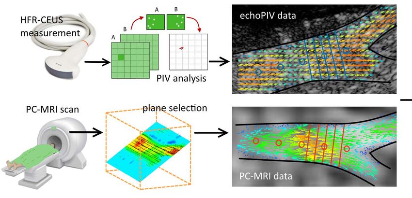
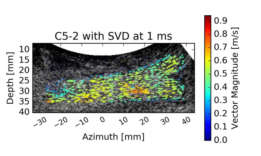
frequency (MHz)Methods: echoPIV and PC-MRI PHYSICS OF FLUIDS.
Ultrafast Contrast Ultrasound
                                                      First in-man

ULTRASOUND PARTICLE IMAGE VELOCIMETRY IN THE ABDOMINAL AORTA: FIRST RESULTS IN HUMANS AND COMPARISON WITH PHASE CONTRAST MAGNETIC RESONANCE IMAGING.
    Stefan A.J. Engelhard, Jason Voorneveld, Hendrik J. Vos, Jos J.M. Westenberg, Frank J.H. Gijsen, Pavel Taimr, Michel Versluis, Nico de Jong, Johan G. Bosch,
    Michel M.P.J. Reijnen, and Erik Groot Jebbink.
    Radiology 289 (1), 119–125 (2018).
HIGH-FRAME RATE CONTRAST-ENHANCED ULTRASOUND FOR VELOCIMETRY IN THE HUMAN ABDOMINAL AORTA.
     J. Voorneveld, S. Engelhard, H.J. Vos, M.M.P.J. Reijnen, F. Gijsen, M. Versluis, E. Groot Jebbink, N. de Jong, and J.G. Bosch.
     IEEE Trans UFFC 65(12), 2245–2254 (2018).
Ultrafast Contrast Ultrasound
First in-man
ULTRASOUND PARTICLE IMAGE VELOCIMETRY IN THE ABDOMINAL AORTA: FIRST RESULTS IN HUMANS AND COMPARISON WITH PHASE CONTRAST MAGNETIC RESONANCE IMAGING.
Stefan A.J. Engelhard, Jason Voorneveld, Hendrik J. Vos, Jos J.M. Westenberg, Frank J.H. Gijsen, Pavel Taimr, Michel Versluis, Nico de Jong, Johan G. Bosch,
Michel M.P.J. Reijnen, and Erik Groot Jebbink.
Radiology 289 (1), 119–125 (2018).
HIGH-FRAME RATE CONTRAST-ENHANCED ULTRASOUND FOR VELOCIMETRY IN THE HUMAN ABDOMINAL AORTA.
J. Voorneveld, S. Engelhard, H.J. Vos, M.M.P.J. Reijnen, F. Gijsen, M. Versluis, E. Groot Jebbink, N. de Jong, and J.G. Bosch.
IEEE Trans UFFC 65(12), 2245–2254 (2018).Ultrafast Contrast Ultrasound
First in-man
ULTRASOUND PARTICLE IMAGE VELOCIMETRY IN THE ABDOMINAL AORTA: FIRST RESULTS IN HUMANS AND COMPARISON WITH PHASE CONTRAST MAGNETIC RESONANCE IMAGING.
Stefan A.J. Engelhard, Jason Voorneveld, Hendrik J. Vos, Jos J.M. Westenberg, Frank J.H. Gijsen, Pavel Taimr, Michel Versluis, Nico de Jong, Johan G. Bosch,
Michel M.P.J. Reijnen, and Erik Groot Jebbink.
Radiology 289 (1), 119–125 (2018).
HIGH-FRAME RATE CONTRAST-ENHANCED ULTRASOUND FOR VELOCIMETRY IN THE HUMAN ABDOMINAL AORTA.
J. Voorneveld, S. Engelhard, H.J. Vos, M.M.P.J. Reijnen, F. Gijsen, M. Versluis, E. Groot Jebbink, N. de Jong, and J.G. Bosch.
IEEE Trans UFFC 65(12), 2245–2254 (2018).beat the single-wave resolution limit.
Ultrafast imaging combined with super-localiza on
Mathias Fink is director of the Langevin Institute at the École Supérieure de Physique et de Chimie Industrielles de la Ville de Paris in
Paris. Mickael Tanter is a research professor in the institute. They, along with six others, founded SuperSonic Imagine in 2005.
LETTER RESEARCH
The human body supports the propagation of many LETTER RESEARCH
Three different types of wave interaction can be ex-
kinds of waves, each of which can provide an image with a ploited in multiwave imaging. In one application, the inter-
specific type of information. For example, ultrasonic 1waves
mm1 mmb
action of one 1kind
mm 1ofmm
wave with tissue can generate a second
aa a a b
reveal a tissue’s density and how it responds to compression kind of wave.
Errico et al,InNature,
thermoacoustic
2015 imaging, for example, ab-
forces, and mechanical shear waves indicate how tissues re- sorbed electromagnetic radiation causes a transient change
spond to shear forces. Low-frequency electromagnetic waves in temperature that radiates an ultrasonic wave through ther-
are sensitive to electrical conductivity; optical waves tell mal expansion (see the article by Stanislav Y. Emelianov, Pai-
about optical absorption. In all those circumstances, physi- Chi Li, and Matthew O’Donnell in PHYSICS TODAY, May 2009,
cists have striven to obtain the best overall contrast and res- page 34).
olution. Now, after decades of work, we are pushing against
the physical limits inherent in each imaging modality. As de-
33 scribed in the box on page 30, that limit is, in many cases, not
determined by wavelength.
22 Physicians quickly realized that for medical imaging and a
diagnosis, one way to overcome the inherent limits of single-
11 mode imaging is to combine different imaging modalities.
The basic idea of multimodality imaging—for example, in the
combination
500 μm
of positron emission tomography and com-
puted tomography—is to associate the high-resolution mor-
500 μm
phological image of a first modality (CT) to an image of the
16 μm
b c 16 μm second
1 modality (PET) that is poorly resolved but that pro-
1 mm 1 mm
b c1 1 c 1 mmd 1 mm
1 vides
2 a clinically interesting
c contrast, revealing metabolic ac- d
2
0.8 3
tivity in this case. A second example of multimodality imag-
Amplitude (a.u.)
0.8 3
Amplitude (a.u.)
0.6 17ing,
μm used for mammography, combines ultrasound and x-ray
0.6 17 μm 9 μm
0.4
images. However,
9 μm multimodality imaging remains extremely
0.4 costly and constrained by the inherent physical limits of each
0.2
0.2
separate imaging mode.
500 μm
500 μm 0
00 20 40 60New approaches
80 100 120
b
0 20 Distance
40 60
(μm) 80 100 120
Is there
Distance (μm) any way to improve diagnostic capabilities other than
d with multimodality imaging? Two scientific communities
d have suggested new research directions. One line of attack,
called molecular imaging, was proposed by chemists and
biologists. It differs from traditional imaging in that biomark-
ers are used to help image particular targets or pathways.
Those biomarkers interact chemically with their surround-
4
ings and thereby increase the contrast.
4
The other approach was proposed independently by var-
ious groups in the physics community. It consists of combin-
Velocity (mm s–1)
ing two different waves—one to provide contrast, another to–14s–1
Velocity (mm
–10 –5 0 5 10 14
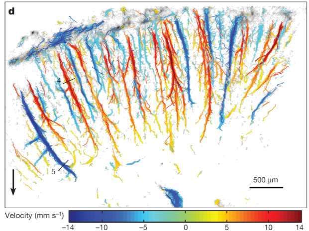
)Figure 1. Conventional versus ultrafast ultrasonic imag-
provide spatial resolution—to
Figure 3 | uULM build a new
of the kind of
rat brain image.a Be-
through thinned –14 skull
–10window
ing. (a)
–5 0or 5 10 14
In conventional ultrasound, 100 or more beams are
cause of the way through
theFigure
wavesthe3are combined,
intact
| uULM skull.
of the multiwave
a, uULM
rat brain imag-
performed
through through
a thinneda thinned
skull skull at aor
window
focused on different locations and the subsequent back-
ing produces a single
coronalimage
through with
section, the best
theBregma
intact −1.5contrast
skull. mm,
a, uULM and
providingreso-
a resolution
performed through of 10 µ m × 8 µskull
a thinned m at a
scattered echoes are processed to generate a single image.
5 lution properties in of depth
the two waves.
andsection,
coronal Multimodality
lateral direction,
Bregma −1.5 mm, imaging,
respectively. c, uULMa performed
providing resolution through
of 10 µ m × 8 µ m
(b) In ultrafast imaging, a plane wave probes the whole
on500
theμmother hand,the
relies
in onskull
intact
depth the
andatanalysis
lateral of
−1twomm.images,
Bregmadirection, Owing toeach
respectively.the c,
attenuation
uULM of the
performed through
5
ultrasound waves in the presence of the bone, the medium
achieved in a single
resolution shot. Again, the backscattered echoes
limited by
500 μm the contrast and resolution properties of the wave
the intact skull at Bregma −1 mm. Owing to the are attenuation of the
processed to produce the ultrasonic image.
tibeat the single-wave resolution limit.
Ultrafast imaging combined with super-localiza on
Mathias Fink is director of the Langevin Institute at the École Supérieure de Physique et de Chimie Industrielles de la Ville de Paris in
Paris. Mickael Tanter is a research professor in the institute. They, along with six others, founded SuperSonic Imagine in 2005.
LETTER RESEARCH
The human body supports the propagation of many LETTER RESEARCH
Three different types of wave interaction can be ex-
kinds of waves, each of which can provide an image with a ploited in multiwave imaging. In one application, the inter-
specific type of information. For example, ultrasonic 1waves
mm1 mmb
action of one 1kind
mm 1ofmm
wave with tissue can generate a second
aa a a b
reveal a tissue’s density and how it responds to compression kind of wave.
Errico et al,InNature,
thermoacoustic
2015 imaging, for example, ab-
forces, and mechanical shear waves indicate how tissues re- sorbed electromagnetic radiation causes a transient change
spond to shear forces. Low-frequency electromagnetic waves in temperature that radiates an ultrasonic wave through ther-
are sensitive to electrical conductivity; optical waves tell mal expansion (see the article by Stanislav Y. Emelianov, Pai-
about optical absorption. In all those circumstances, physi- Chi Li, and Matthew O’Donnell in PHYSICS TODAY, May 2009,
cists have striven to obtain the best overall contrast and res- page 34).
olution. Now, after decades of work, we are pushing against
the physical limits inherent in each imaging modality. As de-
33 scribed in the box on page 30, that limit is, in many cases, not
determined by wavelength.
22 Physicians quickly realized that for medical imaging and a
diagnosis, one way to overcome the inherent limits of single-
11 mode imaging is to combine different imaging modalities.
The basic idea of multimodality imaging—for example, in the
combination
500 μm
of positron emission tomography and com-
puted tomography—is to associate the high-resolution mor-
500 μm
phological image of a first modality (CT) to an image of the
16 μm
b c 16 μm second
1 modalityRAT (PET) that
BRAIN C is poorly resolved but
ORTEX that
ESPCI pro-
PARIS
1 mm 1 mm
b c1 1 c 1 mmd 1 mm
1 vides
2 a clinically interesting
c contrast, revealing metabolic ac- d
2
0.8 3
tivity in this case. A second example of multimodality imag-
Amplitude (a.u.)
0.8 3
Amplitude (a.u.)
0.6 17ing,
μm used for mammography, combines ultrasound and x-ray
0.6 17 μm 9 μm
0.4
images. However,
9 μm multimodality imaging remains extremely
0.4 costly and constrained by the inherent physical limits of each
0.2
0.2
separate imaging mode.
500 μm
500 μm 0
00 20 40 60New approaches
80 100 120
b
0 20 Distance
40 60
(μm) 80 100 120
Is there
Distance (μm) any way to improve diagnostic capabilities other than
d with multimodality imaging? Two scientific communities
d have suggested new research directions. One line of attack,
called molecular imaging, was proposed by chemists and
biologists. It differs from traditional imaging in that biomark-
ers are used to help image particular targets or pathways.
Those biomarkers interact chemically with their surround-
4
ings and thereby increase the contrast.
4
The other approach was proposed independently by var-
ious groups in the physics community. It consists of combin-
Velocity (mm s–1)
ing two different waves—one to provide contrast, another to–14s–1
Velocity (mm
–10 –5 0 5 10 14
)Figure 1. Conventional versus ultrafast ultrasonic imag-
provide spatial resolution—to
Figure 3 | uULM build a new
of the kind of
rat brain image.a Be-
through thinned –14 skull
–10window
ing. (a)
–5 0or 5 10 14
In conventional ultrasound, 100 or more beams are
cause of the way through
theFigure
wavesthe3are combined,
intact
| uULM skull.
of the multiwave
a, uULM
rat brain imag-
performed
through through
a thinneda thinned
skull skull at aor
window
focused on different locations and the subsequent back-
ing produces a single
coronalimage
through with
section, the best
theBregma
intact −1.5contrast
skull. mm,
a, uULM and
providingreso-
a resolution
performed through of 10 µ m × 8 µskull
a thinned m at a
scattered echoes are processed to generate a single image.
5 lution properties in of depth
the two waves.
andsection,
coronal Multimodality
lateral direction,
Bregma −1.5 mm, imaging,
respectively. c, uULMa performed
providing resolution through
of 10 µ m × 8 µ m
(b) In ultrafast imaging, a plane wave probes the whole
on500
theμmother hand,the
relies
in onskull
intact
depth the
andatanalysis
lateral of
−1twomm.images,
Bregmadirection, Owing toeach
respectively.the c,
attenuation
uULM of the
performed through
5
ultrasound waves in the presence of the bone, the medium
achieved in a single
resolution shot. Again, the backscattered echoes
limited by
500 μm the contrast and resolution properties of the wave
the intact skull at Bregma −1 mm. Owing to the are attenuation of the
processed to produce the ultrasonic image.
tibeat the single-wave resolution limit.
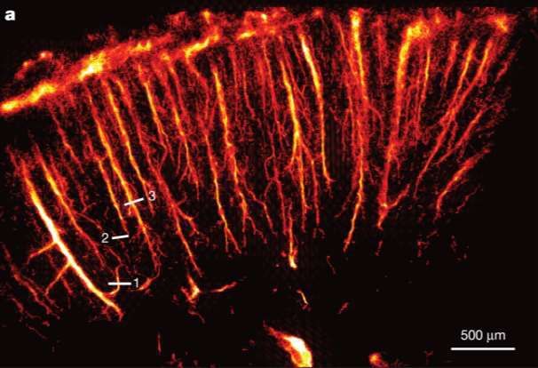
Ultrafast imaging combined with super-localiza on
Mathias Fink is director of the Langevin Institute at the École Supérieure de Physique et de Chimie Industrielles de la Ville de Paris in
Paris. Mickael Tanter is a research professor in the institute. They, along with six others, founded SuperSonic Imagine in 2005.
LETTER RESEARCH
The human body supports the propagation of many LETTER RESEARCH
Three different types of wave interaction can be ex-
kinds of waves, each of which can provide an image with a ploited in multiwave imaging. In one application, the inter-
specific type of information. For example, ultrasonic 1waves
mm1 mmb
action of one 1kind
mm 1ofmm
wave with tissue can generate a second
aa a a b
reveal a tissue’s density and how it responds to compression kind of wave.
Errico et al,InNature,
thermoacoustic
2015 imaging, for example, ab-
forces, and mechanical shear waves indicate how tissues re- sorbed electromagnetic radiation causes a transient change
spond to shear forces. Low-frequency electromagnetic waves in temperature that radiates an ultrasonic wave through ther-
are sensitive to electrical conductivity; optical waves tell mal expansion (see the article by Stanislav Y. Emelianov, Pai-
about optical absorption. In all those circumstances, physi- Chi Li, and Matthew O’Donnell in PHYSICS TODAY, May 2009,
cists have striven to obtain the best overall contrast and res- page 34).
olution. Now, after decades of work, we are pushing against
the physical limits inherent in each imaging modality. As de-
33 scribed in the box on page 30, that limit is, in many cases, not
determined by wavelength.
22 Physicians quickly realized that for medical imaging and a
diagnosis, one way to overcome the inherent limits of single-
11 mode imaging is to combine different imaging modalities.
The basic idea of multimodality imaging—for example, in the
combination of positron emission tomography and com-
Super resolution imaging 16 μm
500 μm
puted tomography—is to associate the high-resolution mor-
500 μm
phological image of a first modality (CT) to an image of the
b c 16 μm second
1 modalityRAT (PET) that
BRAIN C is poorly resolved but
ORTEX that
ESPCI pro-
PARIS
1 mm 1 mm
b c1 1 c 1 mmd 1 mm
1 vides
2 a clinically interesting
c contrast, revealing metabolic ac- d
2
0.8 3
tivity in this case. A second example of multimodality imag-
Amplitude (a.u.)
0.8 3
Amplitude (a.u.)
0.6 17ing,
μm used for mammography, combines ultrasound and x-ray
0.6 17 μm 9 μm
0.4
images. However,
9 μm multimodality imaging remains extremely
0.4 costly and constrained by the inherent physical limits of each
0.2
0.2
separate imaging mode.
500 μm
500 μm 0
00 20 40 60New approaches
80 100 120
b
0 20 Distance
40 60
(μm) 80 100 120
Is there
Distance (μm) any way to improve diagnostic capabilities other than
d with multimodality imaging? Two scientific communities
d have suggested new research directions. One line of attack,
called molecular imaging, was proposed by chemists and
biologists. It differs from traditional imaging in that biomark-
ers are used to help image particular targets or pathways.
Those biomarkers interact chemically with their surround-
4
ings and thereby increase the contrast.
4
The other approach was proposed independently by var-
ious groups in the physics community. It consists of combin-
Velocity (mm s–1)
ing two different waves—one to provide contrast, another to–14s–1
Velocity (mm
–10 –5 0 5 10 14
)Figure 1. Conventional versus ultrafast ultrasonic imag-
provide spatial resolution—to
Figure 3 | uULM build a new
of the kind of
rat brain image.a Be-
through thinned –14 skull
–10window
ing. (a)
–5 0or 5 10 14
In conventional ultrasound, 100 or more beams are
cause of the way through
theFigure
wavesthe3are combined,
intact
| uULM skull.
of the multiwave
a, uULM
rat brain imag-
performed
through through
a thinneda thinned
skull skull at aor
window
focused on different locations and the subsequent back-
ing produces a single
coronalimage
through with
section, the best
theBregma
intact −1.5contrast
skull. mm,
a, uULM and
providingreso-
a resolution
performed through of 10 µ m × 8 µskull
a thinned m at a
scattered echoes are processed to generate a single image.
5 lution properties in of depth
the two waves.
andsection,
coronal Multimodality
lateral direction,
Bregma −1.5 mm, imaging,
respectively. c, uULMa performed
providing resolution through
of 10 µ m × 8 µ m
(b) In ultrafast imaging, a plane wave probes the whole
on500
theμmother hand,the
relies
in onskull
intact
depth the
andatanalysis
lateral of
−1twomm.images,
Bregmadirection, Owing toeach
respectively.the c,
attenuation
uULM of the
performed through
5
ultrasound waves in the presence of the bone, the medium
achieved in a single
resolution shot. Again, the backscattered echoes
limited by
500 μm the contrast and resolution properties of the wave
the intact skull at Bregma −1 mm. Owing to the are attenuation of the
processed to produce the ultrasonic image.
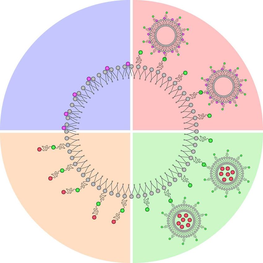
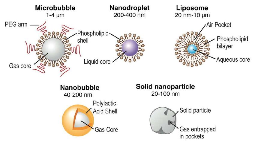
tiBubbles for molecular imaging and targeted drug delivery
Bubbles for molecular imaging and targeted drug delivery

      Jonathan Lindner, OHSU Portland, OR
Bubbles for molecular imaging and targeted drug delivery
Jonathan Lindner, OHSU Portland, OR
ACOUSTIC BEHAVIOR OF MICROBUBBLES AND IMPLICATIONS FOR DRUG DELIVERY (REVIEW).
Klazina Kooiman, Hendrik J. Vos, Michel Versluis, and Nico de Jong. Adv. Drug Deliv. Rev. 72, 28–48 (2014).Bubbles for molecular imaging and targeted drug delivery
Jonathan Lindner, OHSU Portland, OR
Bright eld Bodipy-labeled DiI-labeled Overlay
ACOUSTIC BEHAVIOR OF MICROBUBBLES AND IMPLICATIONS FOR DRUG DELIVERY (REVIEW).
Klazina Kooiman, Hendrik J. Vos, Michel Versluis, and Nico de Jong. Adv. Drug Deliv. Rev. 72, 28–48 (2014).
fiBubbles for molecular imaging and targeted drug delivery
Jonathan Lindner, OHSU Portland, OR
80 kPa, 1000 cycles
Bright eld Bodipy-labeled DiI-labeled Overlay
I. De Cock, G. Lajoinie, M. Versluis, I. Lentacker, S. De Smedt
Biomaterials 83, 294–307 (2016)
fiBubbles for molecular imaging and targeted drug delivery
Jonathan Lindner, OHSU Portland, OR
80 kPa, 1000 cycles
I. De Cock, G. Lajoinie, M. Versluis, I. Lentacker, S. De Smedt G. Lajoinie, Y. Luan, E. Gelderblom, B. Dollet, F. Mastik,
Biomaterials 83, 294–307 (2016) I. Lentacker, H. Dewitte, N. de Jong, and M. Versluis.
Nature Comm. Phys. 1, 22 (2018).Bubbles for molecular imaging and targeted drug delivery
Jonathan Lindner, OHSU Portland, OR
80 kPa, 1000 cycles
I. De Cock, G. Lajoinie, M. Versluis, I. Lentacker, S. De Smedt V. Pereno, M. Arona, O. Vince. C. Mannaris, A. Seth, M. de Saint Victor, G. Lajoinie, Y. Luan, E. Gelderblom, B. Dollet, F. Mastik,
Biomaterials 83, 294–307 (2016) G. Lajoinie, M. Versluis, C. Coussios, D. Carugo, and E. Stride I. Lentacker, H. Dewitte, N. de Jong, and M. Versluis.
Biomicrofluidics 12, 034109 (2018). Nature Comm. Phys. 1, 22 (2018).Cavita on nuclei for ultrasound-triggered drug delivery ti
Cavita on nuclei for ultrasound-triggered drug delivery

        MOLECULAR BODY IMAGING: MR IMAGING, CT, AND US. PART I. PRINCIPLES
        Moritz F. Kircher and Jürgen K. Willmann, Radiology 263(3), 633 (2012).
   ti
Cavita on nuclei for ultrasound-triggered drug delivery
MOLECULAR BODY IMAGING: MR IMAGING, CT, AND US. PART I. PRINCIPLES
Moritz F. Kircher and Jürgen K. Willmann, Radiology 263(3), 633 (2012).
10 μm ACOUSTIC DROPLET VAPORIZATION IS INITIATED BY SUPERHARMONIC FOCUSING.
Oleksandr Shpak, Martin Verweij, Rik Vos, Nico de Jong, Detlef Lohse, and Michel Versluis. PNAS 111, 1697-1702 (2014).
tiCavita on nuclei for ultrasound-triggered drug delivery
a c
a c
b d
10 μm 10 μm
b d
10 μm 10 μm
MOLECULAR BODY IMAGING: MR IMAGING, CT, AND US. PART I. PRINCIPLES
Moritz F. Kircher and Jürgen K. Willmann, Radiology 263(3), 633 (2012).
10 μm ACOUSTIC DROPLET VAPORIZATION IS INITIATED BY SUPERHARMONIC FOCUSING.
Oleksandr Shpak, Martin Verweij, Rik Vos, Nico de Jong, Detlef Lohse, and Michel Versluis. PNAS 111, 1697-1702 (2014).
tiThrombolysis using ultrasound (in real-time) in ‘De Kennis van Nu’ - broadcast Dutch national TV - April 2017
Thrombolysis using ultrasound (in real-time) in ‘De Kennis van Nu’ - broadcast Dutch national TV - April 2017
Aspalatholysis using ultrasound (in real-time) in ‘De Kennis van Nu’ - broadcast Dutch national TV - April 2017
Aspalatholysis using ultrasound (in real-time) in ‘De Kennis van Nu’ - broadcast Dutch national TV - April 2017
  Aspalathus linearis - rooibos [rɔːibɔs] (South-African red bush tea)
Cavita on bubbles
   ti
Cavita on bubbles
   ti
Cavita on bubbles

                    Larry Cru
                    Center for Medical and Industrial Ultrasoun
                    University of Washington, Seattle, USA
m

     ti
          d
Cavita on bubbles
Larry Cru
Center for Medical and Industrial Ultrasoun
University of Washington, Seattle, USA
m
ti
dHow to sink a ship
Therapeu c applica ons of bubbles

                                    14
      ti
               ti
Therapeu c applica ons of bubbles
14
ti
tiTherapeu c applica ons of bubbles
14
SONOPORORATION PHILIPS ULTRASOUND
ti
tiTherapeu c applica ons of bubbles
14
SONOPORORATION PHILIPS ULTRASOUND MEMBRANE PERFORATION AND RECOVERY DYNAMICS IN MICROBUBBLE-MEDIATED SONOPORATION
Yaxin Hu, Jennifer M.F. Wan, and Alfred C.H. Yu, Ultrasound Med. Biol. 39, 2393-2405 (2013)
ti
tiTAA and TriMix mRNA lead to the induction of durable antitumor responses in a
chemorefractory melanoma patient11, 12
. On the basis of these results, we evaluated the
Cancer immunotherapy from an ultrasound perspec ve
potential of simultaneous delivery of TAA mRNA and TriMix via microbubbles and
ultrasound to induce potent antitumor immune responses in mice, as schematically
depicted in Figure 1A.
with Heleen
Figure Dewitte,
1. mRNA Ine De
sonoporation Cock,
of DCs Stefaan
using De Smedt,
mRNA-loaded Ine Lentacker
microbubbles and
ultrasound.
(A) Schematic representation of the use of mRNA-loaded microbubbles, which implode
upon exposure to ultrasound and sonoporate the DCs. As a result, both antigen and
DC modulating proteins are produced by the DC, which can lead to antigen
presentation and T cell activation. (B) Schematic representation of the production of
mRNA-loaded microbubbles. Antigen and TriMix mRNA are premixed and complexed
to biotinylated cationic liposomes. The resulting mRNA-lipoplexes can then be attached
THE POTENTIAL OF ANTIGENtoAND the TRIMIX SONOPORATION
surface USING
of avidinylated lipid MRNA-LOADED MICROBUBBLES FOR
microbubbles.
ULTRASOUND-TRIGGERED CANCER IMMUNOTHERAPY.
H. Dewitte, S.V. Lint, C. Heirman, K. Thielemans, S.C.De Smedt, K. Breckpot, and I. Lentacker,
J. Controlled Release 194, 28 (2014).
126 | C h a p t e r 5
SONOPRINTING AND THE IMPORTANCE OF MICROBUBBLE LOADING FOR THE ULTRASOUND-MEDIATED DELIVERY OF NANOPARTICLES.
Ine De Cock, Guillaume Lajoinie, Michel Versluis, Stefaan C. De Smedt, and Ine Lentacker.
Biomaterials 83, 294–307 (2016).
tiTAA and TriMix mRNA lead to the induction of durable antitumor responses in a
chemorefractory melanoma patient11, 12
. On the basis of these results, we evaluated the
Cancer immunotherapy from an ultrasound perspec ve
potential of simultaneous delivery of TAA mRNA and TriMix via microbubbles and
ultrasound to induce potent antitumor immune responses in mice, as schematically
depicted in Figure 1A.
with Heleen
Figure Dewitte,
1. mRNA Ine De
sonoporation Cock,
of DCs Stefaan
using De Smedt,
mRNA-loaded Ine Lentacker
microbubbles and
ultrasound.
(A) Schematic representation of the use of mRNA-loaded microbubbles, which implode
upon exposure to ultrasound and sonoporate the DCs. As a result, both antigen and
DC modulating proteins are produced by the DC, which can lead to antigen
presentation and T cell activation. (B) Schematic representation of the production of
mRNA-loaded microbubbles. Antigen and TriMix mRNA are premixed and complexed Figure 7. Therapeutic vaccination of E.G7-OVA-beari
to biotinylated cationic liposomes. The resulting mRNA-lipoplexes can then be attached
THE POTENTIAL OF ANTIGENtoAND the TRIMIX SONOPORATION
surface USING
of avidinylated lipid MRNA-LOADED MICROBUBBLES FOR
microbubbles. sonoporated DCs.
ULTRASOUND-TRIGGERED CANCER IMMUNOTHERAPY.
H. Dewitte, S.V. Lint, C. Heirman, K. Thielemans, S.C.De Smedt, K. Breckpot, and I. Lentacker, 10 and 14 days after inoculation of mice with E.G7-OVA lym
J. Controlled Release 194, 28 (2014).
randomized in three treatment groups based on tumor volum
126 | C h a p t e r 5
the
SONOPRINTING AND THE IMPORTANCE OF MICROBUBBLE LOADING FOR THE ULTRASOUND-MEDIATED DELIVERY OF animals received
NANOPARTICLES. therapeutic vaccinations with mRNA s
Ine De Cock, Guillaume Lajoinie, Michel Versluis, Stefaan C. De Smedt, and Ine Lentacker.
Biomaterials 83, 294–307 (2016). show tumor growth as a function of time for mice vaccinate
tiTAA and TriMix mRNA lead to the induction of durable antitumor responses in a
chemorefractory melanoma patient11, 12
. On the basis of these results, we evaluated the
Cancer immunotherapy from an ultrasound perspec ve
potential of simultaneous delivery of TAA mRNA and TriMix via microbubbles and
ultrasound to induce potent antitumor immune responses in mice, as schematically
depicted in Figure 1A.
with Heleen
Figure Dewitte,
1. mRNA Ine De
sonoporation Cock,
of DCs Stefaan
using De Smedt,
mRNA-loaded Ine Lentacker
microbubbles and
ultrasound.
(A) Schematic representation of the use of mRNA-loaded microbubbles, which implode Figure 7. Therapeutic vaccination of E.G7-OVA-bearing mice with mRNA
upon exposure to ultrasound and sonoporate the DCs. As a result, both antigen and
sonoporated DCs.
DC modulating proteins are produced by the DC, which can lead to antigen
presentation and T cell activation. (B) Schematic representation of the production of 10 and 14 days after inoculation of mice with E.G7-OVA lymphoma cells, mice we
mRNA-loaded microbubbles. Antigen and TriMix mRNA are premixed and complexed randomized in three treatment
Figure groups based
7. Therapeutic on tumor volume
vaccination as shown in (A) The
of E.G7-OVA-beari
to biotinylated cationic liposomes. The resulting mRNA-lipoplexes can then be attached the animals received therapeutic vaccinations with mRNA sonoporated DCs. Graph
THE POTENTIAL OF ANTIGENtoAND the TRIMIX SONOPORATION
surface USING
of avidinylated lipid MRNA-LOADED MICROBUBBLES FOR
microbubbles. sonoporated DCs.
ULTRASOUND-TRIGGERED CANCER IMMUNOTHERAPY. show tumor growth as a function of time for mice vaccinated with DCs sonoporate
H. Dewitte, S.V. Lint, C. Heirman, K. Thielemans, S.C.De Smedt, K. Breckpot, and I. Lentacker, 10 and 14 days after inoculation of mice with E.G7-OVA lym
with (B) GFP mRNA (control), (C) OVA mRNA, (D) OVA mRNA and TriMix (DC Tri
J. Controlled Release 194, 28 (2014).
randomized
and (E) OVA in three
mRNA followed by atreatment groups
2h maturation based
with LPS on tumorA volum
(DC OVA/LPS). Kapla
126 | C h a p t e r 5
the
SONOPRINTING AND THE IMPORTANCE OF MICROBUBBLE LOADING FOR THE ULTRASOUND-MEDIATED DELIVERY OF animals Meier survival
received
NANOPARTICLES. curve is shown
therapeutic in (F).
vaccinations with mRNA s
Ine De Cock, Guillaume Lajoinie, Michel Versluis, Stefaan C. De Smedt, and Ine Lentacker.
Biomaterials 83, 294–307 (2016). show tumor growth as a function of time for mice vaccinate
tiTAA and TriMix mRNA lead to the induction of durable antitumor responses in a
chemorefractory melanoma patient11, 12
. On the basis of these results, we evaluated the
Cancer immunotherapy from an ultrasound perspec ve
potential of simultaneous delivery of TAA mRNA and TriMix via microbubbles and
ultrasound to induce potent antitumor immune responses in mice, as schematically
depicted in Figure 1A.
In accordance to the previous experiment, the tumor growth curv
indicate that sonoporation with antigen results in a significant delay of tu
resulting in a 58% increase in median survival. Interestingly, the slow-
growth was markedly shorter-lived in the DC OVA/LPS group com
unstimulated counterparts (DC OVA). This resulted in merely 35% prolonga
survival of animals in the DC OVA/LPS group compared to the DC GFP gro
stimulation of antigen presentation by sonoporation with OVA and TriMix m
in a pronounced effect on tumor growth: median survival was more than
increase), and complete tumor regression was observed in 2/6 animals
with Heleen
Figure Dewitte,
1. mRNA Ine De
sonoporation Cock,
of DCs Stefaan
using De Smedt,
mRNA-loaded Inegroup.
Lentacker
microbubbles and
ultrasound.
(A) Schematic representation of the use of mRNA-loaded microbubbles, which implode Figure 7. Therapeutic vaccination of E.G7-OVA-bearing mice with mRNA
upon exposure to ultrasound and sonoporate the DCs. As a result, both antigen and
sonoporated DCs.
DC modulating proteins are produced by the DC, which can lead to antigen
presentation and T cell activation. (B) Schematic representation of the production of 10 and 14 days after inoculation of mice with E.G7-OVA lymphoma cells, mice we
mRNA-loaded microbubbles. Antigen and TriMix mRNA are premixed and complexed randomized in three treatment
Figure groups based
7. Therapeutic on tumor volume
vaccination as shown in (A) The
of E.G7-OVA-beari
to biotinylated cationic liposomes. The resulting mRNA-lipoplexes can then be attached the animals received therapeutic vaccinations with mRNA sonoporated DCs. Graph
THE POTENTIAL OF ANTIGENtoAND the TRIMIX SONOPORATION
surface USING
of avidinylated lipid MRNA-LOADED MICROBUBBLES FOR
microbubbles. sonoporated DCs.
ULTRASOUND-TRIGGERED CANCER IMMUNOTHERAPY. show tumor growth as a function of time for mice vaccinated with DCs sonoporate
H. Dewitte, S.V. Lint, C. Heirman, K. Thielemans, S.C.De Smedt, K. Breckpot, and I. Lentacker, 10 and 14 days after inoculation of mice with E.G7-OVA lym
with (B) GFP mRNA (control), (C) OVA mRNA, (D) OVA mRNA and TriMix (DC Tri
J. Controlled Release 194, 28 (2014).
randomized
and (E) OVA in three
mRNA followed by atreatment groups
2h maturation based
with LPS on tumorA volum
(DC OVA/LPS). Kapla
126 | C h a p t e r 5
the
SONOPRINTING AND THE IMPORTANCE OF MICROBUBBLE LOADING FOR THE ULTRASOUND-MEDIATED DELIVERY OF animals Meier survival
received
NANOPARTICLES. curve is shown
therapeutic in (F).
vaccinations with mRNA s
Ine De Cock, Guillaume Lajoinie, Michel Versluis, Stefaan C. De Smedt, and Ine Lentacker.
Biomaterials 83, 294–307 (2016). show tumor growth as a function of time for mice vaccinate
tiYou can also read