Development of a pregnant woman phantom using polygonal mesh, for dosimetric evaluations
←
→
Page content transcription
If your browser does not render page correctly, please read the page content below
BRAZILIAN JOURNAL
                                                    OF
                                            RADIATION SCIENCES
            BJRS                                    03-1A (2015) 01-11

            Development of a pregnant woman phantom using
               polygonal mesh, for dosimetric evaluations
                 M.O.M. Cabrala; J.W. Vieirab,c; V. Leal Netoc; F. R.A. Limaa,b,d

       a
           Departamento de Energia Nuclear, Universidade Federal de Pernambuco, 50740-540, Recife-PE, Brasil
                                                    manuela.omc@gmail.com
                              b
                                  Escola Politécnica de Pernambuco, 50720-001, Recife-PE, Brasil
                                                   jose.wilson59@uol.com.br
   c
       Laboratório de Dosimetria Numérica, Instituto Federal de Educação, Ciência e Tecnologia de Pernambuco,
                                                  50740-540, Recife-PE, Brasil
                                                   viriatoleal@yahoo.com.br
                       d
                           Centro Regional de Ciências Nucleares do Nordeste, 50740-540, Recife-PE
                                                      falima@cnen.gov.br

ABSTRACT

Due to the embryo/fetus radiosensitivity the accurate estimation of the absorbed dose distribution in the abdominal
area is an additional problem caused by the exposure of pregnant women to ionizing radiation in medical
applications. This paper reports the construction and insertion of a fetal representation in a female geometry by
means of 3D modeling techniques. In order to characterize an ECM the Grupo de Dosimetria Numerica (GDN) is
using, mainly, simulators emitting gama sources and voxel phantoms coupled to a MC code. The phantoms are
predominantly constructed from stacks of magnetic resonance images (MRI), computed tomography (CT) (obtained
from scans of real patients) or from 3D modeling techniques. Due to the difficulty of obtaining medical images of
pregnant women, 3D objects in several formats (.obj, .max, .blend, etc.) were acquired for anatomical representation
of a non-pregnant adult. To construct a fetal representation, the 3D modeling technique called Poly Modeling
(polygon mesh) was used inside of the software Autodesk 3ds Max 2014 (free student version). Information about
the radiosensibility of organs included in the abdominal area will be used to fit and use the pregnant phantom in
numerical dosimetry. For this, the phantom will be voxelized and the masses of organs of interest will be adjusted
according to data provided by International Commission on Radiological Protection (ICRP). Finally, the phantom
will be coupled to a MC code creating a MCE that will serve as base for the construction of several other models
involving pregnant women submitted to ionizing radiation.

Keywords: Exposure Computational Models, EGSnrc,Voxel phamtoms, Monte Carlo.

Palavras-chave: Exposure Computational Models,Voxel phamtoms, Monte Carlo.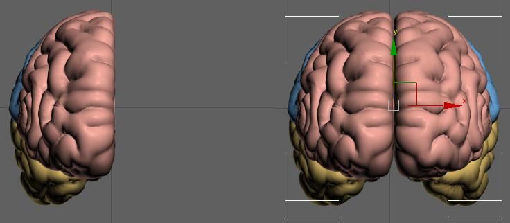
Cabral, et. al. ● Braz. J. Rad. Sci. ● 2015                        2

   1. INTRODUCTION

The dosimetric evaluation in the abdominal area (after the exposure of pregnant women to
ionizing radiation in medical applications) is of great importance due to the radiosensitivity
associated with fetal development. The frequency and magnitude of biological effects arising
from ionizing radiation on the embryo or fetus differ depending on: the type of radiation, the
gestational age at which there was exposure of the patient and the absorbed dose [1].

Modeling techniques for the construction of computational phantoms can be classified into four
categories, depending on the type of construction of anatomical structures: mathematical
phantoms (using simple mathematical surfaces), voxel phantoms (using segmented medical
images), hybrid phantoms (using polygon surfaces and medical images) and mesh phantoms
(using polygon meshes).

Due to the complexity of the human anatomy, the mathematical phantoms are limited in terms of
anatomical realism and therefore have been replaced by more advanced modeling. Among these,
voxel phantoms constructions made predominantly from magnetic resonance images (MRI),
computed tomography (CT) (obtained from scans of real patients) or phantoms mesh from 3D
modeling techniques are most-used manner for dosimetric purposes.

Due to the difficulty of obtaining medical images of pregnant women to manipulate and
construct a voxel phantom, the latest generation of computational mesh phantoms can be
constructed having images of the structures of interest and practical experience in 3D modeling.
Polygon Modeling (or Box-Modeling) is one of the most used techniques for 3D construction [2]
by being intuitive and by being implemented in several softwares available. In this technique, a
solid primitive object (as a cube, for example) serves as the base for creation of any form. For
this purpose, fundamental are the changes in position of the vertices, edges and faces, moreover
the use of specific commands into existing modeling softwares and referential images of the
object to be created.

International Joint Conference RADIO 2014
Gramado, RS, Brazil, August 26-29, 2014
SOCIEDADE BRASILEIRA DE PROTEÇÃO RADIOLÓGICA - SBPR
Cabral, et. al. ● Braz. J. Rad. Sci. ● 2015                                3

The primitive solid objects are parametric, i.e., their sizes are defined by a set of parameters. For
example, an object such as a cuboid has dimensions of width, height and depth, while a sphere is
defined by radious. Therefore, the choice of the object to use as a base, always depend on the
structure that you want to create. If the intention is to build a realistic human geometry, the
dimensions of each object must be changed and the number of faces should be increased to
enable the achievement of smoother surfaces, apparent curves, as shown in Figure 1.

  Figure 1: The same primitive solid object with increased numbers of polygons for smoothness in the
                                                form.

                                      Source: CABRAL, 2015.

However, an object with a large number of polygons is difficult for edition and view, because it
has too much information, resulting negatively with low performance computers. This paper
describes the construction process of a phantom for fetal representation using a minimum
number of polygons required for a realistic topology and easy control. The fetus surface
presented here was constructed through existing references about anatomy of pediatric and
pregnant human phantoms commonly used for internal and external dosimetry[3].

Due to the constant development and therefore the natural anatomical modification of a real
fetus, the final geometry will be posteriorly adjusted according to data provided by the

International Joint Conference RADIO 2014
Gramado, RS, Brazil, August 26-29, 2014
SOCIEDADE BRASILEIRA DE PROTEÇÃO RADIOLÓGICA - SBPR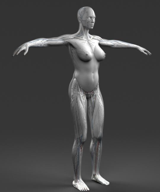
Cabral, et. al. ● Braz. J. Rad. Sci. ● 2015                                4

International Commission on Radiological Protection (ICRP) for fetuses at different gestational
ages and it will be compared with similar computational phantoms existing in the literature.

    2. MATERIALS AND METHODS

In this section the modeling processes for construction and adjustments of the phantom fetus
surface are presented. To construct the surface the anatomical basis of real fetuses with the
wished characteristics were obtained from the literature. For representation of a pregnant
phantom, 3D objects were acquired and adjusted for adaptation and integration of the two
phantoms in a single geometry.

2.1.    Materials

In this paper, the fetal representation was constructed using the following materials: 1) Autodesk
3dsMax 2014 Software (free version for students)[4]; 2) Computer with operating properties
compatible with Autodesk 3dsMax 2014 Software; 3) Sufficiently detailed descriptions about
infant anatomy, contained on atlas and on internet; 4) 3D objects in various formats (.obj, .3ds,
.fbx, .max, .blend, etc.) bought on the internet (http://www.cgshape.com/) to represent
anatomically human organs and tissues.

The methods used are described in the following item, since obtaining primary data until the last
adjustments in the 3D modeling environment.

2.2.    Methods

2.2.1. Modeling of the fetal representation

The 3D modeling is an art that requires much attention to details and technical knowledge of the tools
used in the editing and construction of the desired object. This paper consisted primarily in definition of

International Joint Conference RADIO 2014
Gramado, RS, Brazil, August 26-29, 2014
SOCIEDADE BRASILEIRA DE PROTEÇÃO RADIOLÓGICA - SBPR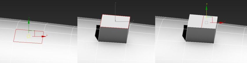
Cabral, et. al. ● Braz. J. Rad. Sci. ● 2015                                5

the scale used for the development of the fetus, which in this case was defined in centimeters. For
referential support in the construction and adjustment of the proportion of each developed structure, there
were created three rectangular frames for head (a), trunk (b) and limbs (c), as shown in Figure 2.

               Figure 2: Rectangular frames to reference head (a), trunk (b) and limbs (c).

                                                     (a)

                                                     (b)

                                                     (c)

                                         Source: CABRAL, 2015.

To start modeling, the primitive object more convenient to work has to be created and constantly
edited in its forms. Therefore, a cuboid with random dimensions was created and it was
converted into editable object to provide access to basic editing tools of the vertices, edges and
faces into the Autodesk 3dsMax. The possibility of manipulating these components separately
makes the control over the shape very flexible and it can be made through extrusion, connecting,
moving, spinning, and other actions that the program allows. The Figure 3 shows an example of
extrusion (a) and connecting edges (b).

       Figure 3: Original polygonal mesh (a) and steps of extrusion (b) and connecting edges (c).

                             (a)                             (b)                              (c)

                                         Source: CABRAL, 2015.

In the construction of the fetal surface, none of the changes made in the object followed the rule
of size because it was assumed that the surface would be symmetric with respect to the sagittal

International Joint Conference RADIO 2014
Gramado, RS, Brazil, August 26-29, 2014
SOCIEDADE BRASILEIRA DE PROTEÇÃO RADIOLÓGICA - SBPR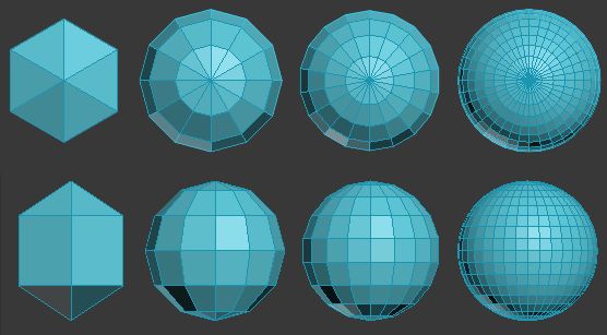
Cabral, et. al. ● Braz. J. Rad. Sci. ● 2015 6
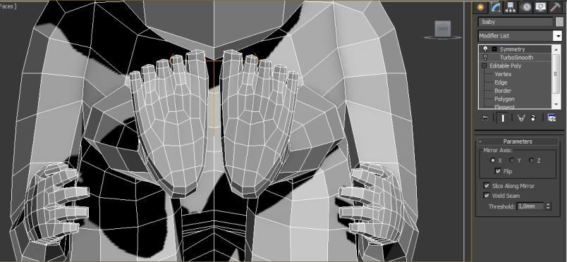
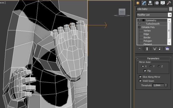
plane, i.e. the plane which divides the object into halves. For this, the Symmetry modifier was
used, creating a mirror effect on the entire side of the body. Thus, only one side has been
modified and all changes from the opposite side were identically and automatically updated, as
shown in Figure 4. The human body does not have a perfect symmetry, so the fine adjustment
will be done after conversion into voxels, which is considered as a future task of this work.
Figure 4: Respectively, step without and with the use of the Symmetry modifier.
Source: CABRAL, 2015.
In general, the process is completed with the application of TurboSmooth modifier, which
performs smoothing out all selected object by rounding forms, through the multiplication of
polygons. The effect of TurboSmooth makes the final object more anatomically realistic and,
equally to the Symmetry modifier, the effect can be disabled at any time if there is any other
modification to do and enable when the developer wants to. The Figure 5 shows the surface of a
heart before (a) and after (b) the use of the TurboSmooth modifier, respectively.
Figure 5: Effect of the TurboSmooth modifier in the surface of a heart.
Source: CABRAL, 2015.
International Joint Conference RADIO 2014
Gramado, RS, Brazil, August 26-29, 2014
SOCIEDADE BRASILEIRA DE PROTEÇÃO RADIOLÓGICA - SBPRCabral, et. al. ● Braz. J. Rad. Sci. ● 2015 7
2.2.2. Modification of the adult representation and integration of surfaces
The 3D files acquired by the GDN contain adult phantoms with internal structures, available in
various formats for use in different modeling environments. The female phantom was chosen to
be used and its internal structures were temporarily discarded. Only the surface of the abdominal
region was modified to accommodate the surface of the fetus previously developed. The Figure 6
shows the original phantom acquired for modification and integration between the two phantoms
surfaces.
Figure 6: Full acquired female phantom for modification and integration of surfaces.
Source: CABRAL, 2015.
3. RESULTS AND DISCUSSIONS
The fetal surface was developed to characterize the end of the second trimester of pregnancy. The
Figure 7 shows the construction stages of the arms by means of extrusions (a) and moving to
choose position (b), respectively. The Figure 8 shows the construction stages of the elbow by
International Joint Conference RADIO 2014
Gramado, RS, Brazil, August 26-29, 2014
SOCIEDADE BRASILEIRA DE PROTEÇÃO RADIOLÓGICA - SBPRCabral, et. al. ● Braz. J. Rad. Sci. ● 2015 8
means of connecting edges (c) and moving (d) in order to adjust it to the desired rotational
position.
Figure 7: Extrusion (a) and movement (b) steps for the arm construction.
Source: CABRAL, 2015.
Figure 8: Connecting edges (c) for the elbow construction and rotation (d) for adjustment of the position.
Source: CABRAL, 2015.
They were finalized with the Symmetry modifier (Figure 9), to automate the creation of the
opposite side of the developed area, and the TurboSmooth (Figure 10) tool for a bigger
smoothness and realism of the form. The Figure 11 shows the original phantom acquired (a) and
the result of the modification and integration between the two phantoms surfaces (b).
International Joint Conference RADIO 2014
Gramado, RS, Brazil, August 26-29, 2014
SOCIEDADE BRASILEIRA DE PROTEÇÃO RADIOLÓGICA - SBPRCabral, et. al. ● Braz. J. Rad. Sci. ● 2015 9
Figure 9: Respectively, step without and with the use of the Symmetry modifier.
Source: CABRAL, 2015.
Figure 10: Effect of the TurboSmooth modifier in the surface of the fetus.
Source: CABRAL, 2015.
Figure 11: Full acquired phantom (a) and integration of mother and fetus surfaces (b).
(a) (b)
Source: CABRAL, 2015.
International Joint Conference RADIO 2014
Gramado, RS, Brazil, August 26-29, 2014
SOCIEDADE BRASILEIRA DE PROTEÇÃO RADIOLÓGICA - SBPRCabral, et. al. ● Braz. J. Rad. Sci. ● 2015 10
4. CONCLUSIONS
It is possible to build realistic phantoms by a solid primitive object, as a cuboid, using the
Autodesk 3dsMax 2014 (Student version) software by means of Poly-Modeling technique. Due
to limitations for coupling a mesh phantom to MC codes, softwares are needed for creation
and/or edition of 3D objects and to convert them into voxels. This article is part of a series of
publications of the Grupo de Dosimetria Numérica and in future the whole internal structure of
the fetus will be modeled and adjusted according to data provided by the ICRP. The phantom
after being voxelized and adjusted, it will be coupled to a MC code creating a MCE that will
serve as base for the construction of several other models involving pregnant women submitted
to ionizing radiation. Dosimetric information about the radiosensitivity of organs and tissues
present in the abdominal region will be stored in a database, which will also be created.
5. ACKNOWLEDGMENTS
To Laboratório de Dosimetria Numérica (LDN/IFPE) for sharing the location for development
of this work. To Departamento de Energia Nuclear (DEN/UFPE), to Centro Regional de
Ciências Nucleares do Nordeste (CRCN-NE) and to Conselho Nacional de Desenvolvimento
Científico e Tecnológico (CNPq) for the financial support.
REFERENCES
1. XU, X. G.; ECKERMAN, K. F. Handbook of Anatomical Models for Radiation
Dosimetry. Series in Medical Physics and Biomedical Engineering. Boca Raton, London,
New York: CRC Press/Taylor & Francis Group, 2009, 760 p.
2. RUSSO, M., “Polygonal Modeling: Basic and Advanced Techniques”. Jones & Bartlett
Publishers, 2005.
International Joint Conference RADIO 2014
Gramado, RS, Brazil, August 26-29, 2014
SOCIEDADE BRASILEIRA DE PROTEÇÃO RADIOLÓGICA - SBPRCabral, et. al. ● Braz. J. Rad. Sci. ● 2015 11
3. ICRP - International Commission on Radiological Protection. Basic anatomical and
physiological data for use in radiological protection reference values. ICRP
Publication 89. Ann. ICRP 32 (3-4), 2002, 279 p.
4. AUTODESK. 3DS MAX 2014 | HELP. Autodesk Creative Commons FAQ. Avaible at:
. Last acess in: 11 out. 2014.
5. CABRAL, M. O. M. Desenvolvimento de um Modelo Computacional de Exposição para
uso em avaliações dosimétricas em gestantes. Masterdegree dissertation, PROTEN,
UFPE, Recife, Pernambuco, 2015, 92 p.
International Joint Conference RADIO 2014
Gramado, RS, Brazil, August 26-29, 2014
SOCIEDADE BRASILEIRA DE PROTEÇÃO RADIOLÓGICA - SBPRYou can also read