JOURNAL OF CLINICAL ORTHODONTICS - June 2021 www.jco-online.com
←
→
Page content transcription
If your browser does not render page correctly, please read the page content below
JOURNAL OF
                     CLINICAL
                     ORTHODONTICS
www.jco-online.com   June 2021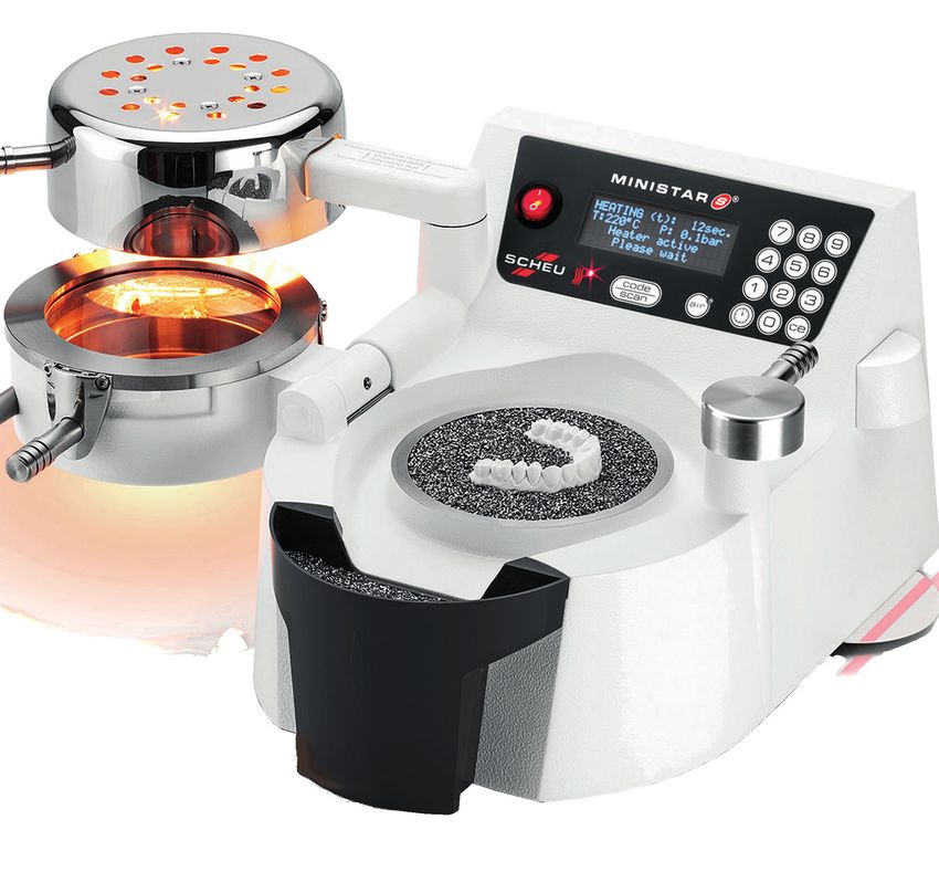
EXPERIENCE
           THE PROVEN DIFFERENCE!
                                                                                                                  The SPEED Appliance
                                                                                     Dramatically Superior Control and Performance!
                                                                                            Decrease in Spring Clip Stiffness During Treatment

                                                                                                                    SPEED                                                        SPEED Imitator
                                                                                                                  Nickel Titanium                                                    Cobalt Chromium
                                                                                                                  0% Decrease1                                                      50% Decrease1

                        Performance and aging of clips significantly depend on the alloy composition 1

                                                                                                   Trillium Compression Hooks
                                                                                                         4X Stronger Than Crimpable Hooks!
                                                                                                    Forces Required to Dislodge Archwire Hooks
                                                                                                                                                                                                       10.26

                                                                                                                      4X Stronger!
                                                                                  11.0
                                                           Mean Pull Force (kg)

                                                                                  8.25
                                                                                  5.50
                                                                                  2.75                                                             2.02                       2.32
                                                                                                0.63                       1.19
                                                                                    0
                                                                                           American Ortho2                TP Ortho2                 Ormco3                   TP Ortho4            Trillium Compression
                                                                                         (Smooth Inner Surface)     (Smooth Inner Surface)   (Smooth Inner Surface)   (Roughened Inner Surface)           Hooks5

                                                                        4X Stronger Than The Closest Competitor 4,5

             Learn more about SPEED and Trillium at www.speedsystem.com,
             or see us at an upcoming show. Ask about our Introductory Kits!

                                                                                                   speedback@speedsystem.com
      SPEED System™                                                                                    Tel: 1-800-267-7333                                                   TRILLIUM
                                                                                                                                                                             HESPELER ORTHODONTICS
        HESPELER ORTHODONTICS
 298 Shepherd Avenue Cambridge Ontario N3C 1V1 Canada                                                  Fax: 1-519-658-6925
Tel:1-800-26-SPEED 1-519-658-2925 Fax:1-519-658-6925
      1. Nikolaos Pandis, Christoph Bourauel, Theodore Eliadesc Changes
                      www.speedsystem.com                                 in the stiffness of the ligating mechanism in retrieved active self-ligating bracketsAm. J. Orthod. Dentofacial Orthop 2007; 132:(6):834–837. 2. A.
      Johal et al, European Journal of Orthodontics, Properties of Crimpable Archwire Hooks: A laboratory Investigation, 21, 1999, pp. 679-683. 3. A. Srivastava et al, Force of Dislodgement of Crimpable Attachments
      with Different Types and Dimensions of Archwire: An In Vitro Study, Orthodontic Cyberjournal, August 2013. 4. A. Johal et al, Journal of Orthodontics, A Clinical Investigation into the Behavior of Crimpable Archwire
      Hooks,Vol. 28, 2001, pp. 203-205. 5. Linder-Aronson Karsten A, Forsberg C-M, Öberg M. The resistance to axial dislodgement of nickel titanium compression arch wire hooks – an in vitro study. Aust Orthod J 2019;
      35: 21-26.
MiniSTAR S with Scan Technology
                              ®

                                      Fabricate precision-fitting appliances
                                         using the exceptional accuracy of
                                        positive pressure thermal forming.

                                  800.828.7626 | GreatLakesDentalTech.com
SMPP767Rev032921

                                  GREAT PEOPLE. GREAT PRODUCTS. GREAT LAKES.
Limited Spots Available! Register Today                                                                                16 CE Hours

THE NORRIS
EXPERIENCE
WHEN: October 1-2, 2021
WHERE: San Antonio, TX

      Jam packed with world-class learning along with
          award winning food and entertainment.
      All Inclusive: Great Food, Lodging, Transportation excludes travel to and from San Antonio

COURSE TOPICS
• How to hire, train, and inspire a winning team.
• Implementing airway-friendly orthodontics into daily practice.
• How to place systems into your office to achieve consistent excellence.
• How to diagnose and treat the most challenging interdisciplinary cases.
• Learn the 6 elements in the diagnosis of gummy smiles.
                                                                                                             Scan The QR Code To
• How to implement the Norris 20/26® system for optimal function and                                        Learn More or Register
  aesthetics in all cases.

                 “One of the best courses I have                                              “The course was fantastic.
                  taken in my orthodontic career.”                                The complexity of the cases presented
                                                                                        were outstanding, learned a ton.
                  Dr. Julian Davila                                                                     Dr. Don Demas
                  Geist Orthodontics                                                          Team Demas Orthodontics

                                          www.dynaflex.com | 800-489-4020
                               Norris 20/26® Bracket System Patent Pending. 050321 © DynaFlex®. All rights reserved.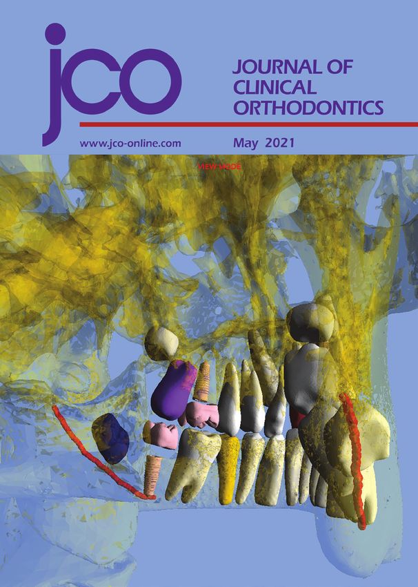
VOLUME LV NUMBER 6                                                                june 2021

                              327                                                A retired orthodontist lists 10 things he would
                                                                                 do today to build a practice in a rapidly changing
                                                                                 business environment.

                                                                                                                359

MASTER CLINICIAN
David B. Kennedy, BDS, MSD, FRCD(C)
DAVID B. KENNEDY, BDS, MSD, FRCD(C)
PETER M. SINCLAIR, DDS, MSD
Dr. Kennedy describes his diagnostic and me-                                     PEARLS
chanical principles for early treatment, with a                                  An Efficient and Ergonomic Device
variety of cases serving as illustrations.
                                                                                 for Easy Elastic Wear
                              343                                                SUMEDH DESHPANDE, MDS
                                                                                 SUSMITA BALA SHENOI, BDS
CASE REPORT                                                                      ROHAN S. HATTARKI, BDS, MDS
Resolution of a Complex Malocclusion                                             The authors demonstrate how to use inex-
Using a Hybrid Aligner Approach                                                  pensive materials to make elastic placers for
                                                                                 patients’ home use.
MARIO PALONE, DDS, MS
FRANCESCA CERVINARA, DDS, MS
SOFIA CASELLA, DDS
                                                                                                   361
GIUSEPPE SICILIANI, MD                                                           THE CUTTING EDGE
LUCA LOMBARDO, DDS, MS                                                           The Diagnostic Advantage of a
This patient was treated with sectional fixed ap-                                CBCT-Derived Segmented STL
pliances after clear aligners alone were unable
to correct a unilateral scissor bite.                                            Rendition of the Teeth and
                                                                                 Jaws Using an AI Algorithm
                355                                                              CHEN LEWIT BOROHOVITZ, DMD
MANAGEMENT & MARKETING                                                           ZEEV ABRAHAM, BDS, MS
                                                                                 W. RONALD REDMOND, DDS, MS
The Golden Age of Orthodontics:
                                                                                 The artificial intelligence capabilities of DICOM-
Already Ended or Just Beginning?                                                 to-STL conversion can have a significant effect
LEON KLEMPNER, DDS                                                               on treatment planning, as two cases show.
The opinions expressed in the Journal of Clinical Orthodontics are those of the writers and do not necessarily reflect the opinions and policies of
JCO. Copyright © 2021 JCO, Inc. Journal of Clinical Orthodontics (USPS 802-120, ISSN 0022-3875) is published monthly for $319 per year (U.S. individual
rate) by JCO, Inc., 5670 Greenwood Plaza Blvd., Suite 506, Greenwood Village, CO 80111-2409. Periodicals postage paid at Englewood, CO, and additional
mailing office. POSTMASTER: Send address changes to JCO, 5670 Greenwood Plaza Blvd., Suite 506, Greenwood Village, CO 80111-2409. Phone: (303)
443-1720; e-mail: info@jco-online.com. The material in each issue of JCO is protected by copyright. Instructions and fees for copying articles from JCO
are available from the Copyright Clearance Center, (978) 750-8400; www.copyright.com.

VOLUME LV NUMBER 6                                                                                                                                321
FOUNDING EDITOR
Eugene L. Gottlieb, DDS
EDITOR-IN-CHIEF
Robert G. Keim, DDS, EdD, PhD
ASSOCIATE EDITORS
Neal D. Kravitz, DMD, MS (South Riding, VA)
online only Birte Melsen, DDS, DrOdont (Aarhus, Denmark)
Ravindra Nanda, BDS, MDS, PhD (Farmington, CT)
Peter M. Sinclair, DDS, MSD (Los Angeles, CA)
Kelton T. Stewart, DDS, MS (Indianapolis, IN)
Bjorn U. Zachrisson, DDS, MSD, PhD (Oslo, Norway)
TECHNOLOGY EDITOR
Marc S. Lemchen, DMD (New York, NY)
WEBINAR EDITOR
Michael C. Meru, DDS, MS (Draper, UT)
CONTRIBUTING EDITORS
CASE REPORT S. Jay Bowman, DMD, MSD (Portage, MI)
First Molar Extractions in a Patient Robert L. Boyd, DDS, MEd (San Francisco, CA)
Vittorio Cacciafesta, DDS, MS, PhD (Milan, Italy)
with Marfan Syndrome Luis Carrière, DDS, MSD, PhD (Barcelona, Spain)
CRISTINA SOLA MARTIN, DDS, MS Mauro Cozzani, DMD, MScD (La Spezia, Italy)
JAIME GIL LÓPEZ-AREAL, DDS, MSc Jorge Fastlicht, DDS, MS (Mexico City, Mexico)
HARRY L. DOUGHERTY JR., DDS, MS Jeremy Fry, DDS, MS (Overland Park, KS)
This is the second in a series of case reports William V. Gierie, DDS, MS (Wilmington, NC)
from the three finalists for the 2021 Eugene L. Gayle Glenn, DDS, MSD (Dallas, TX)
Gottlieb JCO Student of the Year Award. John W. Graham, DDS, MD (Salt Lake City, UT)
Seong-Hun Kim, DMD, MSD, PhD (Seoul, Korea)
Masatada Koga, DDS, PhD (Tokyo, Japan)
Björn Ludwig, DMD, MSD (Traben-Trarbach, Germany)
James Mah, DDS, MS, DMS (Las Vegas, NV)
DEPARTMENTS James A. McNamara, DDS, PhD (Ann Arbor, MI)
The Editor’s Corner���������������������������������326 Elliott M. Moskowitz, DDS, MS (New York, NY)
Jae Hyun Park, DMD, MSD, MS, PhD (Mesa, AZ)
Book Reviews�����������������������������������������342 Jonathan Sandler, BDS, MS, FDS RCPS, MOrth RCS
Guide for Contributors ���������������������������354 (Chesterfield, United Kingdom)
Continuing Education �����������������������������371 Sarah C. Shoaf, DDS, MEd, MS (Winston-Salem, NC)
Georges L.S. Skinazi, DDS, DSO, DCD (Paris, France)
Product News�����������������������������������������373
Michael L. Swartz, DDS (Encino, CA)
Classified�����������������������������������������������374 Flavio Uribe, DDS, MDS (Farmington, CT)
Index of Advertisers�������������������������������375 Executive EDITOR
David S. Vogels III
ASSISTANT EDITOR
Kristy Brunskill
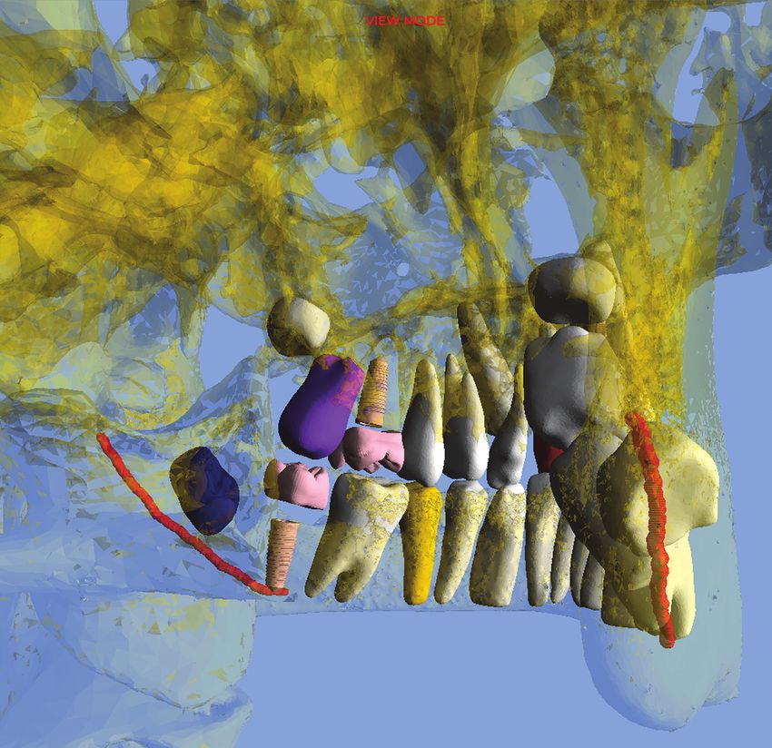
The Cover
EDITORIAL ASSISTANT
Three-dimensional, CBCT-derived segmented imaging Kelly Smith
is illustrated on the cover, as described in The Cutting
Edge by Drs. Lewit Borohovitz, Abraham, and VP MARKETING & BUSINESS DEVELOPMENT
Redmond. Phil Vogels
CUSTOMER SERVICE MANAGER
Address all communications to Journal of Clinical Orthodontics, Ann Marie Bartz
5670 Greenwood Plaza Blvd., Suite 506, Greenwood Village, CO
80111. Phone: (303) 443-1720; fax: (303) 443-9356; e-mail: info@ ART DIRECTOR
jco-online.com. See our website at www.jco-online.com. Irina Lef
322 JCO/june 2021
“After completing Dr. Roncone‘s course, we implemented the PhysioDynamic
System into our practice. We noticed faster leveling, alignment and shorter
treatment times in less visits. The bottom line is that Dr. Roncone‘s Systems
and protocols greatly increased the efficiency in our practice and we also see
an improvement in quality of treatment results.”
                                                              – Aron Dellinger –

Virtual JSOP                                          In-Office TIER V Course
Dates                                                 Course Date
10 monthly sessions of 6 hours each                   Hands on and lecture course

Sat. February 27, 2021                                October 27 – October 31, 2021
Sat. March 27, 2021
Sat. April 17, 2021
Sat. May 22, 2021                                     Topics
Sat. June 12, 2021                                    PDS (2.5 days)
Sat. July 24, 2021                                    TMD (1.5 days)
  No August Date                                      Mini-Lingual (1 day)
Sat. September 18, 2021
Sat. October 16, 2021                                 The TIER V Courses are held in Vista, CA
Sat. November 13, 2021                                at Dr. Roncone’s office.
Sat. December 18, 2021

Registration Fee                                      Registration Fee
JSOP Registration Fee: $4,500                         TIER V Registration Fee: $5,000
The past JSOP sessions will be available to rewatch
on ZOOM replay for those who sign up now.

To register:    Call: 800-758-5836
                or Visit: RonconeROI.com

For more information, visit us at:
www.forestadentusa.com
or call us: 1-800-721-4940

www.forestadentusa.com
Quality Manufacturing.
                     Exceptional Service.

 G&H Orthodontics, proud manufacturer of premium archwires for more than 40 years.
 An archwire is far from a commodity in your practice. G&H knows the integral role the archwire plays in achieving your successful clinical
 outcomes. Every wire alloy demands specific processing techniques to fully express its consistent tooth-moving properties. G&H has been
 manufacturing premium archwires in the U.S.A. for more than 40 years.

                                New to our wires? Try a free sample!
                 Simply contact our customer experience professionals and request an archwire
                                     in the shape and alloy of your choice.

            Visit GHOrthodontics.com to learn more and to place your order today!

                  BRACKETS        |   BANDS       |   TUBES      |   WIRES      |   SPRINGS       |   ELASTOMERICS
                                       Precision engineered and manufactured in the U.S.A.

     Order our full line of products at GHOrthodontics.com or call 800-526-1026 or +1 317-346-6655
MKT.004.Q                                                                                                               © 2021 G&H Orthodontics®
Dolphin
                  MyOrthodontist
                             mobile app for your patients
Put your practice at the center of THEIR lifestyle. MyOrthodontist gives
patients mobile access to information such as appointments; account
balance; questionnaires; patient education videos; practice and staff
info; social media links; and more! It’s entirely customizable with more
than 25 color themes, and can be personalized with your practice
logos, images and even practice videos! For more information visit
www.dolphinimaging.com/myortho.

                                                                    © 2021 Patterson Dental Supply, Inc. All rights reserved.
@2021 JCO, Inc. May not be distributed without permission. www.jco-online.com
THE EDITOR’S CORNER
The Evolution of Orthodontic Radiography
T
he use of radiographs in orthodontics, and in Over the years, a great many authors have
almost every dental and medical field, is pret published new and supposedly improved cephalo
ty much taken for granted nowadays. It is dif metric analyses. To my mind, however, the most
ficult for us even to imagine orthodontic, medical, important recent innovation in orthodontic diag
or dental practice without diagnostic radiography. nostic radiography occurred in 1998, with the
Yet dental radiography was developed in relatively introduction of cone-beam computed tomography
recent times, considering that dentistry has been (CBCT) to dentistry by Mozzo and colleagues.3
practiced as a necessity throughout history and as Its application to orthodontics was a natural pro
a recognized profession since the time of Pierre gression.
Fauchard (1678-1761). The German physicist Wil In this month’s Cutting Edge column, JCO’s
helm Conrad Röntgen is given credit for taking the Technology Editor, Dr. Marc S. Lemchen, presents
first radiograph in 1895—using his wife’s hand, a fascinating article written by Drs. Chen Lewit
presumably because it was the most convenient Borohovitz and Zeev Abraham of Tel Aviv Uni
thing available—with his new imaging invention, versity, Israel, and Dr. Ronald Redmond of the
which was made from a Crookes tube and photo University of the Pacific in San Francisco. With
graphic plates. According to Riaud, the German the challenging title “The Diagnostic Advantage
dentist Otto Walkhoff took the first dental radio of a CBCT-Derived Segmented STL Rendition of
graph, two weeks after the publication of Röntgen’s the Teeth and Jaws Using an AI Algorithm,” this
work.1 Walkhoff, still considered one of the found paper represents another step in the development
ers of modern endodontics, captured the radio of orthodontic diagnostic radiography. Using arti
graph of his own teeth with an exposure time of ficial intelligence, the authors’ technique “seg
about 25 minutes! Fortunately, the technology has ments” individual teeth or groups of teeth out of a
progressed considerably since then. CBCT image for closer analysis. Two cases demon
The early pioneers of dental radiology used strate the direct clinical applicability of this tech
glass photographic plates or roll film to capture nology and its diagnostic benefits for the orthodon
their images. The plates were cut down by the den tist, mainly in allowing the visualization of
tist, wrapped in black paper, and enclosed in rubber structures and anomalies that would not have been
dam material. These glass plates were extremely apparent in standard radiographs. This could be
fragile and quite uncomfortable for the patient. In another game changer. RGK
1903, Kells opened the first dental x-ray laborato
ry in the United States. In 1925, Raper originated REFERENCES
the bite-wing technique, and in 1948, panoramic
radiography was introduced. With specific refer 1. Riaud, X.: First dental radiograph (1896), J. Dent. Health Oral
ence to orthodontics, A.J. Pacini is credited with Disord. Ther. 9:33-34, 2018.
2. History of cephalometric analysis—Using our heads, blog,
taking the first standardized lateral radiograph in March 20, 2017, Cephx.com, cephx.com/reticent-orthodontic-
1922. Also in 1922, Paul Simon of Germany be patients-2-2, accessed June 15, 2021.
came the first to use planes and angles in what was 3. Mozzo, P.; Procacci, C.; Tacconi, A.; Martini, P.T.; and Andreis,
A.: A new volumetric CT machine for dental imaging based on
eventually referred to as “cephalometrics,” now an the cone-beam technique: Preliminary results, Eur. Radiol.
integral part of orthodontic practice.2 8:1558-1564, 1998.
326 © 2021 JCO, Inc. JCO/june 2021@2021 JCO, Inc. May not be distributed without permission. www.jco-online.com
master clinician
David B. Kennedy, BDS, MSD, FRCD(C)
Associate Editor Peter Sinclair conceived this department devoted to recognizing the
Master Clinicians who have made the orthodontic specialty what it is today. Every
few months, Dr. Sinclair will delve into the career story and treatment principles of
one of these seminal figures. We welcome your nominees for future Master Clinicians.
D
r. David Kennedy of the Uni- cialty: early treatment. There are
versity of British Columbia valid arguments regarding early
(UBC) is our featured Master treatment, both pro and con, with
Clinician this month. He addresses highly respected practitioners on
an issue that has been a point of each side of the debate. In this ar-
contention among orthodontists ticle, Dr. Kennedy presents a num-
throughout the history of the spe- ber of treated cases as excellent
examples of what can be accom-
plished.RGK
DR. SINCLAIR Who were your mentors?
DR. KENNEDY I attended two outstanding grad
uate programs where the department chairs—Drs.
Jim Roche, pediatric dentistry at Indiana Universi
ty, and Don Joondeph, orthodontics at the Univer
sity of Washington School of Dentistry—respected
the students, held the highest standards, and led by
example. Both served as directors on their respec
tive American boards. They encouraged their stu
Dr. Kennedy Dr. Sinclair
Dr. Kennedy is a Clinical Professor and Co-Clinic Director, Graduate
dents to become board-certified, something that I
Orthodontics, Faculty of Dentistry, University of British Columbia, did for both specialties and would highly recom
2199 Wesbrook Mall, Vancouver, BC, V6T 1Z3 Canada; e-mail: dbk@
dentistry.ubc.ca. Dr. Sinclair is an Associate Editor of the Journal of
mend; it helped me be more critical of my work.
Clinical Orthodontics and a Clinical Professor, Advanced Orthodontic They were my mentors, along with selected faculty
Program, Division of Endodontics, Oral and Maxillofacial Surgery, and
Orthodontics, School of Dentistry, University of Southern California,
from the University of Washington, including Bob
Los Angeles; e-mail: sinclair@usc.edu. Little, Vince Kokich, and Peter Shapiro.
VOLUME LV NUMBER 6 © 2021 JCO, Inc. 327@2021 JCO, Inc. May not be distributed without permission. www.jco-online.com
MASTER CLINICIAN
a
Fig. 1 Case 1. A. 7½-year-old male patient with anterior crossbite and forward mandibular shift before treatment
(continued on next page).
DR. SINCLAIR What is your philosophy, and how difference, based upon the scientific evidence of
does it guide you? treatment success.
I ask and answer three philosophical ques
DR. KENNEDY When I do a clinical exam, I ask tions when considering early treatment, and I use
myself the following three questions: evidence-based research to determine my deci
1. What do you see? Are the occlusion, dental de sion-making.1
velopment, and eruption sequence normal?
2. What should you see? This requires a compre 1. Can I treat this permanently? Examples are
hensive knowledge of growth and development. anterior and posterior crossbite correction. For
The clinician must be able to recognize normal nonskeletal anterior crossbite correction, a maxil
and abnormal development at various stages of the lary removable appliance can often be used, unless
mixed dentition. significant incisor rotational and torque control are
3. What is the difference? Usually you treat the needed (Fig. 1). Correction is usually accomplished
328 JCO/june 2021@2021 JCO, Inc. May not be distributed without permission. www.jco-online.com
DAVID B. KENNEDY, BDS, MSD, FRCD(C)
b
Fig. 1 (cont.) Case 1. B. Anterior crossbite corrected after nine months of treatment with removable maxillary
appliance; mandibular incisors show spontaneous improvement in alignment.
a b
Fig. 2 Case 2. A. 8-year-old patient
with maxillary lateral incisor cross- b
bite. B. Space created by expansion
with removable maxillary appli-
ance. C. Crossbite corrected after
nine months of treatment. c
VOLUME LV NUMBER 6 329@2021 JCO, Inc. May not be distributed without permission. www.jco-online.com MASTER CLINICIAN Fig. 3 Case 3. A. 9-year-old patient with crossbite before treatment. B. Two years later, showing improve- ment in gingival retreat after cross- bite correction (six months of active treatment). Deciduous canines ex- tracted to relieve crowding; no reten- tion needed. a b Fig. 4 Case 4. 8½-year-old female patient with unilateral left posterior crossbite, mandible shifted to crossbite side, and chin and mandibular dental midlines deflected to left before treatment. Crossbite side shows Class II tendency. 330 JCO/june 2021
@2021 JCO, Inc. May not be distributed without permission. www.jco-online.com
DAVID B. KENNEDY, BDS, MSD, FRCD(C)
Fig. 5 Case 4. After Phase I treatment involving four months of slow maxillary expansion with fixed Hyrax* expander,
followed by six months of retention with same passive appliance. Left Class II tendency improved, with chin and
midlines corrected and mandibular shift eliminated.
in four months of full-time wear. A posterior maxillary incisor alignment, provided adequate
biteplane is used when the vertical overbite ex space is available.
ceeds 2-3mm, to allow bite opening for anterior Bilateral posterior crossbites represent only
crossbite correction; the biteplane is reduced after 10% of mixed-dentition crossbites, with the unilat
the crossbite is corrected to prevent deepening of eral presentation being more common. Unilateral
the overbite. Space must be available for tooth mixed-dentition posterior crossbites often show a
movement to occur; therefore, maxillary expansion Class II tendency on the crossbite side, with the
may sometimes be needed (Fig. 2). Retention is not non-crossbite side being Class I (Fig. 4). The man
needed when the overbite is complete. Any gingi dibular dental and skeletal midlines are deflected
val retreat on the mandibular incisors will improve
after crossbite correction 2 (Fig. 3). Mandibular *Registered trademark of Dentaurum, Inc., Newtown, PA; www.
incisor irregularity often improves secondary to dentaurum.com.
VOLUME LV NUMBER 6 331@2021 JCO, Inc. May not be distributed without permission. www.jco-online.com
MASTER CLINICIAN
a b
Fig. 6 Case 5. A. 11½-year-old patient with bilateral ectopic maxillary canines, missing maxillary right lateral in-
cisor, and small left lateral incisor before extraction of maxillary deciduous canines. Late eruption for chronolog-
ical age is common with ectopic teeth. B. One year later, positions of maxillary permanent canines improved after
extraction of deciduous canines.
a
Fig. 7 Case 6. A. 9-year-old female patient with Class I malocclusion, mild mandibular crowding, and early loss of
mandibular right second deciduous molar before treatment (continued on next page).
332 JCO/june 2021@2021 JCO, Inc. May not be distributed without permission. www.jco-online.com
DAVID B. KENNEDY, BDS, MSD, FRCD(C)
b
Fig. 7 (cont.) Case 6. B. After 15 months of treatment, with mandibular lingual arch in place.
toward the crossbite side, with asymmetry in the In the early mixed dentition, slow maxillary
condyle position before treatment owing to the shift expansion can be used with one-quarter turn every
of the mandible toward the crossbite side. These two to three days. Because the suture is immature,
asymmetries improve secondary to maxillary ex lower force levels are required to obtain skeletal
pansion, which eliminates the shift of the mandible expansion. Rapid maxillary expansion in the young
(Fig. 5). A diagnostic tip relative to the need for child may widen the nasal base, hence the recom
maxillary expansion is that the maxilla shows more mendation for slow expansion. Fixed expanders,
crowding than the mandible. Both bilateral and such as a Haas-type, Hyrax,* or Quad-Helix,** are
unilateral posterior crossbites with functional recommended. About 5-6mm of expansion is usu
shifts require maxillary expansion, but the bilater ally required for correction of unilaterally present
al crossbite presentation requires more expansion. ing crossbites.3
Maxillary constriction is sometimes caused
*Registered trademark of Dentaurum, Inc., Newtown, PA; www.
dentaurum.com. by prolonged digit habits; this can be treated si
**Rocky Mountain Orthodontics, Denver, CO; www.rmortho.com. multaneously by incorporating a habit-breaking
VOLUME LV NUMBER 6 333@2021 JCO, Inc. May not be distributed without permission. www.jco-online.com
MASTER CLINICIAN
crib. Overexpansion of 2mm per side and a mini more often in females and those with small or miss
mum six months of fixed retention yield excellent ing lateral incisors, or with a family history of ec
stability.3 When an open bite exists, retention with topic canines.4-8 Distal crown tipping of the max
the crib should be continued until a positive over illary lateral incisors is a normal development in
bite has been established. the early mixed dentition, which is called the “ugly
duckling” stage. When distal tipping of the lateral
2. Is there something damaging that I cannot incisor persists as the maxillary canines should
leave? Examples are gingival retreat from an an erupt, however, this hints that the maxillary perma
terior crossbite, ectopic canines that can be guided nent canines may be ectopic, which calls for radio
into better positions, and unerupted incisors from graphic assessment (Fig. 6). A panoramic radio
mesiodens or trauma to the deciduous incisors. graph or periapical films are warranted at age 10-11
Palatally displaced ectopic canines occur to check the canine positions, especially when the
a
Fig. 8 Case 6. A. Patient at age 12, showing mandibular premolar rotations prior to start of Phase II. Lingual arch
removed previously, after second molar eruption (continued on next page).
334 JCO/june 2021@2021 JCO, Inc. May not be distributed without permission. www.jco-online.com
DAVID B. KENNEDY, BDS, MSD, FRCD(C)
deciduous canines are not mobile. The maxillary 3. Will early treatment help significantly with fu
deciduous canines should be mobile six months ture Phase II treatment? Will early intervention
after mandibular permanent canine eruption. move the case to nonextraction or make Phase II
Extraction of the maxillary deciduous ca easier or shorter? Examples are early Class III
nines, maxillary expansion, and/or headgear treat treatment, leeway space management, and serial
ment frequently improve the ectopic permanent extraction.
canine positions and encourage normal eruption. Mild to moderate crowding can be resolved
Similarly, space opening improves the likelihood with leeway space management. In 66-70% of cas
that the ectopic permanent canine will erupt with es, 5mm of crowding can be resolved with a
out surgical intervention.9-11 When the tip of the late-mixed-dentition mandibular lingual arch13
maxillary canine has not crossed the midpoint of (Fig. 7). The mandibular second deciduous molars
the lateral incisor, extraction of the deciduous ca may need to be removed to allow the crowding to
nine alone results in an improved permanent ca resolve by distal drifting of the canines and first
nine position 91% of the time.12 premolars into the edentulous space.13 The lingual
b
Fig. 8 (cont.) Case 6. B. After 18 months of Phase II treatment with fixed appliances.
VOLUME LV NUMBER 6 335@2021 JCO, Inc. May not be distributed without permission. www.jco-online.com
MASTER CLINICIAN
arch is left in place until the permanent second Serial extraction is appropriate in 15% of cas
molars erupt, with good long-term stability (Fig. es—those that exhibit severe crowding of 8mm or
8).14 The intermolar angle between the first and more16 (Fig. 9). Indications for serial extraction are
second molars must be evaluated before lingual Class I occlusions with more than 7-8mm of crowd
arch placement, because there is a more of a ten ing per arch. These patients should have protrusive
dency for mandibular second permanent molar dentitions, full faces, shallow overbites or open bites,
impaction when this angle exceeds 24°.15 and no missing or ectopic teeth. Serial extraction is
a
Fig. 9 Case 7. A. 9-year-old female
Class I patient with mixed-dentition
crowding before treatment. B. Pan-
oramic radiograph taken 18 months
earlier, before extraction of decidu-
ous canines. C. Two years later, prior
b c to first premolar extractions.
336 JCO/june 2021@2021 JCO, Inc. May not be distributed without permission. www.jco-online.com
DAVID B. KENNEDY, BDS, MSD, FRCD(C)
contraindicated in patients with flat profiles, short the crowded permanent incisors to spontaneously
lower faces, retrusive incisors, or minimal crowding. align. At age 9-10, the first deciduous molars are
Severely bimaxillary protrusive patients, who re extracted. The purpose is to accelerate the eruption
quire maximum retraction of the incisors, are not of the first premolars. Because normal maxillary
suitable candidates for serial extraction. eruption involves the first premolars erupting
Serial extraction involves deciduous canine ahead of the canines, this is seldom needed in the
extractions as the lateral incisors erupt, allowing maxilla. The first premolars are then extracted
a
Fig. 10 Case 7. A. Patient at age 13, after extraction of first premolars, show-
ing Class I relationship with poor interdigitation, but canines erupting into
attached gingiva. B. After premolar extractions and drifting of teeth. Note
tipping of teeth adjacent to mandibular extractions and difference between
mandibular anterior and posterior occlusal planes, with deepening curve of
b Spee.
VOLUME LV NUMBER 6 337@2021 JCO, Inc. May not be distributed without permission. www.jco-online.com
MASTER CLINICIAN
a
Fig. 11 Case 7. A. Two years later, af-
ter Phase II treatment with fixed appli-
ances. Panoramic radiograph shows
good root parallelism. B. Super
imposition of pre- and post-treatment
cephalometric tracings. Note excel-
lent incisor torque and positioning.
b
338 JCO/june 2021@2021 JCO, Inc. May not be distributed without permission. www.jco-online.com
DAVID B. KENNEDY, BDS, MSD, FRCD(C)
upon eruption, usually at age 11-12 (Fig. 10). One cept”—looking at the anteroposterior, transverse,
modification to be considered is whether the pre vertical, and perimeter planes of space. A family
molars should be enucleated rather than extracted history, especially as it relates to anteroposterior
upon eruption. This is particularly indicated when crowding, missing teeth, and parental treatment, is
the permanent canine is erupting ahead of the first most helpful. Other information, such as the pa
premolar. Some children are apprehensive about tient’s or parents’ chief complaint, anticipated child
extractions; as a result, serial extractions can be cooperation, and occlusal function, will be useful.
come an adverse behavioral issue for needle-phobic Other factors to consider include TMJ, occlusion,
children. After extractions and before braces, there and airway.
is a period of what is commonly called “driftodon Full orthodontic records are necessary. Writ
tics,” in which spontaneous alignment occurs.17 ten diagnostic findings with objectives made from
A major advantage of serial extraction is that the problem list, based on the planes of space data
the crowded permanent canines are not displaced base, will lead the clinician to develop treatment
buccally from the arch, so they erupt into the at alternatives.19 These alternatives should include the
tached gingiva. Crowding tends to improve be advantages and disadvantages, leading to informed
cause of the extractions,18 but we commonly see consent. From there, the treatment goals are select
tipping of the mandibular teeth adjacent to the ex ed, and, lastly, the appliance is chosen—not the
traction sites. The mandibular curve of Spee also other way around. Too often, a clinician takes a
deepens, with a difference between the mandibular continuing education course and wants to use a
posterior and anterior occlusal planes. Comprehen certain appliance. Treatment in the mixed dentition
sive treatment to detail the alignment, close resid should be objective- and not appliance-driven.
ual extraction spaces, and parallel the roots should “Objective-driven” treatment includes ex
be done in the early permanent dentition, with a panding the maxilla to correct a unilaterally pre
reduced treatment time because of the spontaneous senting posterior crossbite by eliminating a func
alignment18 (Fig. 11). Serial extraction cases also tional shift, thus reducing asymmetry. I try to use
have a lower Peer Assessment Rating score com a short duration, 12-15 months or less, to achieve
pared with late premolar extraction cases, making the objectives. Patient fatigue is then reduced
them easier to treat.18 should future Phase II treatment be needed. Par
ents may have limited funds or insurance coverage
DR. SINCLAIR What diagnostic principles do for their children’s orthodontic treatment; respon
you follow? sible clinicians will factor this into their best short-
and long-term decision-making. Extended, expen
DR. KENNEDY Diagnosis is the same for early sive Phase I treatment will unnecessarily increase
and late treatment.19 This is best handled by look patient costs and may reduce future insurance
ing at the face, followed by a “planes of space con coverage if Phase II treatment is needed.
Fig. 12 Mixed-dentition maxillary
Hawley retainer with Adams clasps on
first molars, spurs to control incisors,
and labial bow soldered to Adams
clasps; acrylic cleared away from pre-
molars and canines.
VOLUME LV NUMBER 6 339@2021 JCO, Inc. May not be distributed without permission. www.jco-online.com
MASTER CLINICIAN
DR. SINCLAIR What important mechanical prin DR. SINCLAIR What is your greatest clinical
ciples do you employ? challenge?
DR. KENNEDY Keep it simple. When using slow DR. KENNEDY Choosing the best cases to treat
maxillary expansion, keep the expander in place early. A great place to start is to select mixed-
as its own retainer. After active expansion, tie off dentition cases that present with good mandibular
the expansion screw with a ligature wire. Use slow arches. The clinician is then faced with changing
maxillary expansion and limited fixed appliances the maxillary arch to meet the good mandibular
with coils to open up space for crowded canines arch. The outcomes are usually best when this
when needed. Avoid long spans in 2 × 4 applianc strategy is used. The earlier mixed-dentition
es; deciduous molars can be bonded depending on treatment is started, the more extended will be
their longevity and stability. This reduces the the time available to evaluate its long-term suc
chance of wires coming out with the help of the cess, since both relapse and normal growth can
child’s fingers! For retention, I use a Hawley with occur after the initial treatment. Comprehensive
Adams clasps on the upper first molars and a sol knowledge of the normal growth and development
dered labial bow, and I keep the acrylic away from of the skeleton and dentition is essential.
the erupting permanent premolars and canines There are many other instances of appropri
(Fig. 12). ate early orthodontic treatment that have been
omitted from this article for space reasons. They
DR. SINCLAIR What is your best clinical tip? will be covered in future articles.
DR. KENNEDY Use fixed appliances as much as DR. SINCLAIR Thank you for sharing your clin
possible to eliminate the need for patient compli ical experience with our readers.
ance. When using partial fixed appliances, be
mindful of the deciduous tooth’s longevity. Re ACKNOWLEDGMENTS: Dr. Kennedy gratefully acknowledges
member that the malocclusion is stable, so don’t the assistance of Drs. James Andrews and Abdulraheem Alwafi
(graduate orthodontic students at UBC), who handled the proofread
move teeth beyond the alveolar housing. Maxillary ing, illustrations, and references.
expansion is quite stable, while mandibular expan
sion is often followed by a reduction in intercanine
width and relapse. Therefore, try to avoid lower-
arch expansion.20
340 JCO/june 2021@2021 JCO, Inc. May not be distributed without permission. www.jco-online.com
DAVID B. KENNEDY, BDS, MSD, FRCD(C)
REFERENCES
1. Kennedy, D.B.: Early treatment options, PCSO Bull. 82:19-22, A randomized clinical study, Angle Orthod. 81:370-374, 2011.
2010. 12. Ericson, S. and Kurol, J.: Early treatment of palatally erupting
2. Harrison, R.; Kennedy, D.; and Leggott, P.: Anterior dental maxillary canines by extraction of the primary canines, Eur. J.
crossbite: Relationship between incisor crown length and inci Orthod. 10:283-295, 1988.
sor irregularity before and after orthodontic treatment, Pediat. 13. Gianelly, A.A.: Crowding: Timing of treatment, Angle Orthod.
Dent. 15:394-397, 1993. 64:415-418, 1994.
3. Huynh, T.; Kennedy, D.B.; Joondeph, D.R.; and Bollen, A.M.: 14. Dugoni, S.A.; Lee, J.S.; Varela, J.; and Dugoni, A.A.: Early
Treatment response and stability of slow maxillary expansion mixed dentition treatment: Postretention evaluation of stability
using Haas, Hyrax, and Quad-Helix appliances: A retrospective and relapse, Angle Orthod. 65:311-320, 1995.
study, Am. J. Orthod. 136:331-339, 2009. 15. Sonis, A. and Ackerman, M.: E-space preservation: Is there a
4. Becker, A. and Chaushu, S.: Etiology of maxillary canine im relationship to mandibular second molar impaction? Angle
paction: A review, Am. J. Orthod. 148:557-567, 2015. Orthod. 81:1045-1049, 2011.
5. Johnston, W.D.: Treatment of palatally impacted canine teeth, 16. Proffit, W.R.; Fields, H.W. Jr.; Larson, B.; and Sarver, D.M.:
Am. J. Orthod. 56:589-596, 1969. Early (serial) extraction, in Contemporary Orthodontics, 6th
6. Becker, A.; Smith, P.; and Behar, R.: The incidence of anomalous ed., Mosby Elsevier, Philadelphia, 2018, p. 424.
maxillary lateral incisors in relation to palatally-displaced cus 17. Gönül, N.Y. and Sayinsu, K.: Treatment of a Class II patient with
pids, Angle Orthod. 51:24-29, 1981. four premolar extractions and driftodontics in the lower jaw,
7. Oliver, R.G.; Mannion, J.E.; and Robinson, J.M.: Morphology Turk. J. Orthod. 30:89-100, 2017.
of the maxillary lateral incisor in cases of unilateral impaction 18. O’Shaughnessy, K.W.; Koroluk, L.D.; Phillips, C.; and Kennedy,
of the maxillary canine, Br. J. Orthod. 16:9-16, 1989. D.B.: Efficiency of serial extraction and late premolar extraction
8. Sacerdoti, R. and Baccetti, T.: Dentoskeletal features associated cases treated with fixed appliances, Am. J. Orthod. 139:510-516,
with unilateral or bilateral palatal displacement of maxillary 2011.
canines, Angle Orthod. 74:725-732, 2004. 19. Proffit, W.R.; Fields, H.W. Jr.; Larson, B.; and Sarver, D.M.: Diag
9. Kokich, V.G.: Surgical and orthodontic management of impacted nosis and treatment planning, in Contemporary Orthodontics,
maxillary canines, Am. J. Orthod. 126:278-283, 2004. 6th ed., Mosby Elsevier, Philadelphia, 2018.
10. Olive, R.J.: Orthodontic treatment of palatally impacted max 20. Little, R.M.; Riedel, R.A.; and Stein, A.: Mandibular arch length
illary canines, Austral. Orthod. J. 18:64, 2002. increase during the mixed dentition: Postretention evaluation
11. Armi, P.; Cozza, P.; and Baccetti, T.: Effect of RME and head of stability and relapse, Am. J. Orthod. 97:393-404, 1990.
gear treatment on the eruption of palatally displaced canines:
VOLUME LV NUMBER 6 341@2021 JCO, Inc. May not be distributed without permission. www.jco-online.com
BOOK REVIEWS
Dentofacial Esthetics: From Macro to Micro
DAVID M. SARVER, DMD, MS
512 pages, 2,500+ illustrations. $268. 2020.
Quintessence Publishing Co., Inc.
411 N. Raddant Road, Batavia, IL 60510
(800) 621-0387; www.quintbook.com.
I
t is indeed my pleasure to review Chapter 5 discusses such
the latest text by David Sarver, clinical problems as gummy smile,
whom I have admired as a su hypermobile smile, vertical maxil
perb clinician, author, and speaker. lary deficiency, asymmetrical
This large-format, 512-page book smile, and smile issues related to
is easy to read and profusely illus tooth dimensions. The following
trated with treatment histories. chapter covers micro-esthetic im
Of the eight chapters, the provements involving gingivo
first is an introduction that de plasty for crown lengthening, as
scribes in personal terms the treat well as its timing during treatment.
ment of a patient who severely Several case histories are present
damaged her dentition, compro ed, based on a macro-, mini-, and
mising her smile and facial esthet micro-esthetic evaluation. Chap
ics. The next chapter discusses Dr. ters 7 and 8 describe management
Sarver’s soft-tissue paradigm, of congenitally missing teeth and
elaborating with patient examples interdisciplinary care, including
how problem- and goal-oriented esthetic considerations in ortho
orthodontic treatment planning gnathic and plastic surgery.
can be integrated. Kudos to Dr. Sarver for tack
Chapter 3 offers a detailed description of ling the subject of esthetics, which has often re
macro-, mini-, and micro-esthetics and the com ceived less attention than it deserves. He empha
ponents of balanced facial esthetics. A case sizes that orthodontics is about more than
history-based analysis and strategies are present managing occlusion; “it’s about creating faces and
ed for lifetime esthetics in both adolescent and smiles that are functional and beautiful.” I highly
adult patients. Principles of treatment planning recommend this book to orthodontists and gradu
are summarized in the subsequent chapter, taking ate students, and also to dentists and surgeons who
the reader step-by-step through data acquisition seek to manage smile zone and facial esthetics.
and positioning of teeth based on various compo
nents, including the smile zone. RAVINDRA NANDA, BDS, MDS, PhD
342 © 2021 JCO, Inc. JCO/june 2021@2021 JCO, Inc. May not be distributed without permission. www.jco-online.com
CASE REPORT
Resolution of a Complex Malocclusion
Using a Hybrid Aligner Approach
MARIO PALONE, DDS, MS
FRANCESCA CERVINARA, DDS, MS
SOFIA CASELLA, DDS
GIUSEPPE SICILIANI, MD
LUCA LOMBARDO, DDS, MS
C
lear aligner therapy is often the treatment of choice in today’s ortho-
dontic practice, especially for nonextraction cases of mild to moder-
ate difficulty.1 Aligners offer optimal esthetic properties and patient
comfort, making them particularly suitable for adults.2 In addition, because
they are removable, they are less likely to impact periodontal health.3
Although diagnostic indications for clear conventional biomechanics in complex treatment.6
aligner therapy have broadened, some orthodontic Extrusion or rotation auxiliaries or interarch elas
movements remain unpredictable, even with good tics are therefore often needed.7,8 Another approach
treatment planning.4 Extrusion, intrusion, bodily is to use partial or full fixed appliances to correct
movements, torque, and substantial rotations of the most critical problems before or during clear
rounded teeth are difficult to manage using clear aligner therapy.9
aligners alone, 5 making them less efficient than One such problem, scissor bite, is an alteration
Dr. Palone Dr. Cervinara Dr. Casella Dr. Siciliani Dr. Lombardo
Dr. Palone is a researcher and Dr. Casella is a resident, Department of Orthodontics, University of Ferrara, Via Luigi Borsari 46, Ferrara 44121, Italy.
Dr. Cervinara is in the private practice of orthodontics in Bari, Italy. Dr. Siciliani is Chairman, School of Dentistry, and Dr. Lombardo is Associate
Professor and Chairman, Postgraduate School of Orthodontics, University of Ferrara, Italy. E-mail Dr. Palone at mario.palone88@gmail.com.
VOLUME LV NUMBER 6 © 2021 JCO, Inc. 343@2021 JCO, Inc. May not be distributed without permission. www.jco-online.com RESOLUTION OF COMPLEX MALOCCLUSION USING HYBRID ALIGNER APPROACH in the normal relationship between the cusps and prepubertal patients due to functional displacement fossae of antagonistic teeth. Although the inci of the lower jaw.11 Since there are discrepancies in dence of scissor bite is rare—1.1% of the children both the vertical and buccopalatal dimensions, a investigated in one study10—it can create issues scissor bite is difficult to correct using only clear with masticatory function and growth patterns in aligners.11 Fig. 1 27-year-old female patient with borderline Class I molar and ca- nine relationships, moderate crowd- ing in both arches, and scissor bite involving upper and lower right sec- ond molars before treatment (contin- ued on next page). 344 JCO/june 2021
@2021 JCO, Inc. May not be distributed without permission. www.jco-online.com
PALONE, CERVINARA, CASELLA, SICILIANI, LOMBARDO
Orthodontic miniscrews have made it possi overjet. The lower midline was deviated 1mm to
ble to resolve complex anomalies such as scissor the left of the upper midline, and the curve of Spee
bite without loss of anchorage.12,13 Miniscrews are was accentuated on both sides. Moderate crowding
safe, inexpensive, and minimally invasive and have and malalignment were present in both arches. A
a wide range of clinical applications, owing to their scissor bite of the upper and lower second molars
small size and ease of placement and removal. was observed on the right side.
This report shows a patient with a unilateral The patient did not report any TMJ symp
scissor bite of the upper and lower right second tomatology, as confirmed by the clinical and func
molars who was treated using a hybrid approach— tional exam. All teeth were present on the pan
involving clear aligners and fixed buccal sectional oramic radiograph. Cephalometric analysis (Table
appliances with miniscrew anchorage—after align 1) indicated a moderate biretrusive skeletal Class
ers alone failed to achieve the planned outcome. II (ANB = 5.5°) with a primarily mandibular com
ponent (SNB = 73.8°) and a hyperdivergent pattern
(FMA = 31.5°). Upper and lower incisor inclina
Diagnosis and Treatment Plan
tions seemed to be normal.
A 27-year-old female presented with the chief The main treatment objectives were to main
complaint of unsightly front teeth and traumatic tain the bilateral Class I molar and canine relation
biting in the right posterior cheek region (Fig. 1). ships, resolve the malalignment and crowding in
Extraoral analysis found a convex profile, a long both arches, reduce gingival exposure in smiling,
lower third of the face, and lip strain from the at center the midlines, and improve lip competence.
tempt to achieve lip competence. The patient had An additional objective was to correct the scissor
a gummy smile with complete incisor exposure, bite and resolve the traumatic biting issue in the
bilateral buccal black corridors, and a slight occlu right cheek region.
sal cant. She had borderline Class I molar and ca Because the patient requested esthetic treat
nine relationships with excessive overbite and ment, conventional fixed labial appliances were not
Fig. 1 (cont.) 27-year-old female patient with borderline Class I molar and canine relationships, moderate crowding
in both arches, and scissor bite involving upper and lower right second molars before treatment.
VOLUME LV NUMBER 6 345@2021 JCO, Inc. May not be distributed without permission. www.jco-online.com
RESOLUTION OF COMPLEX MALOCCLUSION USING HYBRID ALIGNER APPROACH
TABLE 1
CEPHALOMETRIC ANALYSIS
Normal Pretreatment Post-Treatment
Horizontal skeletal
SNA 82.0° 79.2° 79.3°
SNB 80.0° 73.8° 73.7°
ANB 2.0° 5.5° 5.7°
A-Na perp 1.0mm −0.5mm −0.9mm
Pg-Na perp −2.0mm −9.3mm −10.6mm
Wits appraisal −1.0mm +6.3mm +4.8mm
Vertical skeletal
FMA (MP-FH) 26.0° 31.5° 32.6°
MP-SN 33.0° 41.9° 42.4°
Palatal-mandibular angle 28.0° 32.2° 32.5°
Palatal-occlusal plane 10.0° 6.1° 8.7°
Mandibular plane-Occlusal plane (PP-OP) 17.4° 26.1° 23.8°
Maxillary-occlusal plane (MxOP-N perp) 95.6° 95.4° 98.7°
Anterior dental
U1 protrusion (U1-APo) 6.0mm 7.7mm 6.1mm
L1 protrusion (L1-APo) 1.0mm 2.6mm 2.3mm
U1-Palatal plane 110.0° 109.0° 106.0°
U1-Occlusal plane 57.5° 64.9° 65.3°
L1-Occlusal plane 72.0° 59.8° 60.0°
IMPA 95.0° 94.1° 96.2°
considered. We evaluated the possibility of using bond lingual brackets to all teeth except the upper
lingual appliances, but the marked lingual inclina and lower right second molars, where buccal tubes
tion of the lower right second molar and the close could be placed using the so-called “crossover
contact between the buccal surface of the lower technique.”14 Despite this viable option, the patient
right second molar and the lingual surface of the preferred an appliance that would be less invasive
upper right second molar would have hindered lin and more comfortable than a full lingual setup.
gual bracket placement on these teeth. A lingual We therefore proposed an esthetic approach
bracket on the upper right second molar would using clear aligner therapy. We recognized that the
likely have been sheared off due to premature con unilateral scissor bite would be more difficult to
tact with the opposing arch. An alternative was to resolve because the posterior section of the aligner
346 JCO/june 2021@2021 JCO, Inc. May not be distributed without permission. www.jco-online.com
PALONE, CERVINARA, CASELLA, SICILIANI, LOMBARDO
is more elastic, and it is therefore less suitable for Digital models were then obtained from a CS
delivering the orthodontic forces and moments 2600* intraoral scanner, and a digital setup was
necessary to resolve such a complex problem. performed. The first phase of treatment involved
a series of 12 individualized F22** clear aligners
per arch (Fig. 2). In the upper arch, grip points
Treatment Progress
The patient was advised to have all four third *Carestream Dental LLC, Atlanta, GA; www.carestreamdental.com.
molars extracted before orthodontic treatment. **Sweden & Martina, Due Carrare, Italy; www.f22aligner.com.
Fig. 2 Virtual setup for F22** aligners.
VOLUME LV NUMBER 6 347@2021 JCO, Inc. May not be distributed without permission. www.jco-online.com
RESOLUTION OF COMPLEX MALOCCLUSION USING HYBRID ALIGNER APPROACH
Fig. 3 Scissor bite unimproved after
two months of treatment.
were positioned on the right first molar (8.8° distal aligner trays on the right side in both arches and
rotation), left first molar (11.7° distal rotation), to end the first phase of therapy, postponing the
right canine (8.1° mesial rotation), left canine correction of the scissor bite to the refinement
(15.9° mesial rotation), and right lateral incisor stage. The patient was instructed to wear each pair
(.6mm extrusion). In the lower arch, grip points of modified aligners for 10 days. This first treat
were placed at the left canine (10.6° mesial rota ment phase lasted four months and 20 days (Fig.
tion), right canine (7.5° distal rotation), right first 4). The crowding and malalignment were resolved,
premolar (16.1° mesial rotation), and right second gingival exposure in smiling was reduced, and the
premolar (18.1° mesial rotation). The patient was midlines were partially centered.
instructed to wear each pair of aligners 22 hours The aims of the second phase were to refine
per day for 15 days, removing them only for meals the occlusion and the alignment, fully center the
and brushing. midlines, and resolve the right posterior scissor
The patient was seen monthly to check prog bite. To improve the predictability and efficiency
ress and to perform the interproximal reduction of the scissor-bite correction, a hybrid approach
(IPR) needed to facilitate the rotation and extru was planned using aligners with fixed buccal sec
sion movements and resolve the crowding. The tional appliances, orthodontic miniscrews, and
amount of IPR prescribed in the upper arch was criss-cross elastics. Brackets*** were bonded to
.2mm from the mesial contact of the right second the upper right first and second molars and the
premolar to the mesial contact of the right central lower right first and second molars, and .016" ×
incisor, and .1mm at the contact points between .022" nickel titanium wires† were placed (Fig. 5).
the left central and lateral incisors and the left ca A 1.5mm × 8mm orthodontic miniscrew‡ was
nine and first premolar. In the lower arch, .3mm manually inserted into the buccal interradicular
of IPR was prescribed from the mesial contact of space between the lower right second premolar and
the left second premolar to the mesial contact of first molar. An .017" × .025" stainless steel sec
the right second premolar. tional wire† was attached from the screw head to
After two months of treatment, the scissor the buccal surface of the lower right first molar to
bite had not improved, and the patient complained obtain indirect anchorage for buccal uprighting of
of difficulty in fitting the aligners over the poste the lower right second molar. Temporary bite tur
rior regions of both arches (Fig. 3). With the pa bos were built with light-cured flowable resin on
tient’s consent, we decided to cut off the remaining the occlusal surfaces of the mandibular first molars
348 JCO/june 2021@2021 JCO, Inc. May not be distributed without permission. www.jco-online.com
PALONE, CERVINARA, CASELLA, SICILIANI, LOMBARDO
Fig. 4 After four months and 20 days of treatment (end of first phase).
Fig. 5 A. Miniscrew‡ inserted in
interr adicular space between right
second premolar and first molar.
B. Miniscrew inserted in interradicu-
lar space between upper right first
and second molars.
a b
***Legend LP tubes, GC America, Inc., Alsip, IL; www.gcamerica.com.
†GC Orthodontics Europe GmbH, Breckerfeld, Germany; www.gcorthodontics.eu.
‡Spider Screw K1 short neck, registered trademark of HDC SRL, Thiene, Italy. Distributed by Ortho Technology, Inc., Lutz, FL; www.ortho
technology.com.
VOLUME LV NUMBER 6 349@2021 JCO, Inc. May not be distributed without permission. www.jco-online.com
RESOLUTION OF COMPLEX MALOCCLUSION USING HYBRID ALIGNER APPROACH
to facilitate scissor-bite correction. porary bite-raising, which might have worsened
After the sectional appliances were placed, the patient’s facial divergence. The combination of
new intraoral scans were taken and a refinement buccal sectional appliances, miniscrews, and criss-
setup was performed, involving eight trays in each cross elastics generated sufficient intrusion forces
arch. Before the refinement aligners were deliv and moments to enable correction of the scissor
ered, a 1.5mm × 8mm palatal miniscrew was in bite within about five months (Fig. 7).
serted in the interradicular space between the A month later, .019" × .025" nickel titanium
upper right first and second molars, and metal orthodontic archwires were placed in the upper and
buttons were bonded to the palatal surfaces of the lower arches, while the criss-cross elastics contin
upper right second molar and the lower right sec ued to be worn. The refinement stage lasted five
ond molar. An elastic chain was connected direct months and 10 days. The miniscrews were then
ly between the miniscrew and the palatal button removed, and removable retainers were delivered.
on the upper right second molar to exert an intru
sive force for the scissor-bite correction (Fig. 6).
Treatment Results
A criss-cross elastic was applied between the
hook on the buccal tube of the upper right second After a total 10 months of treatment, the ob
molar and the lingual button on the lower right jectives had been achieved (Fig. 8). Both the lip
second molar. competence and the anterior dental display in smil
The refinement aligners were delivered to the ing were improved. The Class I molar and canine
patient, who was instructed to wear each pair for relationships were preserved, the anterior crowding
20 days to allow scissor-bite resolution without and malalignment were corrected, the midlines
unwanted extrusion of the other teeth due to tem were fully centered, and satisfactory overjet and
a b
Fig. 6 A. Intrusion of upper right second molar with elastic chain between lingual button and miniscrew. B. Criss-
cross elastic placed between buccal tube hook on upper right second molar and lingual button on lower right sec-
ond molar.
Fig. 7 A. Beginning of refinement
stage, five months after start of treat-
ment. B. One month later, with .019"
× .025" nickel titanium sectional arch-
wires placed in both arches and criss-
a b cross elastics continued.
350 JCO/june 2021You can also read