LA SLEEP APNEA FATTORE DI RISCHIO CARDIOVASCOLARE - Sleep apnea: screening con pacemaker nella pratica clinica - AIAC
←
→
Page content transcription
If your browser does not render page correctly, please read the page content below

LA SLEEP APNEA FATTORE DI
           RISCHIO CARDIOVASCOLARE

Sleep apnea: screening con pacemaker nella pratica clinica

                                      Dott.ssa Marzia Giaccardi
                                    Laboratorio di Elettrofisiologia
                                     UOC Cardiologia I - Firenze
Sia il sonno che l'insonnia, oltre la giusta misura, sono malattie.
                                       (Ippocrate 460-370 a.C.)

    Sleep apnoea is a common, yet
    underestimated, chronic disorder                                   It is becoming recognized as
    with a major impact on morbidity and                               an independent risk factor for
    mortality in the general population.                               cardiovascular impairment.

                                                               Pan Ltd, Macmillan Dictionary for Students Macmillan, p. 936

Jaffe et al. European Heart Journal 2013;34: 809–815
S

                            Prevalence of SDB in cardiovascular patients

Linz et al. Clin Res Cardiol 2015;104:705–718
48%
                                AHI≥15/h

                                           1. 59% of patients with long-term pacing for a spectrum
                                              of indications exhibited sleep-disordered breathing.
                                           2. Obstructive apneas and hypopneas represented the
                                              predominant abnormal respiratory events in paced
                                              patients.
                                           3. In this population of patients, the presence of sleep
                                              apnea could not be predicted by symptoms or
                                              complaints traditionally reported by SAS patients,
                                              although it is severe in many cases.
                                           4. No correlation existed between age, BMI, and
                                              severity of SAS.

 SSS, Bradyarrythmias,                     Consequently, systematic screening of
 Atrioventricular nodal block              these patients should be performed owing to the
                                           potential cardiovascular consequences of SAS.

Roche et al. PACE 2003;26:669–677                     Garrigue et al. Circulation. 2007;115:1703-1709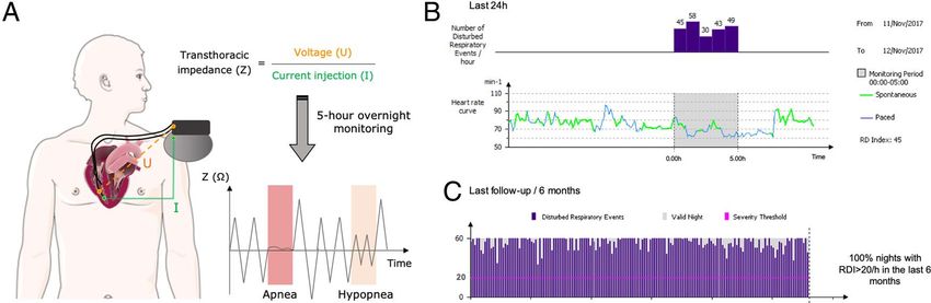
SAM (Sleep Apnea Monitoring)
Some rate-responsive pacemakers have specific algorithms capable of detecting respiratory cycles, recognizing ventilation
pauses and reductions, and deriving indices well correlated with the identification of severe OSA as determined by PSG.
Automatic identification of apnoea and hypopnoea epsiodes for the automatic SAS screening in pacemaker patients (PM and CRT-P).

                                       Period between two respiratory cycles > 10 sec

                         Signal
                         VM (Ω)                                                                            Amplitude VM< 50%
                                                                                                            (for at least 10 sec)*
                                                                                                                      =
                                                                                                              HYPOPNOEA

                                                                                                       t
                                                   Respiratory              Respiratory
                                                   interruption              reduction
                                                    APNOEA                 HYPOPNOEA

                                                                           APNOEA & HYPOPNOEA
     RDI (Respiratory Disturbance index):                                 Number of hours of monitoring

                                RDI defines the severity of Apnoea
The DREAM study showed that a
                                             transthoracic impedance sensor with an
                                             advanced      algorithm,    the  SAM
                                             algorithm, could be used to identify
                                             severe SA in patients with PMs.

                                                  Nasal pressure measurements
                                                  during polysomnography (PSG)
                                                  and corresponding sleep apnea
                                                  monitoring (SAM) recordings of
                                                  transthoracic impedance from the
                                                  minute ventilation sensor .

                                                          Sensitivity Specificity
                                                           88,9%         84,6%
                                                            PPV          NPV
                                                            88,9%       84,6%
Defaye et al. Heart Rhythm 2014;11:842-848
173 patients implanted with an ICD or
cardiac resynchronization therapy
with ICD (CRT-D) endowed with the
ApneaScan diagnostic feature.

In 49 patients the RDI and AHI values
were available for the same night.

At an RDI value of 31 episodes/h, severe SA was detected with 87% (95% CI 72%–
96%) sensitivity and 56% (95% CI 48%–66%) specificity.
           RDI closely correlated with AHI recorded during the same night
                          (r=0.74; 95% CI 0.57–0.84; p
29 pts (20 men, 70 ± 8 years) with indications for implantation of ICD or Pacemaker with ApneaScanTM system.

                                                                Optimal RDI cutoff value to identify
                                                                severe SRBD was 47 episodes/h
                                                                (sensitivity 100%, specificity 100%).

  In the present study, the novel transthoracic impedance-
  based monitoring system ApneaScanTM appeared effective
  in screening PMK and ICD patients for SRBD.
 Della Rocca et al. JCE 2019;56: 327–333
25 pts (24 men, 59.9 ± 14.4 years; LVEF 30.3 ± 6.4%; BMI 25.9 ± 4.2
                                               kg/m²) with indications for implantation of ICD or
                                               CRT-D (INCEPTA; Boston Scientific) were included.

                                                R.O.C. curve. Threshold of 30 for AHI-AS. Sensitivity
                                                is 100%, Specificity 76.47%, PPV 66.67%, NPV 100%.
Defaye et al. Scientific Reports 2019;9:9597
The AIRLESS study shows that a transthoracic impedance sensor
      (ApneaScan algorithm) implemented in the ICDs for SA detection
      through the assessment of transthoracic impedance is a reliable tool
      to identify patients suffering of severe sleep disorders. The
      availability of this new technology might improve the clinical
      management of this patient population. Larger and long-term
      prospective studies are needed to collect data on the clinical impact.

Defaye et al. Scientific Reports 2019;9:9597 doi.org/10.1038/s41598-019-45255-3
Pacemakers were equipped with
the ApneaScan diagnostic feature
(Boston Scientific).
In patients who received a pacemaker,
severe device-detected sleep apnea at the
baseline was independently associated
with a higher risk of AF during follow-up.
Severe sleep apnea on follow-up data
review identified patients who were 2-fold
more likely to experience an atrial
fibrillation episode in the next 3 months.
Mazza et al. Europace 2017;19:1937-1943COSA ABBIAMO IMPARATO?
Case 1, women, 83 years old

               22/5/2015
 Sick sinus syndrome, resistant hypertension,
 obesity, snorer, daytime hypersomnolence, PAF.
Case 4, woman, 83 years
Before CPAP During CPAP training
Giaccardi et al. Clinical Case Reports 2017;5:1465–1467Case 2, man, 82 years old 21/2/2015, FU 23/7/2015
Caso
Case     3, uomo,8985years
     3, women,        anniold
Case 4, women, 75 years old
Case 5, women, 75 years old
Case 6, men, 65 years old
AF and “dangerous liaisons”
Age

                            Alcohol intake abuse               Sleep-disordered
                                                               Breathing

          Chronic diseases
          CAD                                Excessive or incorrect feeding
          Hypertensive heart disease
          Valvular (rheumatic)
          Dilated CMP
          Hypertrophic CMP
          Hyperthyroidism
          Hypertension
          Bronchopulmonary
          disease
          Diabetes
          Congestive HF
          Prior embolic events
The effects of CPAP in OSA patients may even extend as far as affecting the
atrial electrical remodeling: CPAP reduce maximum P-wave dispersion
and maximum P-wave duration presents in moderate to severe OSA,
shortening of signal-averaged P-wave after four or six weeks of therapy.
With CPAP we can treat the
pathophysiological mechanisms
implicated in linking OSA and
AF and can reduce AF episodes
and AF recurrences after
cardioversion and after ablation.
Digby et al . Current Cardiology Reviews 2012; 8: 265-272Case 7, man, 57 years old
Implant FU - 1 month
RDI
PSG results:
OSAS severe
AH Index = 55.6 /hour
t
implant FU - 1 month FU - 4 months
Reduction of
CPAP
compliance
CPAP By courtesy of Dr. J. Marti Almor Hospital del Mar. Barcelona (Sp)Case 8, man, 75 years old
Good CPAP
compliance
Before CPAP CPAPCase 9, men, 70 years old
START CPAPCase 10, men, 79 years old
Atrial FlutterCase 11, men, 70 years old Ablazione efficace di flutter atriale tipico comune, al ripristino del ritmo sinusale riscontro di malattia del nodo del seno. Impianto di pacemaker.
Home Monitoring
COSA POSSIAMO FARE DI PIU’?
Moubarak et al. Heart Rhythm2017;14:359–364
“Sleep apnea Severity SCore” SSSC
    We have collected 103 consecutive pts implanted with single and dual-chamber Microport
    pacemakers with SAM algorithm, irrespective of the type of implantation (first implantation or
    generator replacement) and pacing indication. From January 2015 to October 2017. We classified
    patients according to the percentage of their nights with RDI>20. (Sleep apnea burden).

                      26 pts: 25.2%

                      35 pts: 34.0%

                      13 pts: 12.6%

                      15 pts: 14.6%

                      14 pts: 13.6%

data under revision
“Sleep apnea Severity SCore” SSSC
SSSC 1: no or ‘’mild’’ 34.1 %
OSAS = 59%
SSSC 2: ‘’moderate to
severe’’ OSAS = 41%
data under revisionResults
Sleep apnea burden and mode-switch
SSSC1 SSSC2
8.8 χ² p 0.011
33.3 33.3
14.8
57.9
51.9
Our study confirmed the ability of
specific pacemakers to measure
respiratory disturbances.
The main finding of this study suggests
that the diagnosis of OSA is probably
not binary but introduce the concept of
sleep apnea burden.
Finally confirms the relationship
between SA and AF burden.
data under revisionOur study at first reports a
correlation between CRT
response and sleep apnoea
burden considering gender
differences. In particular, HF-
women responders to CRT
demonstrate a significant
linear decrease in sleep
apnoea burden determined
through a device algorithm,
when compared to a similar
male population.
Mascia et al. Sleep Medicine 2019;64: 106e111Caso
Case 13,3,women,
uomo, 85
80anni
years old
1 month FU 3 months FU
EF 30%
4 months FU 6 months FU
EF 40%A high burden of apneas/hypopneas at night is associated with elevated
NT-proBNP and PCWP values and an increased risk of ADHF over 1 year.
These patients might benefit from early tailored clinical management.
Augusto et al. Am J Cardiol 2019;124:1720−1724Case 12, man, 84 years old
24/8/2015 , FU 16/9/2015
Sick sinus syndrome.Caso
Case 12,3,man,
uomo,
85 85 anniold
years
PSG 07/06/2016CONCLUSION 1
4
4
0
35 16 1.582.000: patients unknow but affected by SDB in Italy
19
About 20 people / for each center / for all days in 1 year
13 219 including Sundays and holidays should be evaluated
8
11
7
3
5
21
1 19
17
4
7
8
Pacemakers have an advantage over
PSG in monitoring sleep every night
and could provide new long-term
17
information on sleep disorders.
Dati aggiornati sito AIPO 2019
A new screening and monitoring tool.CONCLUSION 2
4
4
0
35 16
19
13
8
11
7
3
5
21
1 19
17
4
7
8
17LA SLEEP APNEA FATTORE DI
RISCHIO CARDIOVASCOLARE
GRAZIE PER L’ATTENZIONE
Dott.ssa Marzia Giaccardi
Laboratorio di Elettrofisiologia
UOC Cardiologia I - FirenzeYou can also read