Laparoscopic ovarian cystecomy for a huge ovarian cystic mass : A case report and review of literature
←
→
Page content transcription
If your browser does not render page correctly, please read the page content below
Laparoscopic ovarian cystecomy for a huge ovarian cystic mass : A
case report and review of literature
Case
Report Fady S.Moiety, Osama El Ashkar, Abdel Fattah Agameya
Department of Obstetrics and Gynecology, Faculty of Medicine, Alexandria University,
Alexandria, Egypt.
ABSTRACT
Introduction: Huge ovarian cystic lesions are rarely encountered in modern practice due to the marked development in
health care services and technology on both the diagnostic and therapeutic levels, in addition to the continuous rise of
awareness of women's health issues. Laparoscopic management seems to be the ideal line of intervention.
Case Report: An 18-year-old, virgin female, was presented with abdominal distension. Physical examination and
ultrasonography revealed a huge pelvi-abdominal cystic mass. A laparoscopic ovarian cystectomy was performed. A
follow up for 12 months and was unremarkable. The technique of the operation as well as tips in such a heroic surgery
were described.
Conclusion: Huge ovarian cysts might be successfully and safely treated by laparoscopic excision. There seem to be no
size-related limits for laparoscopic intervention for ovarian cysts; however, experience is a crucial factor.
Key Words: Huge, laparoscopy, ovarian cyst.
Received: 30 January 2018, Accepted: 25 February 2018
Corresponding Author: Fady S.Moiety, Department of Obstetrics and Gynecology, Faculty of Medicine, Alexandria
University, Alexandria, Egypt, Tel.: 00201001455770, E-mail: fmoiety@gmail.com.
ISSN: 2090-7265, February 2018, Vol.8, No.1
INTRODUCTION Vital signs were normal. Abdominal examination
suggested a painless pelvi-abdominal cystic mass, mostly
Ovarian cysts are common and share in many to the left side, reaching up to the left costal margin and
presentations at the gynecology clinic regardless of a few centimeters below the xiphisternum in midline.
the patient's age. They mostly follow a benign course; Laboratory investigations were all normal. Serum
however, some might grow to reach considerable sizes CA-125was 20 U/ml.
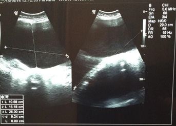
that make surgical intervention inevitable. Huge ovarian Ultrasonographic abdominal scan revealed a huge,
cysts are rare in modern practice due to the outstanding pelvi-abdominal cyst (size: 27 x 16 x 11 cm) reaching up
development in both diagnostic and interventional to the epigastrium, with a single, serous fluid-filled loculus.
technology. Laparoscopic approach is preferred over No papillary, or solid parts nor ascites were detected.
laparotomy in terms of better less blood loss, less (Fig. 1). Right ovary was seen normal. A picture of
pain, shorter hospitalization time and better cosmetic suspected left ovarian benign cystic neoplasm.
outcome[1]. Limitations of laparoscopic route are mainly
related to the size of the mass that if it is above the level
of the umbilicus (i.e. larger than 24 weeks of gestation-
uterine size), it would make laparoscopic intervention
difficult and time-consuming[2]. We present a case of
laparoscopic management of a huge left ovarian cyst. To
our knowledge, only few cases with similar sizes, that were
successfully managed by laparoscopy, were reported.
CASE REPORT
An 18-year-old virgin, was referred to our tertiary
Gynecologic Endoscopy Center, with huge abdominal
Fig. 1: Pre-operative ultrasound image of the cyst.
distension, developing gradually over the past year, causing
pelvic heaviness and disfigured abdomen. She had no past The patient was counseled on the risks and benefits of
medical or surgical history. laparoscopic intervention in such a case and was scheduled
Personal non-commercial use only. EBX copyright © 2017. All rights reserved DOI: 10.21608/ebwhj.2018.5642
138Moiety et al.
for a laparoscopic procedure by the first author. General the mass, the liver, omentum and stomach and taking free
anesthesia and endotracheal intubation was used. Veress fluid samples for cytology, the midline trocar was used to
needle was inserted 3 cm below the costal margin at puncture the cyst, then a suction cannula was introduced
the mid-clavicular line (Palmer's point) for a very short through the same port to aspirate the fluid content which
distance deep to the peritoneum, with caution not to hit was about 6 liters of straw-colored clear fluid, a sample
the mass, to ensure adequate pneumoperitoneum. A five- of which was sent for cytology. The cyst was explored by
mm trocar was then inserted at the same point of entry. A the scope revealing nothing suspicious then widening the
zero degree, 5-mm endoscope was placed after 12 mmHg incision was done by scissors. Excision of the cyst wall
pneumoperitoneum was established. The cystic lesion (stripping technique) was undertaken using traction and
was seen extending from the pelvis to the diaphragm, counter traction by 3 good-grip instruments. Intracorporeal
measuring 28 x 15 x 15 cm. Three ancillary trocars were suturing of the ovarian tissue by Vicryl 0 suture. The cyst
inserted : A 10-mm at the umbilicus, a 5-mm to the left and wall was retrieved via the midline 10-mm port, without the
right of the umbilical port. This was based on the assumption need of a power-morcellator. The procedure duration was
that after aspiration of the fluid content, the cyst size will 98 minutes.
regress to the level below the umbilicus. After exploring Tips for surgery: (Fig. 2)
A: Entry: huge cyst B: Direct trocar puncture
C: Suction from within D: Starting dissection
E: Continuing dissection (stripping) F: Cyst wall almost out
139LAPAROSCOPIC HUGE OVARIAN CYSTECTOMY-CASE REPORT
G: Left ovary back to pelvis H: Cleaning by suction / irrigation
I: Final view
Fig. 2 (A-I): Laparoscopic surgical steps.
A 30-degrees scope would have been a better option The postoperative course was uneventful. There were
for huge cysts; however an experienced camera-driver is no complications. The patient was discharged after 24
needed. hours. Histopathology revealed left ovarian benign serous
cystadenoma. Cytology for peritoneal fluid as well as
Insertion of the Veress needle should be sensible and
cystic fluid aspirates was negative.
slow, for its inadvertent introduction into the cyst would
not only prevent gas insufflation and hence delay the Amazingly, the left ovary restored its normal size and
procedure, but it would also lead to leak of fluid content ultrasonographic texture after 1 month of surgery. Follow
into the peritoneum with potential risk of spread. up at 3, 6 months and at 12 months did not prove any
suspicious clinical, imaging, laboratory or biochemical
Three ancillary trocars should be used, at least one of
markers changes. The young lady is healthy and happy
which should be 10 mm. the level of the ancillary ports
should be at or above the line of the umbilicus and should
be planned according to the estimated size of the cyst after
DISCUSSION
aspiration of the contents.
The best way to minimize recurrence is the stripping Huge ovarian cysts are rare and management of such
technique. Choosing the right plane for cystectomy cases by laparoscopy is difficult and challenging[3]. The
depends on the choice of the right, good-grip, instruments largest ovarian tumor documented weighed 149 kg and
as well as readjusting the 2 instruments at shorter distances removed by Spohn in 1905 by laparotomy[4]. Ishikawa
as we proceed in traction and counter traction. H et al. reported a huge endometriotic cyst managed by
After excision of the cyst, The ovary may be returned laparotomy in 1997, however it was smaller in size than the
to its normal anatomical place and observed irrigated one we are currently reporting[5].
with normal saline solution. Only active bleeding may be The etiology of ovarian cysts varies. Ovarian cysts
controlled by electrosurgery. The ovary usually shrinks may be benign or malignant. The non-neoplastic ovarian
and shows a good self-hemostatic potential. cysts are usually of functional origin. The benign cysts
Suturing the ovarian tissue in such big cysts may be are most frequently endometriotic or simple cysts.
necessary; however, a single purse string suture to pull it Serous and mucinous cystadenomas usually arise from
all together would be enough. neoplastic changes in germinal epithelium. The most
common cystic ovarian neoplasms are serous tumors,
Leave an 18 Fr. drain for 6 – 12 hours. 60% of which are benign, 25% are malignant and 15% are
140Moiety et al.
borderline cases. Clinically, patients with serous tumors CONCLUSION
present with huge abdominal mass with size reaching
even up to 40-45 cm[6]. Most of bigger cysts are benign Huge ovarian cysts might be successfully treated
or of low grade malignancy[5,6]. Small ovarian cysts are by laparoscopic excision. To our knowledge, not many
usually asymptomatic and found incidentally clinically ovarian cysts of that size, managed by laparoscopy, has
or on ultrasound. They may cause pain or discomfort, been reported in the literature. There seem to be no size-
digestive symptoms like nausea and vomiting[9]. Giant limits for laparoscopic intervention for ovarian cysts;
cysts lead to increase in intraabdominal pressure which however, experience is a crucial factor.
may compromise cardiac and respiratory functions. It may
cause supine hypotension secondary to compression of the CONFLICT OF INTEREST
inferior vena cava and aorta[6] which was not reported in
the current case[5]. Ultrasonographic imaging is important There are no conflicts of interest.
in diagnosis. It confirms the ovarian origin of the mass
and provides information on cystic nature and wall
structure[10] and can distinguish between benign and ACKNOWLEDGEMENTS
malignant tumors. Anechoic fluid and thin walls denote
a simple cyst, which in turn signifies a benign tumor. A We would like to thank Alexandria University, Egypt,
malignant cyst is characterized by thick septations and solid for continuous scientific support.
components in the mass[11]. Tagliabue F, reported a similar
huge left ovarian cyst with laparoscopic management,
however, their management entailed extraction of the REFERENCES
cyst through Pfannesteil incision with partial aspiration
of the cyst, which was not our choice of management, 1. Yuen PM, Yu KM, Yip SK, Lau WC, Rogers MS,
as we preferred the entire management to be by Chang A. A randomized prospective study of
laparoscopy[12]. A. Alobaid reported a similar cases, laparoscopy and laparotomy in the management
however, the entry was via open laparoscopy and the cases of benign ovarian masses. Am J Obstet Gynecol
underwent oophorectomy, unlike our management and 1997; 177: 109-14.
approach in the present case[13].
Persistence of the cyst beyond 2 months may justify 2. Knudsen UB, Tabor A, Mosgaard B, Andersen
surgical intervention; however, the majority of the ovarian ES, Kjer JJ, Hahn- Pedersen S, et al. Management
cysts regress spontaneously. Symptomatic or larger of ovarian cysts. Acta Obstet Gynecol Scand
than 7 cm or complex cysts are other indications for 2004; 83: 1012-21.
surgery[13]. Management of giant cysts has traditionally
required a midline laparotomy[14]. This can be accomplished 3. Menahem S, Shvartzman P. Giant ovarian
by enbloc removal of the tumor, with controlled drainage cyst mimicking ascites. J Fam Pract. 1994
of the tumor fluid to decrease the risk of spilling malignant Nov; 39(5): 479-81..
cells[15]. Recently, Laparoscopy became the preferred
approach for ovarian cysts with sizes not exceeding the 4. Spohn AE. Multicystic ovarian tumor weighing 328
umbilicus[16], but only few cases have been reported. lb. Tex State J Med 1905-1906; 1: 273-4.
There is a limit in the working space during laparoscopy
that we could overcome by aspirating the fluid content
within the cyst through the port as described by Salem HA, 5. Ishikawa H1, Taga M, Haruki A, Shirasu K, Minaguchi
who reported laparoscopic management of ovarian cysts H, Hara M. Huge ovarian endometrial cyst: a case
reaching the level of the umbilicus but not up to the size report. Eur J Obstet Gynecol Reprod Biol. 1997
we are reporting[17]. The most significant risk of drainage is Aug;74(2):215-7.
the possibility of contents spillage in the peritoneum with 6. Malkan AD, Singh-Braich P, Panait L, Dudrick SJ.
the subsequent seeding[18]. Although several authors stated Mucinous cystadenoma of the ovary presenting
that giant ovarian cysts are usually benign, there have been as unilateral lower extremity edema. Conn
reports of malignant tumors or tumors of low malignant Med 2009; 73: 517-9.
potential[19, 20]. The excision of giant ovarian cysts by
laparotomy requires lager incision.
7. Bika O, Ola B. Complex serous cystadenoma
The literature, to our knowledge, did not define a with ovarian stroma of the fallopian tube ampulla
maximum size of a cyst to be as contraindication for presentsurgically. Journal of Obstetrics &
laparoscopic intervention as it is mainly dependent on the Gynaecology 2009;29(6):565-566.
experience of the operator. Conservative surgery is always
preferred in young females at reproductive age group[21,22]. 8. Mulayim B, Gurakan H, Dagli V, Mulayim S,
141LAPAROSCOPIC HUGE OVARIAN CYSTECTOMY-CASE REPORT
Aydin O, Akkaya H. Unaware of a giant serous 16. Webb MJ, Decker DG, Mussey E, Williams TJ. Factor
cystadenoma: a case report. Arch Gynaecol Obstet influencing survival in Stage I ovarian cancer. Am J
2006; 273: 381-383 Obstet Gynecol 1973; 116: 222-8.
9. Haspels AA, Zuidema PJ. A giant ovarian cyst in a 17. Salem HA. Laparoscopic excision of large ovarian
Javanese woman. BMJ. 1982; 284: 1410. cysts. J Obstet Gynaecol Res 2002; 28: 290-4.
10. Kaya M, Sakarya MH. Pseudoascites: Report of three
18. Jones DR, Vasilakis A, Pillai L, Timberlake
cases. Turk J Gastroenterol. 2009; 20(3): 224-227.
GA. Giant, benign, mucinous cystadenoma of
the ovary: case study and literature review. Am
11. Mukhopadhyay S. Giant paraovarian cyst. Journal of Surg 1992; 58: 400-3.
Obstet & Gynae India 2006; 56: 352-353.
19. Hunter DJ. Management of a massive ovarian cyst.
12. Tagliabue F, Acquaro P, Confalonieri G, Spagnolo S,
Obstet Gynecol 1980; 56: 254-5.
Romelli A, Costa M. Laparoscopic approach for very
large benign ovarian cyst in young woman. J Min
Access Surg 2009; 5: 75-7 20. Nakamura M, Saitoh M, Miyamoto S, Kubo Y,
Tomita H, Andoh A. Case of a giant mucinous ovarian
13. A. Alobaid, A. Memon, S. Alobaid, and L. Aldakhil, carcinoma with bone metastasis. J Obstet Gynaecol
“Laparoscopic Management of Huge Ovarian Res 2005; 31: 576-8.
Cysts,”Obstetrics and Gynecology International,
vol. 2013, Article ID 380854, 4 pages, 2013. 21. Su-Kon Kim, Jong-Soo Kim, Choong-Hak Park,
doi:10.1155/2013/380854. Jin-Wan Park. A case of giant ovarian cyst managed
successfully through laparoscopic surgery. Korean J
14. Tagge DU, Baron PL. Giant adrenal cyst: management Obstet Gynecol 2012; 55(7): 534-537
and review of the literature. Am Surg 1997; 63: 744-6.
22. Deepak Kumar Singla, Richa Kansal, Isha Bansal,
15. Polat C, Ozacmak ID, Yücel T, Ozmen V. Laparoscopic Gaurav Thami, Nivesh Agrawal. Case report:
resection of giant mesenteric cyst. J Laparoendosc laparoscopic management of a giant ovarian cyst.
Adv Surg Tech A 2000; 10: 337-9. Asian Pac. J. Health Sci., 2014; 1(4S): 43-46
142You can also read