LOPH AND D-MHC IN THE TREATMENT OF FELV ASSOCIATED ACUTE LEUKEMIA IN A CAT - SEER UFRGS
←
→
Page content transcription
If your browser does not render page correctly, please read the page content below
Acta Scientiae Veterinariae, 2021. 49(Suppl 1): 628.

                                                                   CASE REPORT                                                     ISSN 1679-9216
                                                                   Pub. 628

     LOPH and D-MHC in the Treatment of FeLV Associated Acute Leukemia in a Cat

    Rodrigo dos Santos Horta1, Ana Luisa Fajardo Ferreira1, Mariana de Pádua Costa1, Ligia Soares Frossard2,
             Júlia Campero Nimrichter3, Mylena Duarte de Carli3 & Paulo Ricardo de Oliveira Paes1

                                                                ABSTRACT
Background: Bone marrow primary malignancies are denominated leukemias, classified as myeloid or lymphoid, accord-
ing to the cell lineage, and acute or chronic, according to the cell´s state of maturation. In cats, acute lymphoid leukemia
is the most common form, especially in regions endemic for feline leukemia virus and / or feline immunodeficiency virus.
A new treatment protocol for lymphomas, called LOPH, was described for animals with FeLV persistent viremia. This
study aimed to report a case of a cat presenting with FeLV associated acute leukemia and treated with the LOPH protocol,
and, in the rescue phase, a modification of the D-MAC protocol, denominated D-MHC.
Case: A 4-year-old mixed breed intact queen was attended due to lethargy and inappetence. The patient did not present any
relevant abnormalities in the clinical exam and complementary exams were performed including complete blood count,
biochemical profile, SNAP Feline Triple Test, chest radiographs and abdominal ultrasound. Imaging tests and biochemical
values were unremarkable, but the patient presented a reagent result for FeLV and severe leukocytosis due to lymphocyto-
sis. The morphological evaluation of the blood smear revealed the presence of blasts, in a concentration greater than 20%
of the nucleated cells, which allowed the characterization of a leukemic state, probably lymphoid. First-line treatment
was based on the LOPH protocol, including Lomustine, Vincristine, Prednisolone and Doxorubicin, in four-week cycles.
Nevertheless, during the third cycle, 66 days after the institution of this protocol, the patient presented a febrile condition
along with marked leukocytosis due to lymphocytosis, confirming leukemia recurrence. A rescue attempt was performed
with a modification of the D-MAC protocol, originally consisting of the combination of dexamethasone, melphalan, acti-
nomycin-D and cytarabine, but with replacement of actinomycin-D by doxorubicin, and therefore denominated D-MHC.
After the three cycles there was a return of the febrile condition associated with severe pancytopenia and euthanasia was
elected due to poor clinical condition, resulting in a survival time of 124 days. The hematological toxicity of the induction
protocol included anemia and neutropenia, with mainly grade I events, but with the occurrence of a single grade IV event.
The adverse effects of the rescue protocol were similar, but with a greater number of grade IV events.
Discussion: FeLV is considered the most lethal retrovirus of the domestic cat, with a major impact on health and life expec-
tancy. Persistent FeLV antigenemia increases the risk of hematopoietic neoplasms in 62.1 times due to a direct insertional
mutagenesis. In endemic regions, approximately 70% of cats with acute leukemia have persistent FeLV antigenemia, as
the patient in this report. The diagnosis was made through association of retroviral status and the identification of more
than 20% of blasts, possibly lymphoblasts, in the blood stream, sparing the need for a myelogram. Considering the poor
prognosis for acute lymphoid leukemias and the patient’s retroviral status, treatment was initiated with the LOPH protocol,
including lomustine, as a potent agent to induce remission, and doxorubicin, which can result in longer remission interval.
After 66 days free of the disease, the patient presented recurrence of the leukemic condition, starting the rescue protocol
D-MHC. Remission was again obtained with duration similar to the first protocol, however, on the occasion of a new leu-
kemia recurrence, euthanasia was elected. The treatment adopted for the patient in this report resulted in a longer survival
time than reported in other studies. Despite the aggressiveness of the protocols, especially the D-MHC, it was possible to
perform it using the monocyte and granulocyte stimulation factor to reverse neutropenia.
Keywords: oncology, chemotherapy, doxorubicin, lomustine, cytarabine, melphalan.

                                                         DOI: 10.22456/1679-9216.106950
Received: 18 November 2020                                  Accepted: 20 February 2021                                     Published: 13 April 2021
1
 Departamento de Clínica e Cirurgia Veterinárias, Escola de Veterinária, Universidade Federal de Minas Gerais (UFMG), Belo Horizonte, MG, Brazil.
2
 M.V. Autônoma, Belo Horizonte. 3Departamento de Medicina Veterinária, Universidade Vila Velha (UVV), Vila Velha, ES, Brazil. CORRESPONDENCE:
R.S. Horta [rodrigohorta@ufmg.br / rodrigohvet@gmail.com]. Departamento de Clínica e Cirurgia Veterinárias, Escola de Veterinária, Universidade
Federal de Minas Gerais (UFMG). Av. Pres. Antônio Carlos n. 6627. CEP31270-901 Belo Horizonte, MG, Brazil.

                                                                       1
R.S. Horta, A.L.F. Ferreira, M.P. Costa, et al. 2021. LOPH and D-MHC in the Treatment of FeLV Associated Acute Leukemia in
a Cat. Acta Scientiae Veterinariae. 49(Suppl 1): 628.
INTRODUCTION phase, a modification of the D-MAC protocol, deno-
minated D-MHC.
The term leukemia refers to malignant neoplas-
tic proliferation originated in the body´s blood-forming CASE
tissues (i.e., bone marrow), classified based on the cell
A 4-year-old mixed breed intact queen was
lineage in myeloid or lymphoid, and considered acute,
attended at a private practice due to lethargy and
when they involve precursor cells, or chronic, when
inappetence. The physical examination was unrema-
they involve mature cells [3,9]. Lymphoid leukemia,
rkable, however, complementary tests were carried
related to the malignant proliferation of lymphocytes
out and included complete blood count, biochemical
and their precursors, is the most common form in
profile (urea, creatinine, alanine aminotransferase,
humans, dogs and cats, although the real incidence
aspartate aminotransferase, alkaline phosphatase,
remains unknown in Veterinary Medicine [3,6,9,31].
gamma-glutamyl transpeptidase, total protein and
Chronic lymphoid leukemia is the most common in fractions, total bilirubin and fractions, calcium and
dogs, however, in cats, acute lymphoid leukemia, SNAP Feline Triple Test1, chest radiographs and ab-
related to the proliferation of lymphoblasts and/or dominal ultrasound. Imaging exams showed no chan-
pro-lymphocytes, is the most common in endemic ges. Biochemical results were unremarkable, but the
regions for the feline leukemia virus (FeLV) and / or patient presented a reagent result for FeLV and severe
feline immunodeficiency virus (FIV) [12]. leukocytosis (125,500/µL; Ref: 3,000-14,800/µL [18])
A new treatment protocol has been described due to lymphocytosis (95% of the relative count and
for multicentric or mediastinal lymphomas in cats with 119,225/µL; Ref: 1,200-8,000/µL [18], of absolute
FeLV persistent antigenemia. This protocol, called count), associated with normocytemia, with a packed
LOPH, is proposed for an induction phase and consists cell volume (PCV) of 29.2% (Ref: 29-48% [18]) and
of the association of Lomustine, Vincristine (Onco- platelet count within the reference values. In the mor-
vin®), Prednisolone and Doxorubicin (Hydroxydauno- phological evaluation of the blood smear cell types, the
rubicin), showing good tolerance in cats, with complete presence of blast cells was found, in a concentration
response in 81% of cases and an average survival of greater than 20% of the nucleated cells, which allowed
214 days, superior to studies with other protocols in the characterization of a leukemic state, probably lym-
populations with high FeLV infection rates [17]. phoid (Figure 1). As there were no changes in lymph
In cases of disease recurrence, rescue thera- nodes in physical and imaging exams, acute lymphoid
pies can be attempted, with emphasis on the D-MAC leukemia was diagnosed, even in the absence of mye-
protocol, which originally consists of the combination logram. Bone marrow aspirates were not performed
of dexamethasone, melphalan, actinomycin-D and due to the poor clinical condition of the patient and
cytarabine [11]. the need for anesthesia for the procedure.
This study aimed to report a case of a cat With the diagnosis of acute lymphoid leuke-
presenting with FeLV associated acute leukemia and mia, chemotherapy treatment was initiated, and hema-
treated with the LOPH protocol, and, in the rescue tological toxicity was assessed based on the Veterinary
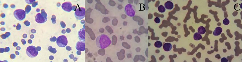
Figure 1. Peripheral blood smear showing blast cells, possibly lymphoid, of medium to large sizes (A, B, C), with frequently irregular or indented nuclei,
with a finely granular chromatin pattern and prominent single or multiple nucleoli (B, C), and moderately basophilic cytoplasm with multivacuolizations
(B, C) [Romanowski; 100x].
2R.S. Horta, A.L.F. Ferreira, M.P. Costa, et al. 2021. LOPH and D-MHC in the Treatment of FeLV Associated Acute Leukemia in
a Cat. Acta Scientiae Veterinariae. 49(Suppl 1): 628.
Table 1. Chemotherapy protocol for acute leukemia in a domestic cat, for induction it was used the LOPH protocol based on the combination of lomus-
tine, vincristine (Oncovin®), prednisolone and doxorubicin (hydroxydaunorubicin) [17] and in a rescue attempt it was used the D-MHC protocol based
on the combination of dexamethasone, melphalan, doxorubicin (hydroxydaunorubicin) and cytarabine [11].
Week Drug
Induction Phase (Loph Protocol)*
Week 1 Lomustine 6 mg/kg PO
Week 2 Vincristine 0,6 mg/m2 IP
Week 3 Doxorubicin 1 mg/kg IV**
Week 4 Vincristine 0,6 mg/m2 IP
Rescue Phase (D-Mhc Protocol)***
Week 1 Doxorubicin 1 mg/kg IV + Cytarabine 300 mg/m2 SC + Dexamethasone 1 mg/kg SC
Week 2 Melphalan 20 mg/m2 PO + Dexamethasone 1 mg/kg SC
*Repeat 5x (total 20 weeks); **Pre-medication with dexamethasone, at a dose of 0.5 mg/kg, intravenously. ***Repeat indefinitely Prednisolone 10 mg
/ cat / day, orally, continuous in the first cycle, followed by a gradual reduction from the second cycle (25% every 2 weeks).
Cooperative Oncology Group-Common Terminology counts (1840/µL) was observed, allowing continuation
Criteria for Adverse Events v.1.1 [33] of the chemotherapy protocol (doxorubicin4). In the
First-line treatment was based on the LOPH following week, as the neutrophils remained close to
protocol [lomustine (magistral), vincristine (Onco- the previous values, chemotherapy was continued, this
vin®)2, prednisolone (Prediderm®)3 and doxorubicin4] time with the concomitant administration of a single
(Table 1). The blood count was repeated 2 days after the dose of filgrastim. The second cycle of chemotherapy
first chemotherapy session (lomustine), with a reduc- and the first session of the third cycle were performed,
tion of leukocytosis and lymphocytosis by more than despite grade I-II neutropenia. Nevertheless, in the
50%, but with the installation of mild anemia (PCV of second session of the third cycle, the patient had a
24%, Ref: 29-48%; red blood cell count of 4.6 x10⁶/µL, fever and chemotherapy was suspended until imaging
Ref: 5.92-9.93x10⁶/µL; and hemoglobin concentration tests were performed, to discard infection. There were
of 7.9 g/dL; Ref: 9.3-15.9 g/dL [18]), normochromic no changes in imaging tests, but another complete
normocytic, with no anisocytosis and polychromasia blood count revealed, once more, marked leukocytosis
and 3% of rubricytes and metarrubriticytes among (71,300/µL; Ref: 3000-14800/µL [18]) due to lym-
total nucleated cells, characteristic of non-regenerative phocytosis (81% of the relative count and 57753/µL;
anemia probably due to bone marrow impairment or Ref: 1200-8000/µL, of absolute count [18]), confirming
myelophitisis. A week after the first chemotherapy ses- leukemia recurrence. Chemotherapy continued with
sion, leukogram was normal, but there was worsening the second session of this cycle (vincristine)2, and in
of the anemia, with reduction of PCV (18.5%; Ref: the following 2 days, the administration of cytarabine
29-48% [18]), red blood cell count (3.01x10⁶/µL; Ref: (Citarax®)7, subcutaneously, at a dose of 600 mg/m2,
5.92-9.93x10⁶/µL [18]) and hemoglobin concentration divided into 4 doses, every 12 h.
(6.0 g/dL; Ref: 9.3-15.9 g/dL [18]), hypochromic With treatment, the febrile condition ceased
macrocytic, with 1% of nucleated erythroid cells. In and the patient’s general condition improved, with
the third week of chemotherapy, a mild recovery from resolution of lymphocytosis, but with the presence
anemia (grade II) was observed, but there was marked of severe neutropenia (820/µL; Ref: 2500-8500/µL
leukopenia and neutropenia (grade IV). Oral antibiotic [18]), requiring, again, institution of filgrastim, which
therapy with marbofloxacin (Marbopet®)5 (3 mg/kg) allowed, in the following week, the institution of the
was initiated and a granulocyte and monocyte stimu- rescue protocol D-MHC [dexamethasone (Dextar®)8
lating factor (i.e., filgrastim (Granulokine®)6), was melphalan (Alkeran®)9, doxorubicin4, and cytarabine7]
administered subcutaneously, at a dose of 5 mcg/kg, for (Table 1), adapted from the D-MAC protocol, with
2 consecutive days. After 48 h, an increase in neutrophil the replacement of actinomycin-D by doxorubicin4
3R.S. Horta, A.L.F. Ferreira, M.P. Costa, et al. 2021. LOPH and D-MHC in the Treatment of FeLV Associated Acute Leukemia in
a Cat. Acta Scientiae Veterinariae. 49(Suppl 1): 628.
(hydroxydaunorubicin), of the same pharmacological [15,20]. In spite of that, the virus remains widespread in
group. The beginning of the rescue protocol was marked underdeveloped countries, as in Brazil, with prevalence
by mild anemia (PCV of 22.1%; Ref: 29-48% [18]), rates ranging from 8% to 63% in the states of São Pau-
which was maintained throughout the treatment (3 lo, Minas Gerais and Rio Grande do Sul [1,5,24,29].
cycles), along with episodes of neutropenia (grade III- In endemic regions, approximately 70% of cats with
-IV). After the 3 cycles there was a return of the febrile acute leukemia have persistent FeLV antigenemia [34],
condition associated with severe pancytopenia (PCV of as in the patient in this report.
10.1%, total leukocyte count of 100/µL and platelets Historically, the diagnosis of acute leukemia
count of 38x103/µL) and euthanasia was elected due is based on the identification of more than 30% of
poor clinical condition. Necropsy was not authorized. blasts in the peripheral blood (leukemic leukemia) or
The disease-free interval since the start of che- in the bone marrow, however, the cutoff point related
motherapy (induction phase) was 66 days and survival to the identification of blasts in the peripheral blood
was 124 days. The LOPH protocol presented packed has been reduced to 20% [6] in dogs, while the World
cell volume and neutrophils toxicity grade I 4 times Health Organization reduced this value to 20% in pe-
and grades II and III once, in addition to grade I and ripheral blood and bone marrow [3], although there is
III hemoglobin toxicity once. The D-MHC protocol no consensus in Veterinary Medicine. In the study by
presented packed cell volume toxicity grade I once, Cotter [8], the diagnosis of acute lymphoid leukemia
grade II 3 times and grade IV twice; hemoglobin was established, in cats, in the presence of more than
toxicity grade I twice and grade II once; neutrophils 50% of lymphoblasts in the bone marrow. However,
toxicity grade III once and grade IV 4 times and other parameters must be considered, including con-
platelets toxicity grade III once. During treatment, in
firmation of the lymphoid origin by flow cytometry
the induction and rescue phases, filgrastim was used
or immunocytochemistry, as well as the occurrence
before 7 treatments (8 applications), in weeks 3, 4, 5,
of anemia, neutropenia and / or thrombocytopenia
8, 9, 11 e 12. Antibiotic therapy with marbofloxacin5
[6,31]. The diagnosis, in the present report, was made
was instituted and maintained whenever the neutrophil
through the association of retroviral status, absence
count was below 1000/µL.
of identifiable lymphadenopathy and, based on the
DISCUSSION current literature [3,6,31], with the identification of
more than 20% of blasts in the blood stream, sparing
FeLV is considered the most lethal retrovirus
the need for a myelogram, whose sample collection
of the domestic cat, with a major impact on the health
and life expectancy [25]. Persistent FeLV antigenemia must be made under general anesthesia, discouraged
reduces the patient’s life expectancy to 2.4 years, with due to the patient’s poor clinical condition.
only 20% of animals surviving after three years [19]. It Although flow cytometry is considered the
is also associated with the occurrence of opportunistic gold standard for differentiation of lymphoid and
infections and damage to target organs, and 15-23% myeloid precursors [9], this technique was not avai-
of cancer development [2,14,24-26]. FeLV infection lable at the time of care for this patient, as for immu-
increases the risk of hematopoietic neoplasms in 62.1 nocytochemistry. Histochemical stains could have been
times, mainly lymphomas, but also leukemias [26]. performed, considering that lymphoid precursors are
The carcinogenic effect might be indirect, related to usually negative for Sudan Black, myeloperoxidase
immunosuppression, with reduction of the antitumor and alkaline phosphatase, however, they have low
response, however it is mainly related to a direct viral specificity and staining with alkaline phosphatase can
effect, characterized by insertional mutagenesis, and occur, albeit weaker, in neoplastic T lymphocytes [28].
promotion of transcription of proto-oncogenes, with Therefore, cell identification was based on morpholo-
emphasis on the MYC [2,14,24,26]. gy. As observed in this report, lymphoblasts are large
FeLV has a worldwide distribution, however, cells with reduced nucleus:cytoplasm ratio basophilic
there has been a drastic reduction in its prevalence due cytoplasm and reduced granulation, regular nuclear
to the institution of preventive measures, including membrane, indented and irregular nucleus, presence of
vaccination, diagnosis and isolation of sick animals, prominent single or multiple nucleoli, being suggestive
in the United States, Canada and most parts of Europe of acute lymphoid leukemia [16].
4R.S. Horta, A.L.F. Ferreira, M.P. Costa, et al. 2021. LOPH and D-MHC in the Treatment of FeLV Associated Acute Leukemia in
a Cat. Acta Scientiae Veterinariae. 49(Suppl 1): 628.
The lymphocytosis (119225/µL; Ref: 1500- a modification of the D-MAC protocol [11], conside-
7000/µL [18]) observed in the patient of this report ring the greater availability and easier acquisition of
exceeds, with a wide margin of difference, that obser- doxorubicin, which contributed to a faster initiation of
ved in reactive lymphocytosis, which rarely exceeds the protocol. Cats have specific polymorphisms in the
30,000/µL [4]. The study by Tomiyasu et al. [31], ABCG2 gene, resulting in the abnormal expression of
included 6 cats with acute lymphoid leukemia, aged their transcriptional product, the breast cancer resistan-
between 1 and 11 years (mean 5.5 years), with anti- ce protein (BCRP), related to anthracyclines resistance,
bodies to FIV identified in 1 cat and 3 others showing with consequent inefficient efflux of their substrates,
FeLV antigenemia. Anemia was identified in 4/6 cats such as doxorubicin [23], which favors, therefore, the
and thrombocytopenia in 5/6, while leukopenia was use of this drug in the induction and rescue protocol
observed in 2/6 cats and leukocytosis in 4/6, 3 of [17]. The patient presented a new clinical remission,
which had lymphocytosis greater than 40,000/µL. The with duration similar to that observed with the first
percentage of lymphoblasts in peripheral blood ranged protocol, but there was a novel recurrence with severe
from 5-97.5%, and 5/6 had lymphoblasts greater than pancytopenia, and euthanasia was elected.
30%. In the present report, despite the magnitude of The prognosis of acute lymphoid leukemia
lymphocytosis, anemia and thrombocytopenia were not in dogs and cats is slightly better than the myloid
observed in the first blood count. However, after the counterpart, however, it remains poor and, although
first chemotherapy session, there was a reduction in 20-40% of patients reach clinical remission with
the lymphocyte count of almost 50%, associated with chemotherapy, this response is usually short, lasting
a reduction in PCV (from 29.2% to 18.5%), attributed 1-3 months [9]. The disease-free interval and survi-
to the reduction in the half-life of erythrocytes, due to val in the present report were, however, higher than
the inflammatory process, possibly associated with described in the literature, especially in cats [9,31].
myelophthisis and reduced bone marrow response. Re- In dogs and cats, the treatment of acute leukemia is
covery on subsequent exams was attributed to clinical usually carried out with the same protocols used for
remission of the disease and bone marrow regeneration high-grade lymphomas, despite the worse prognosis
[13]. A series of hematological abnormalities occurred and the intrinsic differences of each type of leukemia,
as a result of the myelosuppressive effects of chemo- whether lymphoid or myeloid and their subtypes
therapy and disease recurrence. [9,31]. The COP protocol, first described by Cotter [8],
Considering the poor prognosis for acute is the most widespread in cats, with its therapy being a
lymphoid leukemias [9] and the patient’s retroviral combination of cyclophosphamide, vincristine (Onco-
status, treatment was initiated with the LOPH protocol, vin®2) and prednisolone. The treatment of lymphoma
suggested by Horta et al. [17]. This protocol includes results in complete remission in approximately 77% of
lomustine, as a potent agent to induce remission, cases, with a disease-free interval of 150 to 251 days
considering its ineffectiveness in a possible rescue and median survival of 266 days, although the best
approach in cats with lymphoma, which resulted in a results are obtained in populations with reduced FeLV
disease-free interval of only 39 days [10]. The LOPH infection rate, which is related to a worse prognosis
protocol also includes doxorubicin in the induction [8,30,32]. Regarding the viremic status of cats, in a
phase. In underpowered studies, with a small number study with the COP protocol in cats with lymphoma,
of cats with lymphoma, doxorubicin promoted longer with different clinical presentations, a median survival
remission intervals, when included in the induction of only 37 days was observed for cats with persistent
[17,32] or maintenance protocol [22]. FeLV antigenemia [32], while those without detectable
In the present study, after 66 days free of the FeLV had a survival of 170 days. The CHOP protocol
disease, the patient developed a fever that initially and its modifications are based on the addition of do-
raised the suspicion of opportunistic infections, but xorubicin (hydroxydaunorubicin), however, lower rates
with the sequential manifestation of lymphocytosis, of complete remission and survival are observed [7,21].
the recurrence of the leukemic condition was evi- The Wisconsin-Madison protocol, originally described
dent, requiring the rapid establishment of a rescue in 1996 as a CHOP-based protocol, was evaluated by
protocol. For this, the D-MHC protocol was used, as Milner et al. [21], who observed a complete remission
5R.S. Horta, A.L.F. Ferreira, M.P. Costa, et al. 2021. LOPH and D-MHC in the Treatment of FeLV Associated Acute Leukemia in
a Cat. Acta Scientiae Veterinariae. 49(Suppl 1): 628.
rate of only 47% and an average survival of 210 days. with few evidences in the literature. The treatment
The inclusion of L-asparaginase, resulting in the L- of the patient in this report with the LOPH protocol
-CHOP protocol shows similar results with complete resulted in a longer survival than that reported in other
remission rate in 74% of cases and median survival of studies. In addition, the adaptation of the D-MAC pro-
232 days [27]. A study showed that retroviral status tocol, called D-MHC, enabled the rapid institution of
did not influence the survival of cats with acute lym- rescue, which also contributed to the greater survival of
phoid leukemia [31], however the study included only the patient. Both protocols had grade III and grade IV
6 cats and chemotherapy treatment was performed in toxicity events, however, the LOPH protocol presen-
only 5, with a median survival of 55 days. However, ted mainly grade I events, being well tolerated by the
the chemotherapy protocol used was not uniform patient. The D-MHC protocol was shown to be a more
and included the use of prednisolone alone (33-days aggressive protocol for the patient, with more grade
survival), L-CHOP (62-day disease-free interval) and III and IV toxicity events, but with satisfactory results,
the combination of L-asparaginase, cytarabine and when associated with supportive treatments, such as the
nimustine (free interval 106 days of illness). use of filgrastim and prophylactic antibiotic therapy.
The hematological toxicity of the LOPH pro- MANUFACTURERS
tocol included anemia and neutropenia, which reached 1
IDEXX Brasil Laboratórios Ltda. São Paulo, SP, Brazil.
2
Antibióticos do Brasil Ltda. Cosmópolis, SP, Brazil.
grade IV only once, maintaining mainly grade I, as 3
Ouro Fino Saúde Animal Ltda. Cravinhos, SP, Brazil.
reported by Horta et al. [17]. However, it was necessary 4
Eurofarma Laboratórios S.A. São Paulo, SP, Brazil.
to use filgrastim in certain stages of the protocol to 5
Ceva Saúde Animal Ltda. Paulínia, SP, Brazil.
allow continuity of treatment. Although its use in vete-
6
Amgen Biotecnologia do Brasil Ltda. Taboão da Serra, SP, Brazil.
7
Blau Farmacêutica S.A. Cotia, SP, Brazil.
rinary medicine is controversial, it is able to accelerate 8
União Química Farmacêutica Nacional S.A. São Paulo, SP, Brazil.
recovery and reduce the severity of neutropenia in dogs 9
Aspen Pharma Indústria Farmacêutica Ltda. Serra, ES, Brazil.
undergoing chemotherapy with cyclophosphamide
Acknowledgments. Anna Raquel Amaral for being such a
[35]. In the D-MAC rescue protocol, the side effects lovely “mother” for her cat and consenting with this scientific
found were similar, but in a greater number and more publication.
grade IV events.
Declaration of interest. The authors report no conflicts of
Acute leukemia associated with FeLV is a ne- interest. The authors alone are responsible for the content and
oplasm with a poor prognosis and complex treatment, writing of this paper.
REFERENCES
1 Almeida N.R., Danelli M.G.M., Silva L.H.P., Hagiwara M.K. & Mazur C. 2012. Prevalence of feline leukemia
virus infection in domestic cats in Rio de Janeiro. Journal of Feline Medicine and Surgery. 14(8): 583-586. DOI:
10.1177/1098612X12444693.
2 Almeida N.R., Soares L.D.&Wardini A.B.W. 2016. Alterações clínicas e hematológicas em gatos domésticos natu-
ralmente infectados pelo Vírus da Leucemia Felina (FeLV). Revista de Saúde, 7(1): 27-32. DOI: 10.21727/rs.v7i1.85.
3 Arber D.A., Orazi A., Hasserjian R., Thiele J., Borowitz M.J., Le Beau M.M., Bloomfield C.D., Cazzola M. &
Vardiman J.W. 2016. The 2016 revision to the World Health Organization classification of myeloid neoplasms and
acute leukemia. Blood. 127(20): 2391-2405. DOI: 10.1182/blood-2016-03-643544.
4 Avery A.C. & Avery P.R. 2007. Determining the Significance of Persistent Lymphocytosis. Veterinary Clinics: Small
Animal Practice. 37(2): 267-282. DOI: 10.1016/j.cvsm.2006.11.001.
5 Barbosa F.C., Christianine M.P.T. & Waldemarin K.C.A. 2001. Prevalência de Leucemia Felina em Gatos Domé-
stico de Uberlândia – MG. Arquivos de Ciências Veterinárias e Zoologia. 5(2): 207-211.
6 Bennett A.L., Williams L.E., Ferguson M.W., Hauck M.L., Suter S.E., Lanier C.B. & Hess P.R. 2017. Canine acute
leukaemia: 50 cases (1989-2014). Veterinary and Comparative Oncology. 15(3): 1101-1114. DOI: 10.1111/vco.12251.
7 Collette S.A., Allstadt S.D., Chon E.M., Vernau W., Smith A.N., Garret L.D., Choy K., Rebhun R.D., Rodriguez
Jr C.O. & Skorupski K.A. 2016. Treatment of feline intermediate to high-grade lymphoma with a modified university
of Wisconsin-Madison protocol: 119 cases (2004-2012). Veterinary and Comparative Oncology. 14(1): 136-146. DOI:
10.1111/vco.12158.
6R.S. Horta, A.L.F. Ferreira, M.P. Costa, et al. 2021. LOPH and D-MHC in the Treatment of FeLV Associated Acute Leukemia in
a Cat. Acta Scientiae Veterinariae. 49(Suppl 1): 628.
8 Cotter S.M. 1983. Treatment of lymphoma and leukemia with cyclophosphamide, vincristine, and prednisone: II.
Treatment of cats. The Journal of the American Animal Hospital Association. 19(2): 166-172.
9 Dobson J., Villiers E. & Morris J. 2006. Diagnosis and management of leukaemia in dogs and cats. In Practice. 28(1):
22-31. DOI: 10.1136/inpract.28.1.22.
10 Dutelle A.L., Bulman-Fleming J.C., Lewis C.A. & Rosenberg M.P. 2012. Evaluation of lomustine as a rescue agent for
cats with resistant lymphoma. Journal of Feline Medicine Surgery. 14(10): 694-700. DOI: 10.1177/1098612X12448017.
11 Elliot J. & Finotello R. 2018.A dexamethasone, melphalan, actinomycin-D and cytarabine chemotherapy protocol as a
rescue treatment for feline lymphoma. Veterinary and comparative oncology. 16(1): 11-151. DOI: 10.1111/vco.12360.
12 Essex M.E. 1982. Feline leukemia: a naturally occurring cancer of infectious origin. Epidemiologic Reviews. 4: 189-
203. DOI: 10.1093/oxfordjournals.epirev.a036246
13 Fry M.M. & McGavin M.D. 2013. Medula Óssea, Células Sanguíneas e Sistema Linfático. In: Zachary J. & McGavin
M. (Eds). Bases da Patologia em Veterinária. 6.ed. Rio de Janeiro: Elsevier, pp.1831-2026.
14 Hagiwara M.K., Reche Júnior A. & Lucas S.R.R. 1997. Estudo clínico da infecção de felinos pelo vírus da leucemia
felina em São Paulo. Revista Brasileira de Ciência Veterinária. 4(1): 35-38.
15 Hartmann K. 2012. Clinical Aspects of Feline Retroviruses: A Review. Viruses. 4(11): 2684-2710. DOI: 10.4322/
rbcv.2015.065.
16 Harvey J.W. 2001. Atlas of Veterinary Hematology: Blood and Bone Marrow of Domestic Animals. St. Louis: Saunders,
240p.
17 Horta R.S., Souza L.M., Sena B.V., Almeida O.I., Jaretta T.A., Pimenta M.M. & Reche Júnior A. 2020. LOPH: a nov-
el chemotherapeutic protocol for feline high-grade multicentric or mediastinal lymphoma, developed in an area endemic
for feline leukemia vírus. Journal of Feline Medicine and Surgery. 23(2): 86-97. DOI: 10.1177/1098612X20926893.
18 Klaassen J. 1999. Reference Values in Veterinary Medicine. Laboratory medicine. 30(3): 194-197.
19 Levy J.K., Scoot H.M., Lachtara J.L. & Crawford P.C. 2006. Seroprevalence of feline leukemia virus and feline
immunodeficiency virus infection among cats in North America and risk factors for seropositivity. Journal of the
American Veterinary Medical Association. 228(3): 371-376.
20 Lutz H., Addie D., Belák S., Boucraut-Baralon C., Egberink H., Frymus T., Gruffydd-Jones T., Hartmann K.,
Hosie M., Lioret A., Marsilio F., Pennisi M.G., Radford A.D., Thiry E. Truyen U. & Horzinek M.C. 2000. Feline
Leukaemia ABCD guidelines on prevention and management. Journal of Feline Medicine and Surgery. 11(7): 565-574.
DOI: 10.1016/j.fms.2009.05.005.
21 Milner R.J., Peyton J., Cooke K., Fox L.E., Gallangher A., Gordon P. & HesterJ. 2005. Response rates and
survival times for cats with lymhpoma treated with the University of Wisconsin - Madison chemotherapy protocol:
38 cases (1996–2003). Journal of the American Veterinary Medical Association. 227(7): 1118-1122. DOI: 10.2460/
javma.2005.227.1118.
22 Moore A.S., Cotter S.M., Frimberger A.E., Wood C.A., Rand W.M. & L’Heureux D. 1996. A comparison of
Doxorubicin and COP for Maintenance of Remission in Cats with Lymphoma. Journal of Veterinary Internal Medicine.
10(6): 372-375.
23 Ramirez C.J., Minch J.D., Gay J.M., Lahmers S.M., Guerra D.J., Haldorson G.J., Schneider T. & Mealey K.L.
2011. Molecular genetic basis for fluoroquinolone-induced retinal degeneration in cats. Pharmacogennet Genomics.
21(2): 66-75. DOI: 10.1097/FPC.0b013e3283425f44.
24 Reche Júnior A., Hagiwara M.K. & Lucas S.R.R. 1997. Clinical study of acquired immunodeficiency syndrome in
domestic cats in São Paulo. Brazilian Journal of Veterinary Research and Animal Science. 24(3): 152-155.
25 Reinacher M. 1989. Diseases Associated with Spontaneous Feline Leukemia Virus (FeLV) Infection in Cats. Veterinary
Immunology and Immunopathology. 21(1): 85-95. DOI: 10.1016/0165-2427(89)90132-3.
26 Shelton G.H., Grant C.K., Cotter S.M., Gardner M.B., Hardy W.D. & DiGiacomo R.F. 1990. Feline Immuno-
deficiency Virus and Feline Leukemia Virus Infections and their Relationships to Lymphoid Malignancies in Cats: A
Retrospective Study (1968-1988). Journal of Acquired Immune Deficiency Syndromes. 3(6): 623-630.
27 Simon D., Eberle N., Laacke-Singer L. & Nolte I. 2008. Combination chemotherapy in feline lymphoma: treat-
ment outcome, tolerability, and duration in 23 cats. Journal of Veterinary Internal Medicine. 22(2): 394-400. DOI:
10.1111/j.1939-1676.2008.0057.x.
7R.S. Horta, A.L.F. Ferreira, M.P. Costa, et al. 2021. LOPH and D-MHC in the Treatment of FeLV Associated Acute Leukemia in
a Cat. Acta Scientiae Veterinariae. 49(Suppl 1): 628.
28 Stokol T., Schaefer D.M., Shuman M., Belcher N. & Dong L. 2015. Alkaline phosphatase is a useful cytochemical
marker for the diagnosis of acute myelomonocytic and monocytic leukemia in the dog. Veterinary Clinical Pathology.
44(1): 79-93. doi:10.1111/vcp.12227
29 Teixeira B.M., Rajão D.S., Haddad J.P., Leite R.C. & Reis J.K.P. 2007. Ocorrência do vírus da imunodeficiência
felina e do vírus da leucemia felina em gatos domésticos mantidos em abrigos no município de Belo Horizonte. Arquivo
Brasileiro de Medicina Veterinária e Zootecnia. 59(4): 939-942. DOI: 10.1590/S0102-09352007000400019.
30 Teske E., Straten G., Noort R. & Rutterman G.R. 2002. Chemotherapy with cyclophophamide, vincristine, and
prednisolone (COP) in cats with malignant lymphoma: new results with an old protocol. Journal of Veterinary Internal
Medicine. 16(2): 179-186. DOI: 10.1892/0891-6640(2002)0162.3.co;2.
31 Tomiyasu H., Doi A., Chambers J.K., Goto-Koshino Y., Ohmi A., Ohno K. & Tsujimoto H. 2018. Clinical and
clinicopathological characteristics of acute lymphoblastic leukaemia in six cats. Journal of Small Animal Practice.
59(12): 742-746. DOI: 10.1111/jsap.12917.
32 Vail D.M., Moore A.S., Ogilvie G.K. & Volk L.M. 1998. Feline Lymphoma (145 Cases): Proliferation Indices, Cluster
of Differentiation 3 Immunoreactivity, and Their Association with Prognosis in 90 Cats. Journal of Veterinary Internal
Medicine. 12(5): 349-354. DOI: 10.1111/j.1939-1676.1998.tb02134.x.
33 Veterinary cooperative oncology group. 2016. Common terminology criteria for adverse events (VCOG-CTCAE)
following chemotherapy or biological antineoplastic therapy in dogs and cats v1.1. Veterinary Comparative Oncology.
14(4): 417-446. DOI: 10.1111/vco.283.
34 Weijer K. & Daams J.H. 1976. The presence of leukaemia (lymphosarcoma) and feline leukaemia virus (FeLV) in
cats in The Netherlands. Journal Small Animal Practice. 17(10): 649-659. DOI: 10.1111/j.1748-5827.1976.tb06925.x.
35 Yamamoto A., Fujino M., Tsuchiya T. & Iwata A. 2011. Recombinant canine granulocyte colony-stimulating factor
accelerates recovery from cyclophosphamide-induced neutropenia in dogs. Veterinary immunology and immunopathol-
ogy. 142(3-4): 271-275. DOI: 10.1016/j.vetimm.2011.05.021.
CR628
http://seer.ufrgs.br/ActaScientiaeVeterinariae
8You can also read