MAGNESIUM ALLOYS FOR BIOMEDICAL APPLICATIONS - A REVIEW - UNIVERSITA' DEGLI STUDI DI ROMA
←
→
Page content transcription
If your browser does not render page correctly, please read the page content below
UNIVERSITA’ DEGLI STUDI DI ROMA
           TOR VERGATA

 MAGNESIUM ALLOYS FOR
BIOMEDICAL APPLICATIONS
          A REVIEW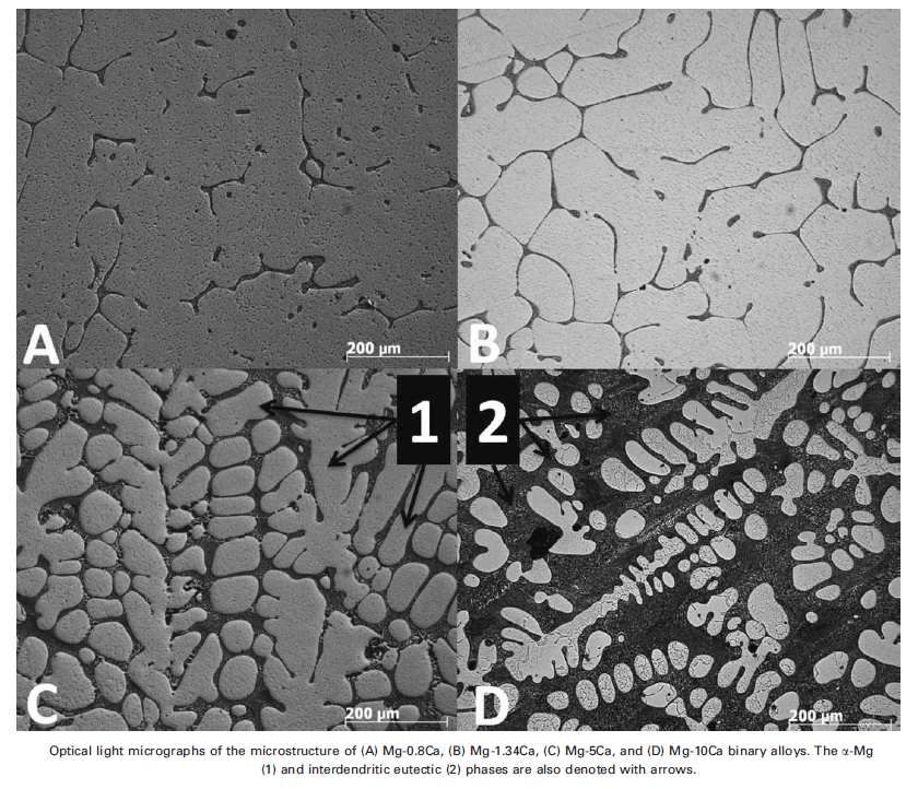
OUTLINE

Magnesium and its alloys

Biodegradable/bioresorbable materials

Toxicity

Characterization

Applications
IMPURITY ELEMENTS

Iron, nickel and copper are extremely deleterious because of low solubility limits and
provide active cathodic sites.
At the same concentration, the detrimental effect of these elements decreases as
follows: Ni, Fe, Cu
Tolerance limits are influenced by the presence of third elements: the iron tolerance
limit for magnesium-aluminum alloys depends on the Mn concentration
IMPURITY ELEMENTS

IRON
• Galvanic coupling
• Al-Fe compound is cathodic to the Mg matrix
• 7% Al → 5wt-ppm Fe
• 10% Al → too low to be determined

NICKEL
IMPURITY ELEMENTS

MANGANESE
•   Mn enhances ductility
•    Mn does not improve corrosion resistance, but reduces the harmful effect of
    impurities
•   1% Mn sharply decreased the corrosion rate of Mg when Fe And Cu impurity
    contents exceed their tolerance limits
        Two possible routes
       1. Mn combines with Fe in the molten Mg alloy and forms an intermetallic
        compound which settles to the melt bottom, lowering Fe content
       2. Mn encapsulates Fe particles, making them less active as local cathodes
ALLOYING ELEMENTS ALUMINUM • Solid solution • Mg17Al12 • Al improves the corrosion resistance (passivating element)
ALLOYING ELEMENTS

ZINC
• Zn can increase the tolerance limits and reduce the effect of impurities once the
tolerance limit has been exceeded
• The addition of 1% Zn to pure Mg raises the tolerance limit for Ni
• Zn improved the tolerance for Mg-Al alloys for Fe, Ni, Cu

CALCIUM
• Ca contributes to solid solution strengthening and precipitation strengthening
• Grain refining agent
• In binary Mg-Ca alloys Mg2Ca is formed while in Al containing alloys Al2Ca forms
first. Both phases improve creep resistance due to solid solution strengthening and
precipitation strengthening
•It is well tolerated in the human body since it is an essential cation
ALLOYING ELEMENTS

LITHIUM
• Li is able to change the lattice structure from h.c.p. to b.c.c.
• It can be used to enhance ductility and formability of Mg alloys but it has a negative
effect on strength

RARE EARTH ELEMENTS (RE)
RE elements are chemically classified by their ionic radii in three groups:
    1. Light RE elements (from La to Pr)
    2. Medium Re elements (from Nd to Gd)
    3. Heavy RE elements (from Tb to Lu)
•Large solid solubilities in Mg: Y, Gd, Tb, Dy, Ho, Er, Tm, Yb, Lu
• Limited solubility in Mg: Nd, La, Ce, Pr, Sm, Eu
• RE is kept in solid solution → solid solution strenghtening
• RE can form complex intermetallic phases with Al or Mg → obstacles for dislocation
movements
Mg ALLOYS FOR BIOMEDICAL APPLICATIONS
Mg ALLOYS FOR BIOMEDICAL APPLICATIONS

The purpose of biodegradable imlants and coatings is to support tissue regeneration
and healing in a specific application by material degradation and concurrent implant
replacement through the surronding tissue
Biodegradable materials have an advantage over existing biodegradable materials
such as polymers, ceramics or bioactive glasses in load bearing applications that
require a higher tensile strenght and a Young’s modulus that is closer to bone
Mg2+ is an essential element and present in large amounts (the fourth most
abundant cation) in the human body
Mg ALLOYS FOR BIOMEDICAL APPLICATIONS

Rapid corrosion is an intrinsic response of magnesium alloys to chloride containing
solutions, including the human body fluid or blood plasma
The degradation of magnesium alloys leads to hydrogen evolution and alkalization
In the human body, the evolved hydrogen bubbles from a corroding magnesium
implant can be accumulated in gas pockets next to the implant, which will delay
healing of the surgery region and lead to necrosis of tissues, because the gas
pockets can cause separation of tissue layers
In the worst case when large hydrogen bubbles are present in the blood circulating
system, there will be a risk that the bubbles may block the blood stream, causing
death of a patient
Mg ALLOYS FOR BIOMEDICAL APPLICATIONS

The local alkalization can unfavorably affect the pH dependent physiological
reaction balances in the vicinity of the magnesium implant and may even lead to an
alkaline poisoning effect if the local in vivo pH value exceeds 7.8 in that region.
A strategy to solve these problems is to slow down the biodegradation (i.e.
corrosion) of magnesium alloys, so Mg2+ ions, H2 bubbles and OH- ions will be
generated more slowly, which will allow the human body to gradually adjust or deal
with the biodegradation products
A corrosion resistant coating can significantly delay the initiation of biodegradation.
A delayed degradation process is critical to a biodegradable implant, as the implant
needs to fully function for a certain period of time before the surgery region start
healing
The corrosion resistant film formed on a magnesium implant should also be wear
resistant, so the film will not be damaged by scratching during implanting
Mg ALLOYS FOR BIOMEDICAL APPLICATIONS

Bulk properties dictate the mechanical properties of biomaterials

Tissue–biomaterials interactions are surface phenomena and are governed by
surface properties
Mg ALLOYS FOR BIOMEDICAL APPLICATIONS
INFLUENCE OF THE BIOLOGICAL ENVIRONMENT

•The presence of biological macromolecules can influence the rate of corrosion by
interferring in some way with the anodic or cathodic reactions

•The biological molecules could upset the equilibrium of the corrosion reactions by
consuming one or other of the products of the anodic or cathodic reaction. For
example, proteins can bind to metal ions and transport them away from the implant
surface: this will upset the equilibrium across the charged double layer allow further
dissolution of the metal

•The stability of the oxide layer depends on the electrode potential and the pH of the
solution. Proteins often have electron-carrying roles and thus can affect the electrode
potential, and bacteria can alter the pH of the local environment through the
generation of acidic metabolic products
INFLUENCE OF THE BIOLOGICAL ENVIRONMENT

•The stability of the oxide layer is also dependent on the availability of oxygen.
•The adsorption of proteins onto the surface of materials could limit the diffusion of
oxygen to certain regions of the surface. This could cause preferential corrosion of
the oxygen-deficient regions and lead to the breakdown of the passive layer

•The cathodic reaction often results in the formation of hydrogen.
• In a confined region, the buildup of hydrogen tends to inhibit the cathodic reaction
and thus restricts the corrosion process. If the hydrogen can be eliminated, then the
active corrosion can proceed.
•It is possible that bacteria in the vicinity of an implant could utilize the hydrogen and
thus play a crucial role in the corrosion process
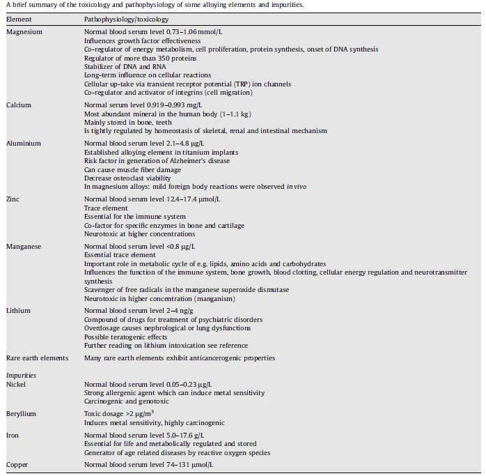
TOXICOLOGY
TOXICOLOGY
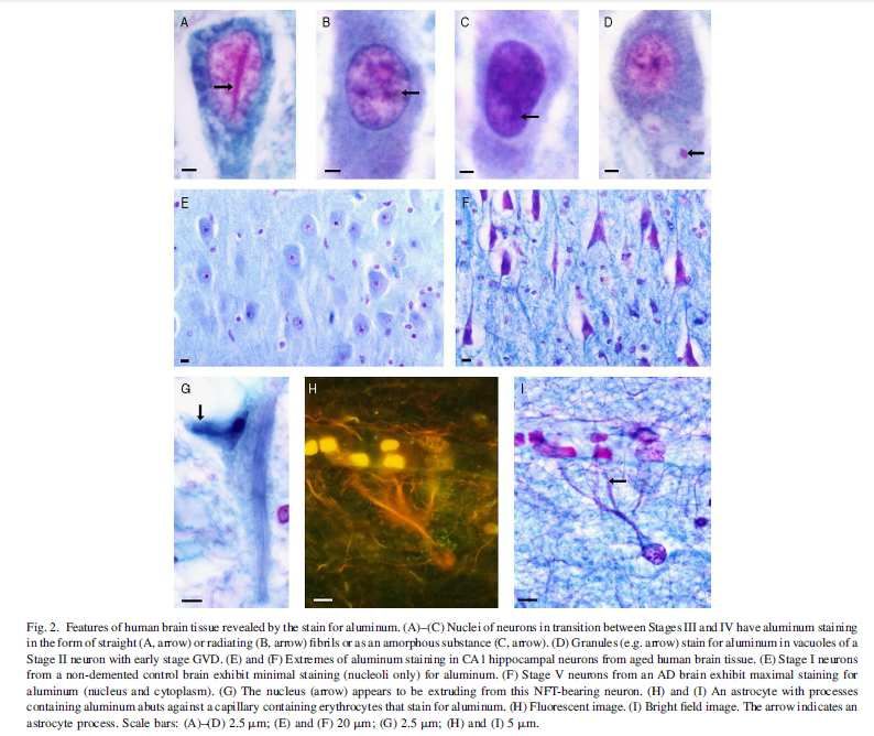
Aluminium
The presence of aluminium is generally regarded as a risk factor, being implicated in
the onset of different degenerative pathologies, e.g. Alzheimer’s disease (AD),
muscle fiber damage, and decreased osteoclast viability
The linkage between aluminium and AD is controversially discussed, starting a
debate whether aluminium is deposited in brain as a result of AD or whether it acts as
its inducer or accelerator and concluding that is neurotoxic and cannot be
disregarded as a factor in AD
On the other hand, binary Mg-Al alloy did not show negative effects on the viability of
blood vessel related cells, human umbilical vein endothelial cells and rodent vascular
smooth muscle cells
Open pore AZ91D scaffolds implanted in rabbits showed a good biocompatibility and
reacted in vivo with an appropriate inflammatory host responseTOXICOLOGY
RE Elements
It should be underlined that RE alternative option is not fully investigated and the mid-
and long-term effects of these elements need to be clarified.
RE metals can be expected to exhibit a slower degradation than the major alloy
component magnesium.
It can be expected, that they remain at the implantation site even after complete
degradation of magnesium. Because of this local accumulation the remaining rare
earth metals may exhibit adverse effects on the surrounding cells.
The incidence of RE on bone marrow cells needs a detailed investigation because
the clearance from the bone is known to be very slow
Ionic RE easily form colloid in blood and the resulting colloid material is taken by
phagocytic cells of the liver and spleen
RE ions cause haemolysis at very low concentrations, in the range 3 - 17•10-7 M/L, by
inducing domain and pore formation of the erythrocyte membraneTOXICOLOGY
RE Elements
Light RE elements are known to be hepatotoxic
Pr is the most toxic element leading to animal death in comparable concentrations
used for Ce, probably due to the low clearing rate
Pr and Nd induce chromosome aberrations in mice in vivo
Short-term effects of RE on primary cells and cell lines revealed that La and Ce
showed the highest cytotoxicity
The investigation of RE metals used in magnesium-based vascular stents revealed
no major adverse effects on the proliferation of smooth muscle cells when added as
low concentrated alloying elements, while led to the upregulation of inflammatory
genes at high concentrations
A coronary magnesium stent weighs about 10 mg
If the rare earth concentration is assumed with 5–10% and the total degradation time
is anticipated within three months (90 days), the daily amount of released metal ions
is calculated to result in 6–12 µg assuming linear degradation kinetics.BIOLOGICAL EVALUATION OF MEDICAL DEVICES
Tests for in vitro cytotoxicity
Extracting conditions should attempt to simulate or exaggerate the clinical use
conditions so as to determine the potential toxicological hazard without causing
significant changes in the test sample.
The choice of the extraction vehicle(s) taking into account the chemical
characteristics of the test sample shall be justified and documented. For mammalian
cell assays one or more of the following vehicles shall be used:
a) culture medium with serum;
b) physiological saline solution;
c) other suitable vehicle.
EN ISO 10993-5BIOLOGICAL EVALUATION OF MEDICAL DEVICES
Tests for in vitro cytotoxicity
The extraction shall be conducted under one of the following conditions and shall be
applied according to the device characteristics and specific conditions for use:
a) (24 ± 2) h at (37 ± 1) °C;
b) (72 ± 2) h at (50 ± 2) °C;
c) (24 ± 2) h at (70 ± 2) °C;
d) (1 ± 0,2) h at (121 ± 2) °C.
Extraction conditions described above, which have been used to provide a measure
of the hazard potential for risk estimation of the device or material, are based on
historical precedent. Other conditions, e.g. prolonged or shortened extraction times
at 37 °C, which simulate the extraction that occurs during clinical use or provide an
adequate measure of the hazard potential, may be used, but shall be justified and
documented. For medical devices that are in short-term contact (no greater than 4 h
cumulative contact duration) with intact skin or mucosa and that are not implanted,
this may include extraction times of less than 24 h but no less than 4 h, as given in
a) to c).
Cell culture medium with serum should only be used in accordance with a) because
extraction temperatures greater than (37 ± 1) °C can adversely impact chemistry
and/or stability of the serum and other constituents in the culture medium.INFLUENCE OF TEST SOLUTIONS
INFLUENCE OF TEST SOLUTIONS
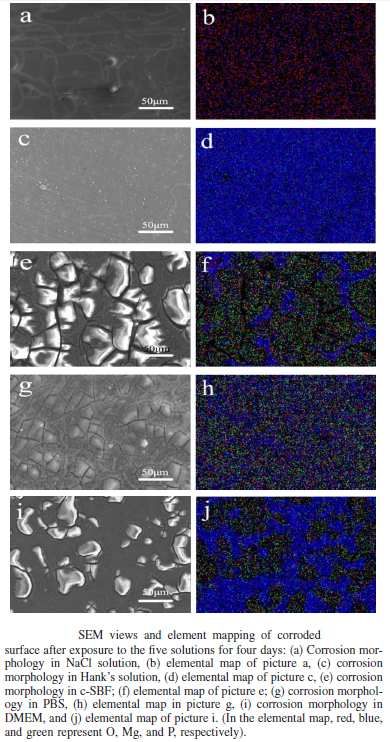
The selection of the suitable simulated biological fluid to determine the accurate
degradation performance of magnesium alloys during initial exposure is crucial to
the understanding of cell response and in vivo behaviour
The body fluid is a system with good buffering capability

Hank’s solution contains similar contents of inorganic ions as body plasma, the
concentrations of hydrocarbonates in Hank’s solution (~4.2 mM/L) is much lower
compared to that in plasma (~27 mM/L). Hydrocarbonates consitute one of the most
important buffers in body fluids and provide about 53% buffering capability of
plasma.
DEGRADATION MEASUREMENT IN HANK’S SOLUTION IS NOT CONVINCED

The buffering capability of DMEM seems too high

The degradation rates in c-SBF and DMEM are similar but ten times higher than
those in Hank’s solution, 0.9% NaCl solution and PBS
In vitro investigations carried out to predict in vivo degradation behavior by testing
Mg in inorganic-based SBF at ambient temperature and without pH control have
failed
The pH of human serum is kept constant by the complex biochemistry of the
physiological system. In contrast, a corrosion cell is a confined static environment of
variable pH as corrosion proceeds.
OH- ions are released during the corrosion of Mg, thereby raising the pH, which in
turn reduces the corrosion rate
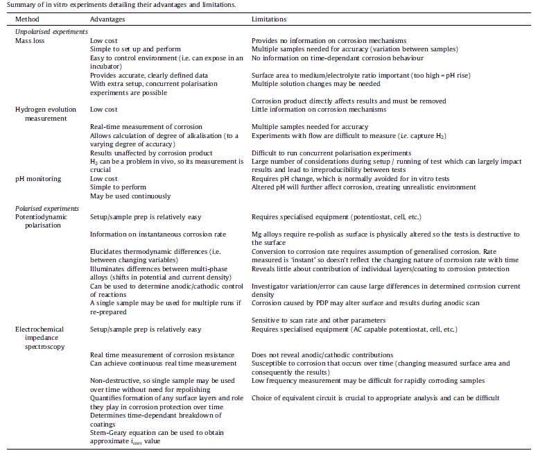
METHODOLOGIES AND LIMITATIONS
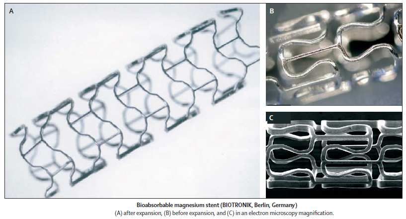
STENT

Stent metallici (acciaio inossidabile, nitinol…)
Drug eluting stents (DES)
Stent bioriassorbili
1. Too rapid degradation rates exceeding the achievable clearance rate of the alloy
   components.
2. Release and accumulation of hydrogen due to the corrosion of magnesium
   leading to vascular damage.
3. Local alkalosis due to the corrosion of the magnesium alloy leading to vascular
   damage.
4. Inferior mechanical properties or inadequate stent designs contributing to early
   stent fatigue fractures leading to mechanical negative interference with the
   vessel wall.
5. Toxic effects of the alloy compounds.
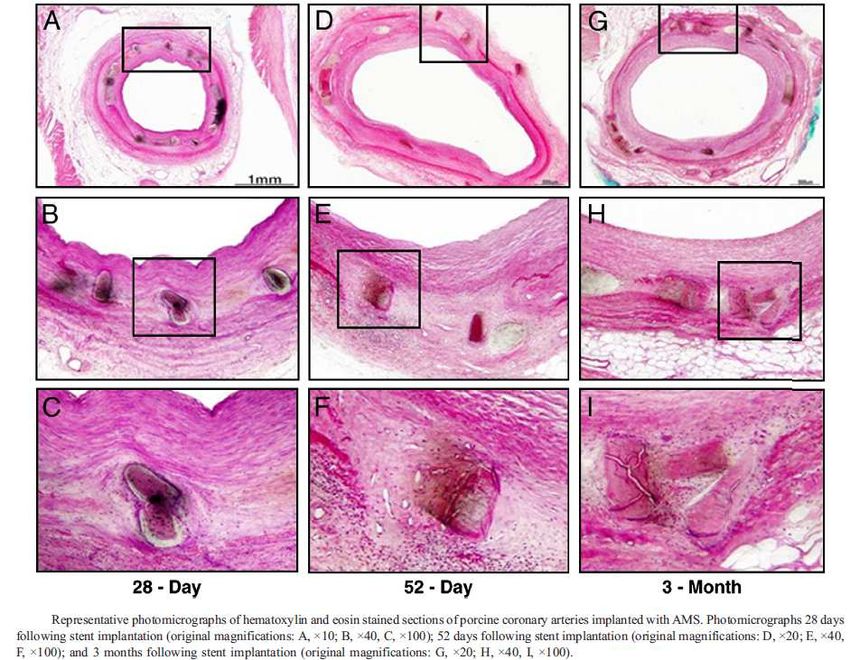
MAGNESIUM ALLOY STENTS

Di Mario et al. J Interv Cardiol 2004;17:391-5
Integrity of the magnesium alloy stent at 3 days
Reendothelialization starts to occur at 3 days postimplantation, and potentially
prevents stent particles from embolizing distally
Magnesium alloy or the degraded products of the magnesium alloy do not cause
any more inflammation than stainless steel stents.
There is a significant improvement in both percentage area stenosis and percentage
diameter stenosis (10%), suggesting that positive remodeling of the vessels is
taking place in the magnesium alloy stented vessels (3 months)
Degradation studies with the magnesium alloy stent demonstrated that at 56 days,
once the stent completely degraded, the vessel area was actually larger compared
to the 28-day time point, suggesting the capability of the vessel to remodel positively
Garg et al. J Am Coll Cardiol 2010;56:S43-78
WE43
Stents
BONE

Natural bone is a composite material made up of collagen fiber matrix stiffened by
hydroxyapatite (HAP) (Ca10(PO4)6(OH)2) crystals that account for 69% of the weight
of the bone.
The inorganic phase, HAP, is present in the form of small crystallites of dimensions
5 × 20 × 40 nm.
The organic phase is composed of type I collagen.
The elastic modulus of bone (17 GPa in tension in human femur) is intermediate
between that of apatite and collagen.
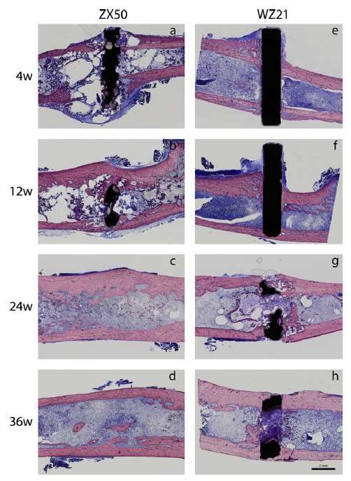
BONE

• Bone-implant interface strength and osteointegration are significantly greater for
magnesiun than conventional titanium materials
• Using biodegradable materials can avoid subsequent surgical intervention for
implant removal: morbidity related to repeated surgery is reduced
•Temporary implants are attractive in pediatric patients
•For Mg alloys to be used as viable implant materials, degradation rates should not
exceed the healing rate of the affected tissue. For adults they should maintain their
mechanical integrity at least for 12–18 weeks, while in pediatric trauma patients a
shorter presence in the bone is tolerated
•The corrosion process depends not only on the element composition and its
processing, but also on the corrosive environment to which the magnesium alloys
are subjected
WZ21 implants maintain their integrity for 4 weeks and corrode subsequently with
0.5% volume loss per day; ZX50 alloys commence the degradation process
immediately after implantation and degrade with 1.2% daily volume loss.

WZ21 alloys generate enhanced bone neoformation around the implant and give
evidence for good osteoconductivity and osteoinductivity of magnesium.

Bone recovers after complete degradation of the magnesium implant, even in the
case of massive gas formation (ZX50) and corresponding alterations of the bone.
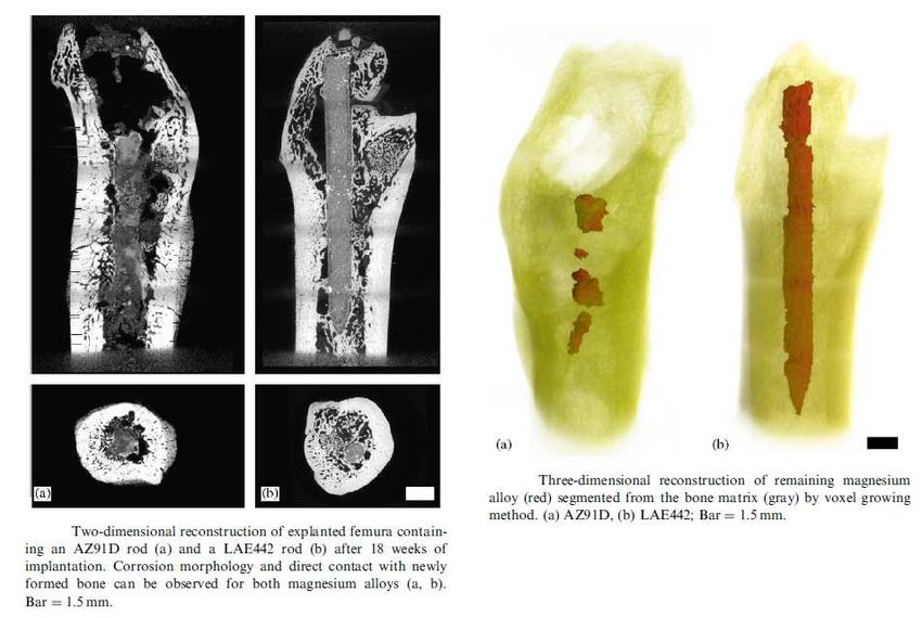
Alloy      Mg         Al       Zn         Li       RE
            AZ91D     Balance      9         1
            LAE442 Balance         4                   4         2

The sample rods were implanted intramedullary into the femora of the guinea pigs
after predrilling with a 1.5mm hand-operated drill (18 weeks).

While the AZ91D was more corrosion resistant than LAE442 in in vitro corrosion
tests, the LAE442 proved to be more corrosion resistant than AZ91D in in vivo
experiments. Both magnesium alloys revealed corrosion rates in vivo that where
about four orders of magnitudes lower than those acquired from in vitro tests.
Acceleration of corrosion could be due to different conditions existing along the implant surface The corrosion product Mg(OH)2 as a main component of the corrosion layer is not stable in aqueous solutions, especially not in chloride containing environments Local changes in electrochemical conditions could also be caused by locally passivated areas that are covered by newly formed bone. These locally different corrosive environments could cause local anodic and cathodic sites which could lead to locally accelerated corrosion rates following the morphology of pitting corrosion. A further parameter influencing the corrosion rate could be proteins that tend to adhere to almost all solid surfaces in vivo. This may explain the faster corrosion of AZ91D in vivo but it does not explain the more uniform corrosion morphology of LAE442 in vivo
References
Witte F. The history of biodegradable magnesium implants: a review. Acta Biomater. 2010;6(5):1680-92
Witte F et al. Degradable biomaterials based on magnesium corrosion. Curr Opin Solid St Mater Sci 2008;12:63-72
Mueller WD, Nascimento ML, Fernandez Lorenzo de Mele M. Critical discussion of the results from different corrosion studies of Mg and Mg
  alloys for biomaterial applications. Acta Biomater. 2010;6(5):1749-55
Hermawan H, Dubè D, Mantovani D. Developments in metallic biodegradable stents. Acta Biomater. 2010;6(5):1693-7
Song GL, Atrens A. Corrosion mechanism of magnesium alloys. Adv Eng Mater. 1999;1:11-33
Xin Y, Hu T, Chu PK. Influence of test solutions on in vitro studies o biomedical magnesium alloys. J Electrochem Soc 2010;157:C238-43
Katti KS. Biomaterials in total joint replacement. Colloids Surf B Biointerfaces. 2004;39(3):133-42
Kraus T et al. Magnesium alloys for temporary implants in osteosynthesis: in vivo studies of their degradation and interaction with bone. Acta
  Biomater. 2012;8(3):1230-8.
Witte F et al. In vitro and in vivo corrosion measurements of magnesium alloys. Biomaterials. 2006;27(7):1013-8
Kirkland NT et al. In-vitro dissolution of magnesium-calcium binary alloys: clarifying the unique role of calcium additions in bioresorbable
   magnesium implant alloys. J Biomed Mater Res B Appl Biomater. 2010;95(1):91-100.
Gu X et al. In vitro corrosion and biocompatibility of binary magnesium alloys. Biomaterials. 2009;30(4):484-98.
Witte F et al. Biodegradable magnesium scaffolds: Part I: Appropriate inflammatory response. J Biomed Mater Res A. 2007;81:748–56.
Hirano S e t al. Exposure, metabolism, and toxicity of rare earths and related compounds. Environ Health Perspect. 1996;104 Suppl 1:85-95.
Nakamura Y, et al. Differences in behaviour among the chlorides of seven rare earth elements administered intravenously to rats. Fundam
  Appl Toxicol. 1997;37(2):106-16.
Waksman R, Pakala R, Kuchulakanti PK, Baffour R, Hellinga D, Seabron R, Tio FO, Wittchow E, Hartwig S, Harder C, Rohde R, Heublein B,
  Andreae A, Waldmann KH, Haverich A. Safety and efficacy of bioabsorbable magnesium alloy stents in porcine coronary arteries.
  Catheter Cardiovasc Interv. 2006 Oct;68(4):607-17.
Kirkland et al.. Assessing the corrosion of biodegradable magnesium implants: A critical review of current methodologies and their limitations.
   Acta Biomater 2012;8:925-36.
Hornberger H, Virtanen S, Boccaccini AR. Biomedical coatings on magnesium alloys - a review. Acta Biomater. 2012 Jul;8(7):2442-55
You can also read


