Managing Melanoma In Situ - Kristen L. Toren, MD, and Eric C. Parlette, MD
←
→
Page content transcription
If your browser does not render page correctly, please read the page content below
Managing Melanoma In Situ
Kristen L. Toren, MD, and Eric C. Parlette, MD†
Melanoma is a highly aggressive skin cancer with an increasing incidence. Melanoma in
situ is an early, non-invasive form in which the tumor is confined to the epidermis.
Treatment of melanoma in situ is challenging due to the frequent subclinical microscopic
spread and to the presentation on the head and neck in cosmetically sensitive areas with
chronic sun damage. Optimizing tumor eradication is imperative to reduce the potential
progression into invasive disease and metastasis, all while maintaining cosmesis. Multiple
treatment regimens have been implemented for managing difficult melanoma in situ tu-
mors. We provide a thorough review of surgical, and non-surgical, management of mela-
noma in situ which can pose therapeutic dilemmas due to size, anatomic location, and
subclinical spread.
Semin Cutan Med Surg 29:258-263 © 2010 Elsevier Inc. All rights reserved.
M elanoma is a highly aggressive form of skin cancer with
an increasing incidence.1 Melanoma in situ (MIS) is an
early form of melanoma in which the malignancy is confined
ciated with a greater risk of melanoma, with the exception of
lentigo maligna. Lentigo maligna, unlike other melanomas,
has a greater association with nonmelanoma skin cancers.3
to the epidermis. According to the American Cancer Society,
an estimated 68,720 new cases of malignant melanoma were
reported in 2009, and 53,120 new cases of melanoma in situ.
Diagnostic Criteria
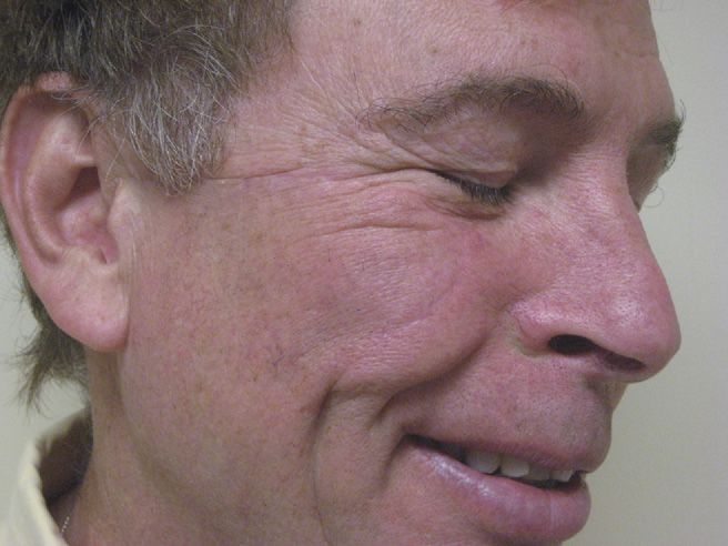
Lentigo maligna is a subtype of MIS found on sun-exposed Melanoma in situ can have a highly variable presentation,
areas and accounts for approximately 80% of all MIS tu- from a well-demarcated, small brown macule on healthy-
mors.2 With its increasing incidence and being a precursor to appearing skin to an asymmetric, variably pigmented large
invasive melanoma, the treatment of MIS, in particular len- patch on grossly actinically damaged skin (Fig. 1). It can even
tigo maligna, is a topic of increasingly significant interest. The present as a nondescript pink patch, especially on fair skin.
ideal management of MIS is openly debated. Clinical appearance along with history of change, new onset,
or any symptoms, such as itch or pain may prompt a biopsy.
Histologic examination of the entire lesion is critical to
Etiology and Epidemiology diagnosis of melanoma in situ. Even when the clinically dark-
Melanoma is a malignant tumor arising from melanocytes. est or “most suspicious” part of a pigmented lesion is biop-
Melanoma is an aggressive, heterogeneous cancer with both sied, there is a risk of missing the histologically most signifi-
host and environmental risk factors for development.1 Both cant area. Partial biopsy may show only MIS while there is an
rare high-risk susceptibility genes and common polymorphic unidentified invasive component elsewhere. Melanoma in
genes have been linked to an increased risk.1 Exposure to situ presents with atypical melanocytes confined to the epi-
ultraviolet radiation remains the predominant environmental dermis. Features consistent with a diagnosis of MIS include a
risk factor for melanoma. However, the history of significant predominance of single atypical melanocytes; multiple single
sunburns rather than chronic ultraviolet exposure seems to melanocytes greater in the epidermis instead of in the basal
more greatly increase the risk for development of melanoma.1,3 layer; and confluent, broad, irregularly sized, and distributed
The use of tanning beds has also contributed to the increased nests of melanocytes. The epidermal component is often
incidence of melanoma, especially in the younger popula- poorly demarcated with single melanocytes that tend to trail
tion. The presence of multiple nevi (greater than 50) is asso- off. Although many view lentigo maligna as a form of in situ
melanoma, it remains somewhat controversial whether len-
tigo maligna should be regarded as a melanocytic dysplasia as
*Department of Dermatology, Walter Reed Army Medical Center, Washing- opposed to in situ melanoma.4 Histopathologically, lentigo
ton, DC.
†Dermatology Associates, Winchester, MA. maligna is characterized by atypical melanocytes, singly and
Address reprint requests to Eric C. Parlette, MD, Dermatology Associate, in nests, usually confined to the basal layer and with little
Winchester, MA. E-mail: ecparlette@hotmail.com pagetoid invasion of the epidermis as opposed to other mel-
258 1085-5629/10/$-see front matter © 2010 Elsevier Inc. All rights reserved.
doi:10.1016/j.sder.2010.10.002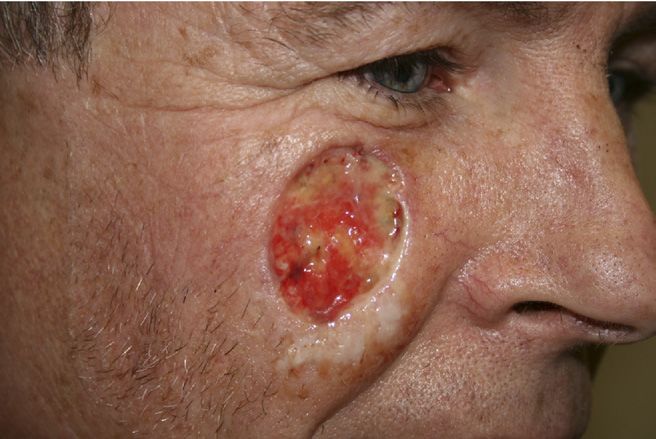
Managing melanoma in situ                                                                                                          259

                                                                     are employed for managing melanoma in situ, each with their
                                                                     individual strengths and weaknesses. We will provide an
                                                                     overview of the various treatment options to delineate the
                                                                     preferred regimens.

                                                                     Excisional Surgery
                                                                     Surgical excision of melanoma in situ has long been the treat-
                                                                     ment of choice. Excision ensures removal of periadnexal me-
                                                                     lanocytes and allows for thorough histologic assessment
                                                                     identifying any potentially previously undetected invasive
                                                                     component. The standard 5-mm margin for melanoma in
                                                                     situ was established at the 1992 National Institutes Health
                                                                     consensus conference and supported by the American Acad-
                                                                     emy of Dermatology’s 2001 guidelines for treatment of mel-
Figure 1 Classic clinical appearance of lentigo maligna in sun-ex-   anoma.7,8 Unfortunately, the 5-mm margin is inadequate for
posed area. Photo courtesy of H.L. Parlette, III, MD.
                                                                     many MIS lesions, especially those on the head and neck and
                                                                     sun-damaged skin.4,9-15 Recurrence rates after excision with 5
                                                                     mm margins range from 6% to 20%.16-19 Multiple studies
anoma in situs.4 Occasionally, multinucleate melanocytes
                                                                     have confirmed the unsatisfactory clearance of MIS tumor
with prominent dendritic processes are present in the basal
                                                                     with routine 5 mm margin excisions.11,12,14,15,20-30 The need
layer.4 Biopsies of lentigo maligna also typically reveal evi-
                                                                     for larger margins and/or better margin control has been
dence of chronic actinic damage, such as solar elastosis.
                                                                     recognized.13
    Multiple stains may be implemented to facilitate the diag-
                                                                        Various staged excisional techniques with better margin
nosis of MIS, including S-100, HMB-45, Mel-5, and MART-
                                                                     control have been devised and revised to optimize tissue
1/Melan-A. S-100 is an acidic Ca2⫹- and Zn2⫹-binding pro-
                                                                     analysis and reduce recurrence rates. In 1990, Dhawan et al31
tein that stains melanomas as well as benign melanocytic
                                                                     first described a modified staged surgery allowing for margin
lesions, dendritic cells, histiocytes, Schwann cells, muscle,
                                                                     control in the treatment of lentigo maligna. The technique
chondrocytes, and eccrine and apocrine cells.5 S-100 is use-
                                                                     consists of excision and mapping of the tumor similar to
ful in identifying the dermal component of melanomas as
                                                                     standard Mohs micrographic surgery. Rushed permanent
well as desmoplastic melanomas.5 HMB-45 is a mouse mono-
                                                                     sections are then examined by a dermatopathologist and sub-
clonal antibody that recognizes melanosome-associated
                                                                     sequent stages taken as necessary to clear the tumor.31 This
sialated glycoprotein seen in malignant melanocytes.5 Mel-5
                                                                     technique is now referred to as the “slow Mohs” procedure.
recognizes gp75, a glycoprotein abundantly present in mela-
                                                                     Arguments against “slow Mohs” include a potentially pro-
nocytes.5 It briskly stains melanomas but also many other
                                                                     longed opened wound, leading to a greater infection risk and
nonmelanocytic lesions.5 MART-1/Melan-A is a cytoplasmic
                                                                     the formation of granulation tissue during the wait time (Fig.
melanosome-associated melanocyte differentiation antigen
                                                                     2) .13 Rush permanent sections reduce wait time while main-
present in 80-100 percent of melanomas.5 Most recently,
                                                                     taining high-quality histology.23 Prophylactic oral antibiotics
fluorescence in situ hybridization test has been used to dis-
                                                                     are used to reduce infection risks with delayed closures.
tinguish between benign nevi and malignant melanoma in
                                                                     Wound granulation may actually benefit and accelerate heal-
histologically ambiguous melanocytic neoplasms. The fluo-
rescence in situ hybridization (FISH) test is an assay that uses
DNA probes hybridized to the melanocytic lesion and iden-
tifies multiple recurrent chromosomal copy number changes
seen in more than 95% of melanomas. The fluorescence in
situ hybridization test may be used as an ancillary tool with
difficult histology.6

Management of
Melanoma In-Situ
Management of melanoma in situ can often pose a therapeu-
tic dilemma. Ill-defined clinical margins, especially with len-
tigo maligna, frequently yields unsatisfactory cure rates with
standard excision. The frequent occurrence of MIS, espe-
cially lentigo maligna, on the head and neck in cosmetically
sensitive areas warrants optimal margin control. Further-            Figure 2 Two days status post completion of slow Mohs excision for
more, the presentation of MIS in nonsurgical candidates              melanoma in situ, clear margins after second stage. Early granula-
raises management questions. Multiple treatment modalities           tion tissue formation evident.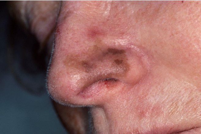
260                                                                                                           K.L. Toren and E.C. Parlette

                                                                      81%.43 Bene et al found that only 95.1% of MIS lesions con-
                                                                      sidered clear on frozen section analysis were truly clear when
                                                                      analyzed with subsequent permanent sections.39 Interpreta-
                                                                      tion of melanocytic lesions with frozen sections can be very
                                                                      challenging. Vacuolated keratinocytes can be difficult to dif-
                                                                      ferentiate from melanocytes and dermal inflammatory cells
                                                                      may obscure melanocytes.9,41
                                                                         Malignant melanocytes must also be differentiated from
                                                                      benign melanocytic hyperplasia, frequently found in sun-
                                                                      damaged skin. Weyers et al44 identified criteria indicative of
                                                                      malignant melanoma compared with benign melanocytes.
                                                                      The greatest diagnostic value is the presence of melanocytic
                                                                      nests. Irregular distribution of pigment and melanocytes, ad-
                                                                      nexal extension, and pagetoid spread are additional findings
                                                                      suggestive of malignancy.44
Figure 3 Good cosmetic result 6 months status post rotation flap         Immunostaining of frozen sections has been studied to deter-
closure following a staged slow Mohs excision.                        mine its utility in better identifying atypical melanocytes. Several
                                                                      stains have been used in frozen section processing, including
                                                                      S-100, HMB-45, Mel-5, and MART-1/Melan-A. Comparative
                                                                      studies have found MART-1/Melan-A to be the most sensitive
ing of the final closure as the wound healing process has             and specific immunostain for identifying melanoma in frozen
already started (Fig. 3).                                             section.24,25,28,45 Protocols for MART-1 staining techniques
   Alternative staged excision methods have been developed,           with frozen sections provide high efficacy detection of MIS.46
including analyzing small strip, 2-mm peripheral margins, 1           Many advocate the use of immunostains in preparation of
week before primary tumor excision to guide margin con-               frozen sections for MIS. Limitations include additional pro-
trol.12,15,20,23,26,29,32,33 The reported benefit of the peripheral   cessing time, skill level of the histotechnician, and the possi-
rim preanalysis is to avoid prolonged open wounds. Disad-             bility of false-positive margins caused by the staining of pig-
vantages with this technique include delay in tumor excision          mented actinic keratoses and actinically damaged skin.47
and an unsuspected invasive component may not be identi-
fied until after closure of the defect.13
   Recurrence rates for the staged excisions range from 0% to         Topical Imiquimod
5% with variable follow-up.11,12,15,21-23,26-28,30,32-39 Total mar-   Topical imiquimod has reported efficacy for melanoma in
gin control by the use of staged excisions with permanent             situ and lentigo maligna. Imiquimod is a synthetic imidazo-
sections offers a simple, effective treatment for MIS tumors          quinoline amine that stimulates immune activity. The innate
with indistinct margins, eliminating the concerns of inade-           immune system is activated, binding toll-like receptors 7 and
quate margins and higher recurrence rates. A good working             8, leading to synthesis and release of multiple cytokines,
relationship with the dermatopathologist is imperative for            including interferon-␣ and tumor necrosis factor-␣. The re-
success. Long-term follow-up with prospective studies is still        sult is apoptosis and suppression of tumor genesis.9,48
needed to more thoroughly evaluate the efficacy of staged                Imiquimod is currently approved by the Food and Drug
excisions with permanent sections.                                    Administration for the treatment of external genital warts,
                                                                      actinic keratoses, and superficial basal cell carcinomas. The
Mohs Micrographic Surgery                                             use for lentigo maligna was first reported in 2000 for a large
Compared with standard excision, Mohs micrographic sur-               scalp lesion on an elderly male. He remained clear at
gery (MMS), like staged excisions, provides the advantages of         9-months follow-up.49 Subsequent reports and studies have
complete margin evaluation, tissue conservation, and greater          shared protocols for clearance of MIS with response rates
cure rates for MIS and lentigo maligna.9,40 The main advan-           ranging from 66% to 100%.9,49-68,69,70,61
tage over the staged excision techniques is immediate recon-             Despite a positive response to imiquimod, the optimal
struction. MMS involves tangential excision of the tumor allow-       treatment regimen has yet to be defined. Furthermore, re-
ing for examination of 100% of the peripheral margins.9,41            sponse to therapy and tumor clearance are difficult to assess
Despite the aforementioned advantages, controversy exists re-         post treatment, leading to the concern for recurrence or, even
garding the use of MMS for the treatment of MIS because of            more worrisome, invasive disease. One large case series
significant difficulties in recognizing malignant melanocytic         showed only 30 of 33 cases to be histologically clear of tumor
cells on frozen sections.9,24,28,39,41,42 Zitelli et al42 reported    when judged clinically clear after 3 months of therapy.68 The
100% sensitivity and 90% specificity of frozen section in the         use of topical imiquimod for a superficial, but potentially
detection of atypical melanocytes at the margins of melanoma          very aggressive malignancy is risky when there is potential of
based on comparison with paraffin-embedded specimens.                 an incorrect initial diagnosis as melanoma in situ due to
Subsequent investigations have reported lower accuracy.               failure to detect an invasive component on initial biopsy. As
Barlow et al report a sensitivity of only 59% and specificity of      many as 22% of pigmented lesions believed to be MIS or
Managing melanoma in situ 261
lentigo maligna on initial biopsy have invasive components ratory able to adequately perform the necessary special im-
identified histologically after complete excision.9 Patients munostains. The limitations of Mohs for MIS are the limited
have developed invasive melanoma after treatment with imi- number of Mohs surgeons capable and/or comfortable per-
quimod for lentigo maligna.50,51 Imiquimod represents an forming Mohs for melanoma in situ. This is due to the diffi-
alternative treatment option for MIS and lentigo maligna that culty in reading melanocytic histology on frozen sections, the
are particularly large and/or are on cosmetically sensitive ar- lack of skilled technicians, and the high liability associated
eas in elderly and/or poor surgical candidates. with recurrence.
Alternative treatments for melanoma in situ include radi-
Radiation Therapy ation therapy and topical imiquimod. Radiation therapy has a
Radiation therapy (XRT) is a noninvasive, destructive treat- longer history of use and follow-up but with greater tissue
ment option for MIS and lentigo maligna. Treatment with destruction and scarring. Topical imiquimod has variable
radiation is appealing for elderly patients and for poor surgi- predictability in responsiveness and clearance but with ex-
cal candidates with large MIS lesions on the head and neck. A cellent cosmetic results. Both treatments may be considered
95% clearance is reported with the Miescher technique, de- for nonsurgical candidates or large, inoperable tumors. Ad-
livering high-dose Grenz ray or soft x-rays (12-50 kV) with ditionally, imiquimod may be considered for unique scenar-
surface doses of 20 Gy once weekly for 4 to 5 weeks.71 Con- ios in cosmetically sensitive areas.
ventional radiotherapy is reportedly effective as a treatment
modality with an 86% clearance rate at 5 years.18,72-74 References
Radiation therapy is a good second-line treatment best 1. Tucker MA: Melanoma epidemiology. Hematol/Oncol Clin North Am
suited for nonsurgical candidates. The nonselective tissue 23:383-395, 2009
destruction is a significant side effect. XRT may yield a poor 2. Swetter SM, Boldrick JC, Jung SY, et al: Increasing incidence of lentigo
cosmetic outcome with skin pallor, atrophy and telangiecta- maligna melanoma subtypes: northern California and national trends
1990-2000. J Invest Dermatol 125:685-691, 2005
sias involving the entire treatment field.72 3. Gaudy-Marqueste C, Madjlessi N, Guillot B, et al: Risk factors in elderly
people for lentigo maligna compared with other melanomas: a double
Laser Treatment case-control study. Arch Dermatol 145:418-423, 2009
4. Weedon D: Lentigines, nevi and melanoma, in Skin Pathology London,
Multiple lasers, to include the argon, carbon dioxide, Q- Elsevier, 2002
switched ruby, Q-switched alexandrite, and Q-switched neo- 5. Carucci JA: Mohs’ micrographic surgery for the treatment of melanoma.
dymium-doped yttrium-aluminum-garnet, have been used Dermatol Clin 20:701-708, 2002
for management of MIS.75-80 Although reports proclaiming 6. Gerami P, Jewell SS, Morrison LE, et al: Fluorescence in situ hybridiza-
tion (FISH) as an ancillary diagnostic tool in the diagnosis of melanoma.
short treatment duration, minimal postoperative care, and
Am J Surg Pathol 33:1146-1156, 2009
excellent cosmesis exist, the use of lasers for management of 7. National Institutes of Health: Consensus Development Conference
melanoma in situ is associated with high recurrence rates and Statement on diagnosis and treatment of early melanoma, January 27-
is still below the standard of care for most tumors. Both 29, 1992. Am J Dermatopathol 15:34-43, 1993
inadequate margin control and inadequate laser targeting of 8. Sober AJ, Chuang TY, Duvic M, et al: Guidelines/Outcomes Commit-
tee. Guidelines of care for primary cutaneous melanoma. J Am Acad
the tumor lead to high recurrence rates. Atypical cells may
Dermatol 45:579-586, 2001
extend down appendageal structures or may be amelanotic 9. Erickson C, Miller SJ: Treatment options in melanoma in situ: topical
and, thus, elude laser destruction.80 Laser therapy may offer and radioation therapy, excision and Mohs surgery. Int J Dermatol
an excellent option in the future, but is currently not a rec- 49:482-491, 2010
ommended therapy for MIS. 10. Zitelli JA: Surgical margins for lentigo maligna. Arch Dermatol 104:
607-608, 2004
11. Agarwal-Antal N, Bowen GM, Gerwels JW: Histologic evaluation of
lentigo maligna with permanent sections: implications regarding cur-
Conclusions rent guidelines. J Am Acad Dermatol 47:743-748, 2002
The incidence of melanoma in situ, and particularly lentigo 12. Bosbous MW, Dzwierzynski WW, Neuburg M: Staged excision of len-
tigo maligna and lentigo maligna melanoma: a 10-year experience. Plast
maligna, continues to increase. It is imperative to understand
Reconstr Surg 124:1947-1955, 2009
the multiple treatment options, as well as the associated risks 13. Clark GS, Pappas-Politis E, Cherpelis BS, et al: Surgical management of
and benefits, to best guide our patients’ therapy. Excision of melanoma in situ on chronically sun damaged skin. Cancer Control
melanoma in situ remains the treatment of choice. Given the 15:216-224, 2008
location, tumor characteristics, surgical candidacy, and pro- 14. Raziano RM, Clark GS, Cherpelis BS, et al: Staged margin control tech-
niques for surgical excision of lentigo maligna. G Ital Dermatol Vene-
vider capabilities, treatment may vary. Routine surgical exci-
reol 144:259-270, 2009
sion with standard 5-mm margins may be sufficient for small, 15. Moller MG, Pappas-Politis E, Zagar JS, et al: Surgical management of
well-demarcated tumors on less actinically damaged skin. melanoma-in-situ using a staged marginal and central excision tech-
“Slow-Mohs” with permanent section tissue analysis is pre- nique. Ann Surg Oncol 16:1526-1536, 2009
ferred for less discrete lesions, especially lentigo malignas, on 16. Pitman GH, Kopf AW, Bart RS, et al: Treatment of lentigo maligna and
lentigo maligna melanoma. J Dermatol Surg Oncol 5:727-737, 1979
actinically damaged skin. Mohs micrographic surgery could
17. Coleman WP 3rd, Davis RS, Reed RJ, et al: Treatment of lentigo maligna
be the treatment of choice for MIS provided a there is a Mohs and lentigo maligna melanoma. J Dermatol Surg Oncol 6:476-479,
surgeon skilled in reading melanocytic neoplasms on frozen- 1980
tissue sections, a highly skilled histotechnician, and a labo- 18. Tsang RW, Liu FF, Wells W, et al: Lentigo maligna of the head and262 K.L. Toren and E.C. Parlette
neck: results of treatment by radiotherapy. Arch Dermatol 130: 43. Barlow RJ, White CR, Swanson NA: Mohs’ micrographic surgery using
1008-1012, 1994 frozen sections alone may be unsuitable for detecting single atypical
19. Osborne JE, Hutchinson PE: A follow-up study to investigate the effi- melanocytes at the margins of melanoma in situ. Br J Dermatol 146:
cacy of initial treatment of lentigo maligna with surgical excision. Br J 290-294, 2002
Plast Surg 55:611-615, 2002 44. Weyers W, Bonczkowitz M, Weyers I, et al: Melanoma in situ versus
20. Johnson TM, Headington JT, Baker SR, et al: Usefulness of the staged melanocytic hyperplasia in sun-damaged skin. Assessment of the sig-
excision for lentigo maligna and lentigo maligna melanoma: the nificance of histopathologic criteria for differential diagnosis. Am J
“square” procedure. Dermatol Surg 37:758-764, 1997 Dermatopathol 18:560-566, 1996
21. Zitelli JA, Brown C, Hanusa BH: Mohs micrographic surgery for the 45. Davis DA, Kurtz KA, Robinson RA: Ultrarapid staining for cutaneous
treatment of primary cutaneous melanoma. J Am Acad Dermatol 37: melanoma: study and protocol. Dermatol Surg 31:753-756, 2005
236-245, 1997 46. Kelley LC, Starkus L: Immunohistochemical staining of lentigo maligna
22. Zitelli JA, Brown C, Hanusa BH: Surgical margins for excision of pri- during Mohs micrographic surgery using Mart-1. J Am Acad Dermatol
mary cutaneous melanoma. J Am Acad Dermatol 37:422-429, 1997 46:78-84, 2002
23. Cohen LM, McCall MW, Zax RH: Mohs micrographic surgery for len- 47. Shabrawi-Caelen LE, Kerl H, Cerroni L, et al: Not a helpful marker in
tigo maligna and lentigo maligna melanoma: a follow-up study. Der- distinction between melanoma in situ on sun-damaged skin and pig-
matol Surg 24:673-677, 1998 mented actinic keratosis. Am J Dermatopathol 26:364-366, 2004
24. Zalla MJ, Lim KK, Dicaudo DJ, et al: Mohs micrographic excision of 48. Kang HY, Park TJ, Jin HS: Imiquimod, a toll-like receptor 7 agonist,
melanoma using immunostains. Dermatol Surg 26:771-784, 2000 inhibits melanogenesis and proliferation of human melanocytes. J In-
25. Albertini JG, Elston DM, Libow LF, et al: Mohs micrographic surgery vest Dermatol 129:243-246, 2009
for melanoma: a case series, a comparative study of immunostains, an 49. Ahmed I, Berth-Jones J: Imiquimod: a novel treatment for lentigo ma-
informative case report, and a unique mapping technique. Dermatol ligna. Br J Dermatol 143:843-845, 2000
Surg 28:656-665, 2002 50. Fisher GH, Lang PG: Treatment of melanoma in situ on sun-damaged
26. Bub JL, Berg D, Slee A, et al: Management of lentigo maligna and lentigo skin with topical 5% imiquimod cream complicated by the develop-
maligna melanoma with staged excision: a 5-year follow-up. Arch Der- ment of invasive disease. Arch Dermatol 139:945-947, 2003
matol 140:552-558, 2004 51. Naylor MF, Crowson N, Kuwahara R, et al: Treatment of lentigo maligna
27. Huilgol SC, Selva D, Chen C, et al: Surgical margins for lentigo maligna with topical imiquimod. Br J Dermatol 149:66-69, 2003 (suppl 66)
and lentigo maligna melanoma: the technique of mapped serial exci- 52. Chapman MS, Spencer SK, Brennick JB: Histologic resolution of mela-
sion. Arch Dermatol 140:1087-1092, 2004 noma in situ (lentigo maligna) with 5% imiquimod cream. Arch Der-
28. Bricca GM, Brodland DG, Zitelli JA: Immunostaining melanoma frozen matol 139:943-944, 2003
sections: the 1-hour protocol. Dermatol Surg 30:403-408, 2004 53. Epstein E: Extensive lentigo maligna clearing with topical imiquimod.
29. Hazan C, Dusza SW, Delgado R, et al: Staged excision for lentigo Arch Dermatol 139:944-945, 2003
maligna and lentigo maligna melanoma: a retrospective analysis of 117 54. Flemming CJ, Bryden AM, Evans A, et al: A pilot study of treatment of
cases. J Am Acad Dermatol 58:142-148, 2008 lentigo maligna with 5% imiquimod cream. Br J Dermatol 151:485-
30. Jejurikar SS, Borschel GH, Johnson TM, et al: Immediate optimal re- 488, 2004
construction of facial lentigo maligna and melanoma following total 55. Munoz CM, Sanchez JL, Martin-Garcia RF: Successful treatment of
peripheral margin control. Plast Reconstr Surg 120:1249-1255, 2007 persistent melanoma in situ with 5% imiquimod cream. Dermatol Surg
31. Dhawan SS, Wolf DJ, Rabinovitz HS, et al: Lentigo maligna: the use of 30:1543-1545, 2004
rush permanent sections in therapy. Arch Dermatol 126:928-930, 56. Powell AM, Russell-Jones R: Amelanotic lentigo maligna managed with
1990 topical imiquimod as immunotherapy. J Am Acad Dermatol 50:792-
32. Mahoney MH, Josephy M, Temple CLF: The perimeter technique for 796, 2004
lentigo maligna: an alternative to Mohs micrographic surgery. J Surg 57. Powell AM, Russell-Jones R, Barlow RJ: Topical imiquimod immuno-
Oncol 91:120-125, 2005 therapy in the management of lentigo maligna. Clin Exp Dermatol
33. Walling HW, Scupham RK, Bean AK, et al: Staged excision versus Mohs 29:15-21, 2004
micrographic surgery for lentigo maligna and lentigo maligna mela- 58. Kupfer-Bessaguet I, Guillet G, Misery L, et al: Topical imiquimod treat-
noma. J Am Acad Dermatol 57:659-664, 2007 ment of lentigo maligna: clinical and histologic evaluation. J Am Acad
34. Clayton BD, Leshin B, Hitchcock MG, et al: Utility of rush paraffin- Dermatol 51:635-639, 2004
embedded tangential sections in the management of cutaneous neo- 59. Michalopoulis P, Yawalkar N, Bronnimann M, et al: Characterization of
plasms. Dermatol Surg 26:671-678, 2000 the cellular infiltrate during successful topical treatment of lentigo ma-
35. Anderson KW, Baker SR, Lowe L, et al: Treatment of head and neck ligna with imiquimod. Br J Dermatol 151:903-906, 2004
melanoma, lentigo maligna subtype: a practical surgical technique. 60. Wolf IH, Cerroni L, Kodama K, et al: Treatment of lentigo maligna
Arch Facial Plast Surg 3:202-206, 2001 (melanoma in situ) with the immune response modifier imiquimod.
36. Bienert TN, Trotter MJ, Arlette JP: Treatment of cutaneous melanoma of Arch Dermatol 141:510-514, 2005
the face by Mohs micrographic surgery. J Cutan Med Surg 7:25-30, 61. Noel B, Kunzle N: Image in clinical medicine: lentigo maligna. N Engl
2003 J Med 353:2176, 2005
37. Bhardwaj SS, Tope WD, Lee PK: Mohs micrographic surgery for lentigo 62. Ray CM, Kluk M, Grin CM, et al: Successful treatment of malignant
maligna and lentigo maligna melanoma using Mel-5 immunostaining: melanoma in situ with topical 5% imiquimod cream. Int J Dermatol
University of Minnesota experience. Dermatol Surg 32:690-696, 2006 44:428-434, 2005
38. Temple CL, Arlette JP: Mohs micrographic surgery in the treatment of 63. Van Meurs T, van Doorn R, Kirtschig G: Recurrence of lentigo maligna
mentigo maligna and melanoma. J Surg Oncol 94:287-292, 2006 after initial complete response to treatment with 5% imiquimod cream.
39. Bene NI, Healy C, Coldiron BM: Mohs micrographic surgery is accu- Dermatol Surg 33:623-626, 2007
rate. 95.1 % of the time for melanoma in situ: a prospective study of 167 64. Spenny ML, Walford J, Werchniak AE, et al: Lentigo maligna (mela-
cases. Dermatol Surg 34:660-664, 2008 noma in situ) treated with imiquimod cream 5%: 12 case repots. Cutis
40. Dawn ME, Dawn AG, Miller SJ: Mohs surgery for the treatment of 79:149-152, 2007
melanoma in situ: a review. Dermatol Surg 33:395-402, 2001 65. Mahoney MH, Joseph MG, Temple C: Topical imiquimod therapy for
41. Stevenson O, Ahmed I: Lentigo maligna: prognosis and treatment op- lentigo maligna. Ann Plast Surg 61:419-424, 2008
tions. Am J Clin Dermatol 6:151-164, 2005 66. deTroya-Martin M, Frieyro-Elicegui M, Funez Liebana R, et al: Lentigo
42. Zitellii JA, Moy RL, Abell E: The reliability of frozen sections in the maligna managed with topical imiquimod and dermoscopy: report of
evaluation of surgical margins for melanoma. J Am Acad Dermatol two cases. Dermatol Surg 34:1561-1566, 2008
24:102-106, 1991 67. Buettiker UV, Yawalkar NY, Braathen LR, et al: Imiquimod treatment ofManaging melanoma in situ 263
lentigo maligna: an open-label study of 34 primary lesions in 32 74. Harwood AR: Conventional fractionated radiotherapy for 51 patients
patient. Arch Dermatol 144:943-945, 2008 with lentigo maligna and lentigo maligna melanoma. Int J Radiat Oncol
68. Cotter MA, McKenna JK, Bowen GM: Treatment of lentigo maligna with Biol Phys 9:1019-1021, 1983
imiquimod before staged excision. Dermatol Surg 34:147-151, 2008 75. Arndt KA: Argon laser treatment of lentigo maligna. J Am Acad Derma-
69. Ramsdell AM, Zeitouni N: Long-term follow-up of a hemifacial lentigo tol 10:953-957, 1984
maligna treated using 5% imiquimod. Dermatol Surg 35:287-290, 2009 76. Kopera D: Treatment of lentigo maligna with the carbon dioxide laser.
70. Van Meurs T, van Doorn R, Kirtschig G: Treatment of lentigo maligna Arch Dermatol 131:735-736, 1995
77. Thissen M, Westerhof W: Lentigo maligna treated with ruby laser. Acta
with imiquimod cream: a long-term follow-up study of 10 patients.
Dermatol Venereol 77:163, 1997
Dermatol Surg 36:853-858, 2010
78. Orten SS, Waner M, Dinehart SM, et al: Q-switched neo-dymium:
71. Harwood AR, Cummings BJ: Radiotherapy for malignant melanoma: a
yttrium-aluminum-garnet laser treatment of lentigo maligna. Otolaryn-
reappraisal. Cancer Treat Rev 8:271-282, 1981 gol Head Neck Surg 120:296-302, 1999
72. Dancourt F, Harwood AR, Fitzpatrick PJ: The radiotherapy of lentigo 79. Iyer S, Goldman M: Treatment of lentigo maligna with combination
maligna and lentigo maligna melanoma of the head and neck. Cancer laser therapy: recurrence at 8 months after initial resolution. J Cosmet
45:2279-2283, 1980 Laser Ther 5:49-52, 2003
73. Harwood AR: Conventional radiotherapy in the treatment of lentigo 80. Madan V, August PJ: Lentigo maligna— outcomes of treatment with
maligna and lentigo maligna melanoma. J Am Acad Dermatol 6:310- Q-switched Nd: YAG and alexandrite lasers. Dermatol Surg 35:607-
316, 1982 611, 2009You can also read