MBMSS Maine Biological and Medical Sciences Symposium April 27-28, 2018 MDI Biological Laboratory Maren Conference Center - The MDI Biological ...
←
→
Page content transcription
If your browser does not render page correctly, please read the page content below
MBMSS Maine Biological and Medical Sciences Symposium April 27-28, 2018 MDI Biological Laboratory Maren Conference Center
2018 MBMSS
The 45th Annual Maine Biological and Medical Sciences
Symposium (MBMSS) is a state-wide gathering of scientists
and students. It is an opportunity to share research results,
exchange ideas, promote collaboration, and network with Maine
scientists in a variety of disciplines.

Organizing Committee
Judi Alexander | MDI Biological Laboratory
James Coffman, Ph.D. | MDI Biological Laboratory
Patsy Dickinson, Ph.D. | Bowdoin College
Jane Disney, Ph.D. | MDI Biological Laboratory
Jean Doty, Ph.D. | University of Maine at Farmington
Markus Frederich, Ph.D. | University of New England
Lynn Hannum, Ph.D. | Colby College
Ellen Hostert, Ph.D. | University of Maine at Machias
Steven Munger, Ph.D. | The Jackson Laboratory
Chris Petersen, Ph.D. | College of the Atlantic
Paula Schlax, Ph.D. | Bates College
Chris Smith | MDI Biological Laboratory
Rob Wheeler, Ph.D. | University of Maine

Program cover image shows polyploidy in a map of Maine.
Credit: K. Gjelsvik, Losick Lab and A. Farrell, Community Lab

   Maine’s IDeA Network of Biomedical Research Excellence is supported
     by grants from the National Institute of General Medical Sciences
              (P20GM103423), National Institutes of Health

                                                                         2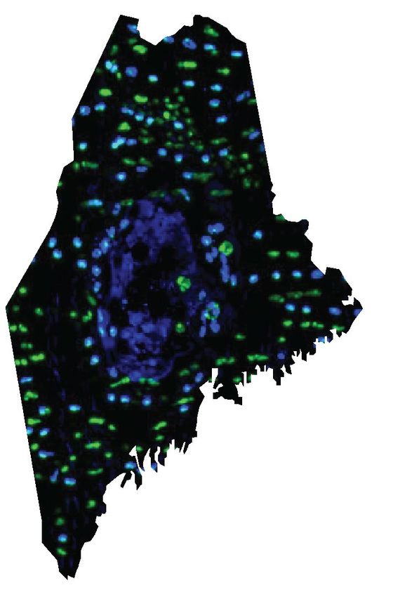
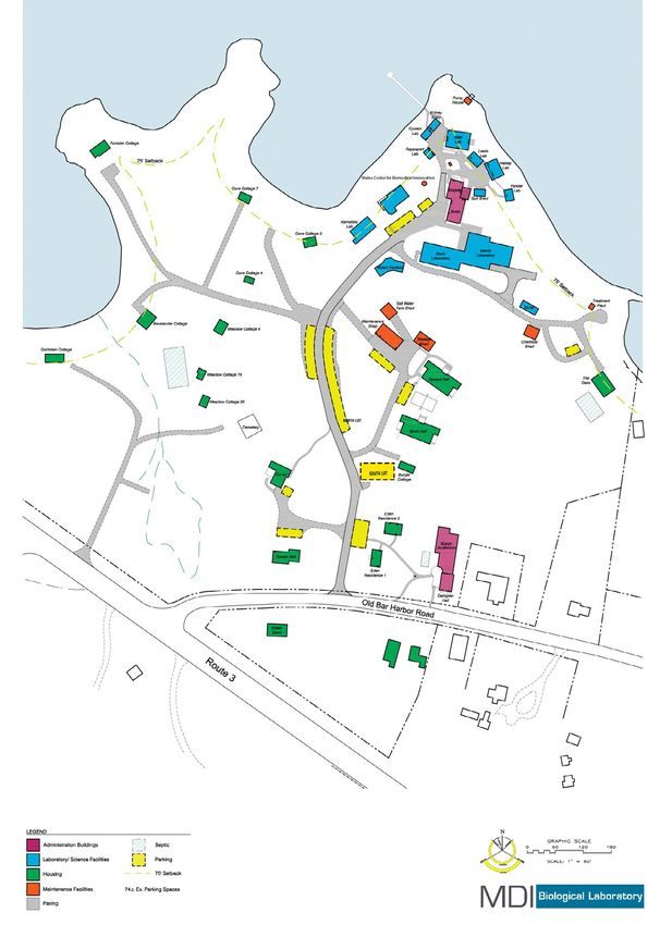
Campus Map

                                 3

                     2
                                     1

     Maren Conference Center and Auditorium
1.   (Session talks and keynote)

2.   Dining Hall

     Maine Center for Biomedical Innovation
3.   (Poster sessions A and B)

                                              3
Friday, April 27TH

 11:00 AM - 4:00   Symposium Registration
       PM          Maren Conference Center

    11:30 AM       Lunch
                   MDI Biological Laboratory Dining Hall

    12:50 PM       Symposium Welcome and Introduction
                   Maren Auditorium
                   Jane Disney, Ph.D., Director of Education,
                   MDI Biological Laboratory

 1:00 - 2:15 PM    Session 1: Evolutionary Biology/Ecology/
                   Regeneration
                   Maren Auditorium
                   Session Chair: Vicki Losick, Ph.D., MDI
                   Biological Laboratory

 1:00 - 1:15 PM       MaryLynn FitzSimons, University of Maine
                      GSBSE, Graduate Student
                      Crosstalk between the epicardium and
                      cardiomyocytes – PGE2 signaling directed
                      by miR-101a promotes cardiomyocyte
                      proliferation during zebrafish heart
                      regeneration

 1:15 - 1:30 PM       Dave Angelini, Ph.D., Colby College, Assistant
                      Professor
                      Insulin signaling manipulation phenocopies
                      evolution of a host-associate polyphenism

 1:30 - 1:45 PM       Patricia Jones, Ph.D., Bowdoin College,
                      Assistant Professor
                      Impacts of alcohol in floral nectar on foraging
                      behavior in bumblebees

                                                                   4
Friday, April 27TH

 1:45 - 2:00 PM      Suegene Noh, Ph.D., Colby College, Assistant
                     Professor
                     Molecular signatures of cheating and altruism
                     in wild social amoebas

 2:00 - 2:15 PM      Cameron Fudge, University of Maine,
                     Undergraduate Student
                     Defining the role of IFT88 during zebrafish
                     caudal fin regeneration

 2:15 - 2:30 PM   Break

 2:30 - 3:45 PM   Session II: Genetics/Bioinformatics/
                  Interdisciplinary
                  Maren Auditorium
                  Session Chair: Benjamin King, Ph.D., University of
                  Maine

 2:30 - 2:45 PM      Kayla Gjelsvik, M.S., MDI Biological
                     Laboratory, Research Assistant
                     The mechanics of polyploidy in wound repair

 2:45 - 3:00 PM      Nishad Jayasundara, Ph.D., University of
                     Maine, Assistant Professor
                     Chronic kidney disease, pollutant mixtures,
                     and sentinel species

 3:00 - 3:15 PM      Ryan Tewhey, Ph.D., The Jackson Laboratory,
                     Assistant Professor
                     High-throughput screens for the identification
                     of polymorphisms modulating cis-regulatory
                     activity

                                                                   5
Friday, April 27TH

  3:15 - 3:30 PM      Karissa Tilbury, Ph.D., University of Maine,
                      Assistant Professor
                      Stromal alterations in ovarian cancers via
                      wavelength dependent Second Harmonic
                      Generation microscopy and optical
                      scattering

  3:30 - 3:45 PM      Samuel Beck, Ph.D., MDI Biological
                      Laboratory, Assistant Professor
                      Implications of CpG islands on
                      chromosomal architectures

  3:45 - 4:00 PM   Break

  4:00 - 4:05 PM   Keynote Speaker Welcome and Introduction
                   Dustin Updike, Ph.D., MDI Biological
                   Laboratory

  4:05 - 5:00 PM   Keynote
                   Cassandra Extavour, Ph.D., Harvard University
                   Reproductive Capacity Evolves in Response
                   to Ecology through Common Developmental
                   Mechanisms

  5:00 - 6:30 PM   Social Hour and Dinner*
                   MDI Biological Laboratory Dining Hall
                   *Please note there will be two dinner seatings
                   to ease congestion at the dining tables: 5:00
                   p.m. and 5:45 p.m. Check your name tag for
                   your seating.

  6:30 - 8:00 PM   Poster Session A, Career Expo, and Dessert
                   Evolutionary Biology, Ecology, Regeneration,
                   Genetics, Bioinformatics, and Interdisciplinary
                   Science
                   Maine Center for Biomedical Innovation
                                                                     6
Saturday, April 28TH

  8:00 - 9:00 AM    Continental Breakfast
                    MDI Biological Laboratory Dining Hall

  8:30 - 9:00 AM    Poster Session B Set Up
                    Maine Center for Biomedical Innovation,
                    second floor

 9:00 - 10:30 AM    Poster Session B and Career Expo
                    Neurology, Physiology, Immunology, and
                    Developmental and Cellular Biology
                    Maine Center for Biomedical Innovation
                    Poster presenters must take their posters away
                    at the end of the session.

 10:45 - 12:00 PM   Session III: Neuroscience/Physiology/
                    Immunology
                    Maren Auditorium
                    Session Chair: Melissa Maginnis, Ph.D.,
                    University of Maine

 10:45 - 11:00 AM      Andrew Kennedy, Ph.D., Bates College,
                       Assistant Professor
                       Targeting the TET enzymes to enhance
                       cognition

 11:00 - 11:15 AM      Ashley Soucy, University of Maine,
                       Undergraduate Student
                       Regulation of JCPyV infection by IP3R-
                       mediated ER Ca2+ release

 11:15 - 11:30 AM      Timothy Breton, Ph.D., University of Maine
                       at Farmington, Assistant Professor
                       A fish’s tale to be male: Understanding
                       gene expression patterns during sex
                       change in black sea bass (Centropristis
                       striata)
                                                                    7
Saturday, April 28TH
11:30 - 11:45 AM Emily Oleisky, Bowdoin College,
Undergraduate Student
Differential effects of neuropeptides
and their modifications on motor and
pacemaker neurons that control the heart
of the lobster, Homarus americanus
11:45 - 12:00 PM Kristy Townsend, Ph.D., University of Maine,
Assistant Professor
Investigating adipose tissue neural
innervation: Plasticity and neuropathy
12:00 - 1:00 PM Lunch
MDI Biological Laboratory Dining Hall
1:00 - 2:15 PM Session IV: Developmental and Cellular
Biology
Maren Auditorium
Session Chair: Tariq Ahmad, Ph.D., Colby
College
1:00 - 1:15 PM Elisabeth Marnik, Ph.D., MDI Biological
Laboratory, Postdoc
Using CRISPR to determine the role of
GLH’s protein motifs in C. elegens
1:15 - 1:30 PM Sarah McCarthy, University of Maine
GSBSE, Graduate Student
Generation and characterization of a novel
primary renal interstitial cell line
1:30 - 1:45 PM Sarah Alamer, University of Maine GSBSE,
Graduate Student
Characterization of G-protein membrane
clustering by super resolution imaging
8Saturday, April 28TH
1:45 - 2:00 PM William Simke, University of Maine,
Graduate Student
Dynamic regulation of G-protein signaling in
S. cerevisiae
2:00 - 2:15 PM Elizabeth Coffey, University of Maine,
Graduate Student
Regulation of laminin expression in aging
muscle
2:15 PM Symposium Conclusion
Please take a moment and fill out our
evaluation survey. Your feedback is greatly
appreciated and will be used to improve the
quality of this and other symposia.
https://www.surveymonkey.com/r/MBMSS2018
Thank you!
9Poster Session A Presenters
Aaminah Aleem Connor Murphy
Dakota Archambault Hector Orellana
Emily Bacon Michael Palopoli
Alan Baez Loryn Porter
Jessie Bolduc Jesse Rochester
Fern Calkins Gabriella Shpilsky
Dexter Canning Kodey Silknitter
Maria-Anna Chrysovergi Sarai Smith
Anthony Cirrincione Grace Smith
Emily Cooper Ashley Smith
Jaycee Cushman Arielle Spalla
Daniel D'Alessio Ryan Tebo
Christina Dykeman Cody Theriault
Larry Feinstein Elizabeth Whitmore
Jackson Foley Benjamin Williams
Tristan Fong Xiaoyue Zheng
Emma Freeman
Jeremy Grant
Emily Haggett
Corey Halliday
Danielle Harmer
Travis Haysley
Xiaojie Ji
Kesuma Laizer
Nicholas Leclerc
Claudia Maynard Use the QR code to access
Sarah McCallister the session abstracts directly
Hannah Melotto on your smart phone.
Jay Moore
10Poster Session B Presenters
Ruslan Abdukalikov Kashif Mehmood
Erin Bailey Jacob Montgomery
Bailey Blair Sarah Nichols
Andrea Boitnott Kathryn Patenaude
Kyle Bond Laura Paye
Kristin Burkholder Judith Roe
Mason Crocker Danielle Smith
Keanna Daniels Savannah Sojka
Melyssa Demers Cara Sullivan
Emily Disler Benjamin Tero
Heather Duquette Brittany White
Jeanne Dushane Michael Wilczek
Genesis Escalante Jake Willows
Lindsey Fitzsimons Chenhao Yang
Ian Gans Christian Zwirner
Caleb Gordon
Christine Hale
Matthew Hartmann
Joshua Havelin
Ross Heinrich
Sarah Holbrook
Audrey Hoyle
Alyssa Jones
Taaniel Kiidli
Anna Landry
Noelle Leon-Palmer Use the QR code to access
the session abstracts directly
Matthew Maguire
on your smart phone.
Colleen Mayberry
Sari Mayhue
11Keynote Speaker
Cassandra Extavour, Ph.D.
Harvard University
Cassandra Extavour is a native of Toronto,
where she attended the University of
Toronto Schools and went on to obtain an
Honors BSc at the University of Toronto
with a specialist in Molecular Genetics and
Molecular Biology, a Major in Mathematics
and a Minor in Spanish. She obtained her PhD with Antonio Garcia
Bellido at the Severo Ochoa Center for Molecular Biology at the
Autonomous University of Madrid. She performed postdoctoral work
first with Michalis Averof at the Institute for Molecular Biology and
Biotechnology in Crete, Greece, and subsequently with Michael Akam
at the University of Cambridge. At Cambridge she received a BBSRC
Research Grant and became a Research Associate in the Department
of Zoology. In 2007 she established her independent laboratory as an
Assistant Professor in the Department of Organismic and Evolutionary
Biology at Harvard University, where she was promoted to Associate
Professor in 2011 and to Full Professor in 2014.
Dr. Extavour has received numerous honors and awards, including
the NSERC Canada, Trinity College and Edward Blake Admissions
Scholarships and the Robert Philips Award for Excellence in Spanish
as an undergraduate student; a graduate training fellowship of the
Spanish Ministry of Science and Research as a graduate student;
the EMBO Short Term Fellowship as a postdoctoral researcher, and
the Ellison Medical Foundation New Scholar in Aging Award as an
Assistant Professor. For her teaching and mentoring activities, she
has been nominated for the Joseph R. Levenson Memorial Teaching
Prize and the Harvard Graduate Women in Science and Engineering
Mentoring Award.
Dr. Extavour began working on germ cell development in graduate
school. In her Ph.D. thesis, she used classical Drosophila genetics to
explore the genetic requirements of germ cells during development.
12Using clonal analysis, she showed that primordial germ cells engage
in cell-cell competition prior to gametogenesis, revealing a level of
natural selection that operates not only pre-zygotically, but in the very
precursors of gametes themselves. This means that allele frequencies
can potentially be changed from one generation to the next, not only
by natural selection operating on sexually mature adult individuals,
but also on the cells responsible for producing the gametes that
will ultimately give rise to those individuals. Because of the critical
role of germ cells not only in development but also in evolution,
her subsequent work has focused on germ cell development in a
comparative context.
The Extavour laboratory is interested in understanding early
embryonic development, the genes that control this development,
the evolutionary origins of these genes and how their functions have
changed over evolutionary time. The lab is particularly interested in
the development and evolution of reproductive systems, including
both germ cells, which are cells that make eggs and sperm in sexually
reproducing animals, and somatic gonad cells, which create the
structures to house and protect the germ cells, and regulate egg and
sperm production.
Outside the lab, Dr. Extavour has been a musician and performer
since the age of five, and a professional classical singer since her
undergraduate days in Toronto, when she was a member of the
Tafelmusik Chorus. She currently performs with the Handel and
Haydn Society and Emmanuel Music in Boston.
13Keynote Abstract
Reproductive Capacity Evolves in Response to Ecology through Common
Developmental Mechanisms
Sarikaya, D.P.1,2, Church, S. H.1, Lagomarsino, L.M.3, Magnacca, K.4,
Montgomery, S.5, Price, D.P.6,7, Kaneshiro, K.Y.8, Extavour, C.G.1,9
1
Department of Organismic and Evolutionary Biology, Harvard University,
Cambridge, MA, 2Department of Ecology and Evolution, University of California
Davis, Davis, CA, 3Shirley C. Tucker Herbarium, Louisiana State University,
Baton Rouge, LA, 4O’ahu Army Natural Resources Program, Schofield
Barracks, HI, 5Waipahu, HI, 6Department of Biology, University of Hawai’i at
Hilo, Hilo, HI, 7University of Nevada Las Vegas, Las Vegas, NV, 8Department
of Biology, University of Hawai’i at Manoa, Honolulu, HI, 9Department of
Molecular and Cellular Biology, Harvard University, Cambridge, MA
extavour@oeb.harvard.edu
Evolution by natural selection requires heritable variation in traits conferring
fitness. One such trait is lifetime reproductive capacity. In Drosophila,
reproductive capacity in females is determined in large part by the number
of ovarioles, the egg-producing subunits of the ovary. Ovariole number is
highly variable across Drosophila, and is regulated genetically and also
by environmental conditions, including nutritional input. In the Hawaiian
Drosophila, ovariole number can range from one to over 100 ovarioles per
ovary. Here we report novel insights into the developmental mechanisms
regulating ovariole number and its evolution among Hawaiian Drosophila.
We find evidence that the same developmental mechanisms control ovariole
number in laboratory and wild populations. Further, we demonstrate a
trade-off between ovariole number and egg size, that convergent reductions
in ovariole number evolve with shifts to specific food sources, and that
interspecific ovariole number variation is best explained by adaptation to
specific ecological niches.
14Abstracts Session I
Crosstalk between the epicardium and cardiomyocytes
PGE2 signaling directed by miR-101a promotes cardiomyocyte proliferation
during zebrafish heart regeneration
FitzSimons, M.1,2, Beauchemin, M.1,3, Yin, V.1,2
1
Graduate School of Biomedical Sciences and Engineering, University of Maine,
Orono, ME 2MDI Biological Laboratory, Kathryn W. Davis Center for Regenerative
Biology and Medicine, Salisbury Cove, ME 3Current affiliation: University of New
England, Department of Biomedical Sciences, Biddeford, ME
mfitzsimons@mdibl.org
Previous studies have demonstrated that the lipid-derived signaling molecule,
Prostaglandin E2 (PGE2), promotes multiple pro-regenerative processes;
however, a role for PGE2 signaling, and the genetic circuitry governing this
pathway, remain under-examined in the context of heart regeneration. Using
the zebrafish, which demonstrate a remarkable regenerative capacity, we
have found that after ventricular resection, PGE2 is elevated at 3 days-post-
amputation (DPA), while cox2a, an enzyme required for PGE2 synthesis, and
multiple PGE2 receptors, are also upregulated. Furthermore, in response
to injury, primary cox2a expression shifts from endothelial/endocardial
to epicardial cells, while the PGE2 receptor ptger2a is upregulated in
cardiomyocytes. Importantly, pharmacologic inhibition of Cox2 activity
suppresses PGE2, and inhibits cardiomyocyte proliferation at 3 DPA,
demonstrating the necessity of PGE2 signaling to the early regenerative
response. MicroRNAs are powerful post-transcriptional regulators of gene
expression. Our lab has previously documented a dramatic downregulation of
the miRNA miR-101a at 3 DPA. Interestingly, in silico analysis has identified
zebrafish cox2a and ptger2a as predicted miR-101a targets. In vivo sensor
assays confirmed that both these PGE2 pathway members are indeed direct
targets of miR-101a. Together, these studies suggest that miR-101a regulates
crosstalk between the epicardium and cardiomyocytes to optimize PGE2
signaling, thereby promoting regeneration in the injured zebrafish heart.
Insulin signaling manipulation phenocopies evolution of host-associated
polyphenism in the soapberry bug Jadera haematoloma
Angelini, D.R.1, Swart, J.S. 1, Fawcett, M.M. 1, Parks, M.C. 1, Tibbetts, A.E. 1,
Simmons, W.R. 1,2, Richards, E.M. 1, Vanegas, J.C.1, Steele, J.L. 1, Hou, W. 1,
Crowley, L. 1,3 Cenzer, M.4
1
Colby College, Department of Biology, 5734 Mayflower Hill, Waterville, ME
04901
2
National Human Genome Research Institute, 49 Convent Drive, Bethesda, MD
20892
3
Columbia University Medical Center, Department of Genetics & Development,
15Abstracts Session I
1130 St. Nicholas Ave, room 208B, New York, NY 10032
4
University of California, Davis, Department of Entomology, One Shields Ave,
Davis, CA 95616
dave.angelini@colby.edu
Plasticity, the capacity of an organism to respond to its environment, is
thought to evolve through changes in development altering the integration
of environmental cues. In polyphenism, a discontinuous plastic response
produces two or more phenotypic morphs. Our lab studies environmental
inputs on development and the evolution of this process in populations of the
red-shouldered soapberry bug, Jadera haematoloma, using a combination of
geometric morphometrics, transcriptome comparisons, controlled crosses,
cross rearing, functional genetic tests, and studies of fecundity. We have
found significant differences in the wing polyphenism and its underlying
developmental regulation in natural populations of J. haematoloma adapted to
different host plants. Morphs differ in fecundity and in the host ecotypes differ
in the degree of sexual conflict presented by the polyphenism. Expression of
insulin signaling components, among other pathways, differs in the gonads.
Further, the plastic response of bugs can be shifted to resemble the reaction
norm of the other host ecotype by manipulation of the insulin pathway.
These results suggest that changes in insulin signaling may be involved in
the evolution of this polyphenism, allowing adaptation to a novel nutritional
environment. This work has been supported by funds from the Colby College
Division of Natural Sciences, Maine INBRE and NSF grant IOS-1350207.
Impacts of alcohol in floral nectar on foraging behavior in bumblebees
Jones, P.1
Bowdoin College, Department of Biology, Brunswick, ME 04011
1
Pjones3@bowdoin.edu
Yeasts frequently occur in flower nectar, and are likely generating low alcohol
concentrations, but the impacts of floral alcohol on bee foraging behavior has
received little investigation. We found that bees showed avoidance and reduced
consumption of sucrose solution with alcohol concentrations above 5% by
volume but not below 5% by volume. Bumblebees foraging on flowers with
1% alcohol by volume added to a sucrose solution showed less preference for
the floral color they had been trained to than control bees foraging on flowers
containing sucrose alone. Bees foraging on flowers with alcohol made more
floral visits total, and more visits to novel flower colors, but visits were shorter
than control bees feeding on sucrose alone. Therefore, while bumblebees do
not appear to be deterred by alcohol in floral nectar at low concentrations it
does change their behavior, in ways that are likely to decrease pollination rates
16Abstracts Session I
for plants.
Molecular signatures of cheating and altruism in wild social amoebas
Noh, S.1
1
Department of Biology, Colby College, Waterville, ME
suegene.noh@colby.edu
Microbes engage in social behaviors and experience cooperation and conflict
just as visible organisms do. But because their invisible interactions are more
abstract, it is difficult to interpret how important such social behaviors may be
in their evolution. Individuals of the social amoeba Dictyostelium discoideum
aggregate and interact in groups of tens of thousands during their multicellular
cycle. The outcome of this interaction determines which individuals survive to
reproduce as spores rather than stalk. We used RNA-seq to identify genes that
are differentially expressed in the presence of other genotypes in wild strains of
amoebae during this stage. We then used signatures of molecular evolution to
determine whether these genes show significantly different patterns of evolution
compared to the genomic background. Our results suggest that inducible
adaptations that can change the relative position of a cell within an aggregate
during multicellular development are important for social amoebae evolution.
Defining the Role of IFT88 during Zebrafish Caudal Fin Regeneration
Fudge, C.C.1,2,3, Smith, A.M.1 and Yin, V.P.1
1
MDI Biological Laboratory, Davis Center for Regenerative Biology and
Medicine, Bar Harbor, ME
2
Southern Maine Community College, South Portland, ME
3
University of Maine, Orono, ME
cameron.fudge@maine.edu
Zebrafish possess a natural ability to fully regenerate many internal and external
tissues after damage. For example, in response to appendage amputation,
zebrafish regenerate a near duplicate copy of the missing appendage such
that it is indistinguishable from the uninjured form. This regenerative process
is driven by the blastema, a tissue of dedifferentiated proliferative cells that
give rise to regenerating tissues. In this study, we assess the impact of the
protein coding gene associated with the formation of cilium, Intraflagellar
Transport homolog 88 (IFT88) during blastema formation. How and to what
extent IFT88, and associated cilium, may control the directionality and
positioning of blastemal cells during regeneration has not been assessed. IFT88
mRNA is upregulated during adult zebrafish appendage regeneration and
antibody staining reveal IFT88 protein is restricted to a proximal mesenchymal
domain adjacent to the amputation plane. Importantly, IFT88 depletion with
17Abstracts Session II
an antisense locked-nucleic-acid oligonucleotide inhibited migration of the
wound epidermis, resulting in complete abrogation of blastema formation and
regenerative outgrowth. These results indicate that cilium plays a fundamental
role in coordinating the early events of the regenerative process. Understanding
the signaling circuitry that regulates tissue regeneration in zebrafish may lead
to the development of novel approaches that enhance the limited regenerative
capacity of mankind.
The mechanics of polyploidy in wound repair
Gjelsvik K.1 and Losick, V.P.1
1
MDI Biological Laboratory, Kathryn W. Davis Center for Regenerative Biology
and Medicine, Bar Harbor, ME
kgjelsvik@mdibl.org
Polyploidy (>2n) frequently arises in response to injury, disease, and age-
related tissue degeneration. Despite its prevalence, major gaps exist in
our understanding of how polyploid cells emerge and alter tissue function.
Multinucleated, polyploid cells form during wound healing in the adult
Drosophila epithelium, like in vertebrates. Taking advantage of the biophysical
and genetic tools in Drosophila, we have found that the mechanosensor non-
muscle myosin II is activated (phosophorylated) and upregulated during wound-
induced polyploidization (WIP). Inhibition of myosin activity delays wound
closure causing misregulation of WIP. The upregulation and phosphorylation
of myosin are known to correlate with enhanced tissue tension, suggesting
that polyploid cell growth alters tissue’s mechanics. Indeed, we found that WIP
enhances relative tissue tension using a laser microsurgery approach. Taken
together, these results and our on-going studies will provide the first insights
into how polyploid cell growth is regulated and alters tissue function by affecting
the tissue’s mechanics.
Chronic kidney disease, pollutant mixtures and sentinel species
Jayasundara, N.1
1
University of Maine
nishad.jayasundara@maine.edu
Chemical pollutant mixtures are a significant global public health concern.
A mysterious chronic kidney disease (termed as CKDu) prevalent in certain
farming communities in South Asia is a characteristic example of a disease
attributed to chemical mixture exposure. Notably, this disease is a major
epidemic in Sri Lanka, affecting over 20% of adults. Interactive effects of heavy
metals and pesticides are thought to contribute to CKDu, but the precise role
of chemical mixtures underlying this disease remains to be tested. To that
18Abstracts Session II
end, we used zebrafish Danio rerio to evaluate toxicity of chemical mixtures
derived from lakes and drinking-water wells from CKDu regions. We examined
exposure effects on survival rates, teratogenicity, oxidative stress response,
kidney development, mitochondrial function, and behavior. Results suggest
that mixture-effects on mitochondrial integrity are contributing to CKDu. Using
laboratory studies and sentinel species, ongoing studies are investigating
mechanisms of mitochondrial toxicity and mitochondrial etiology of CKDu.
High-throughput screens for the identification of polymorphisms modulating
cis-regulatory activity.
Tewhey, R.1
1
The Jackson Laboratory, Bar Harbor, ME,
ryan.tewhey@jax.org
The past decade has seen a transformational change in our understanding
of the human genome and the role it plays in influencing disease risk.
Genome-wide association studies (GWAS) have implicated genetic variation
at thousands of loci in various human diseases and traits. Nevertheless,
improved understanding of these diseases is significantly hindered by the
difficulty of pinpointing the causal alleles in each disease-associated region
of the genome. This shortcoming is largely due to our inability to directly test
the effects of non-coding variation, which includes the majority of disease-
associated variants. To address this challenge, we have adapted massively
parallel reporter assays (MPRA) to identify non-coding variants that impact gene
regulation. One version of our assay tests a variants ability to directly modulate
gene expression. Variants identified by MPRA have strong correlations between
existing measures of regulatory function, demonstrating MPRA’s capabilities for
pinpointing causal alleles. Furthermore, we have modified the assay to perform
high resolution saturation mutagenesis to identify specific binding motifs as well
as a 3’ UTR MPRA for polymorphisms impacting post-transcriptional processes.
Our work illustrates the promise of using high-throughput experimental systems
for comprehensively interrogating how non-coding polymorphisms impact gene
regulation and human biology.
Stromal alterations in ovarian cancers via wavelength dependent Second
Harmonic Generation microscopy and optical scattering
Tilbury, K.1
1
University of Maine
karissa.tilbury@maine.edu
Ovarian cancer remains the deadliest gynecological cancer with a poor
aggregate survival rate, however the specific rates are highly dependent on the
19Abstracts Session II
disease stage at diagnosis. Current screening and imaging tools are insufficient
to detect early lesions. As an alternative, we utilized collagen-specific Second
Harmonic Generation (SHG) imaging microscopy and optical scattering
measurements to probe the structural differences in the extracellular matrix
(ECM) of normal stroma, benign tumors, endometrioid tumors, and low and
high-grade serous tumors. The SHG signatures of the emission directionality
and conversion efficiency as well as the optical scattering are related to the
organization of collagen on the sub-micron size. The wavelength dependence
of these readouts adds additional characterization of the size and distribution
of collagen fibrils/fibers relative to the interrogating wavelengths and is related
to significant structural differences consistent with the dualistic classification
of type I and II serous tumors. A linear discriminant model using SHG metrics
and optical scattering accurately discriminated (>90%) high-grade serous
tumors from other tissue types. High-grade serous tumors account for ~70% of
ovarian cancers and this delineation has potential clinical applications in terms
of supplementing histological analysis, understanding the etiology, as well as
development of an in vivo screening tool.
Implications of CpG islands on chromosomal architectures.
Beck S.1
1
MDI Biological Laboratory, Kathryn W. Davis Center for Regenerative Biology
and Medicine, Bar Harbor, ME
sbeck@mdibl.org
Three-dimensional (3-D) chromatin architecture changes dynamically under
a variety of conditions including cell fate determination, differentiation and
development. Each cell type of our body has a unique chromatin architecture
that reflects its own gene expression program. The disorganization of chromatin
architecture is associated with various human diseases, thus understanding the
underlying mechanisms is critical for human health. Through computational
analyses using over 4,000 NextGen sequencing data and experimental
validations, we found the critical roles that CpG islands (CGIs) have in shaping
chromatin architecture in mammalian systems. We first identified that CGI-
containing (CGI+) and CGI-less (CGI−) genes are non-randomly clustered
within the genome, which reflects CGI-dependent spatial gene segregation in
the nucleus and corresponding gene regulatory modes. Regardless of their
transcriptional activities, CGI+ genes are mainly located at the nuclear center
and encounter frequent long-range chromosomal interactions. Meanwhile,
nuclear peripheral CGI− genes forming heterochromatin are activated and
internalized into the nuclear center by local enhancer–promoter interactions.
Our findings demonstrate the crucial implications of CGIs on chromosomal
architectures and nuclear gene positioning, linking the significance of CGIs in
20Abstracts Session III
determining distinct mechanisms of global gene regulation in three-dimensional
space in the nucleus.
Targeting the Tet Enzymes to Enhance Cognition
Kennedy A.J.1
1
Bates College
akennedy@bates.edu
The storage and retrieval of a memory is driven by a series of biochemical
reactions that ultimately alter the expression of plasticity-regulating genes
in the neurons that encode the experience. Epigenetic mechanisms, such
as DNA methylation, can regulate levels of gene expression and have been
associated with memory function. Specifically, active DNA methylation has
been demonstrated to be necessary for the formation and consolidation of
long-term memory. We have been investigating the action of the Tet family of
dioxygenase enzymes that drive active cytosine DNA de-methylation in the
CNS as targets for enhancing memory formation and storage. By the selective
knock-out or knock-down of the different Tet enzymes, we examined their
efficacy at altering the methylation and expression of plasticity related genes in
the hippocampus. Next, we determined whether these Tet-driven changes were
sufficient to enhance spatial and context-associated long-term memory. Finally,
we examined whether targeting Tet isoforms is sufficient to treat the cognitive
deficits associated with Pitt Hopkins Syndrome (PTHS), a monogenetic
autism-spectrum disorder caused by haploinsufficiency of the Tcf4 gene that
is associated with reduced DNA methylation in the hippocampus. Altogether,
our results indicate a therapeutic potential for Tet enzyme inhibition to improve
learning and memory in diseases and disorders of memory.
Regulation of JCPyV Infection by IP3R-Mediated ER Ca2+ Release
Soucy A.N.1,2, DuShane J.K.2, Maginnis M.S.2
1
Honors College, 2Department of Molecular and Biomedical Science, The
University of Maine
ashley.n.soucy@maine.edu
JC polyomavirus (JCPyV) infects the majority of the human population and
persists as an asymptomatic infection in the kidney of healthy individuals.
Upon severe immunosuppression, JCPyV migrates to the central nervous
system (CNS) and can cause the fatal demyelinating disease progressive
multifocal leukoencephalopathy (PML). There are no effective therapies for
either JCPyV infection or PML. JCPyV infection is mediated by interactions
with host cell receptors including the serotonin receptors (5-HT2Rs). 5-HT2Rs
activation via ligand binding induces inositol triphosphate (IP3) binding to its
21Abstracts Session III
receptor (IP3R) on the endoplasmic reticulum (ER). This interaction results in
intracellular calcium (Ca2+) release to activate signaling pathways. Inhibition
of Ca2+ release reduces JCPyV infection demonstrating that intracellular Ca2+
is required for infection. Current studies focusing on the regulation of Ca2+
release suggest that JCPyV infection directly induces Ca2+ flux immediately
following infection. These findings demonstrate that JCPyV-mediated Ca2+
release is essential for the infectious process.
A fish’s tale to be male: understanding gene expression patterns during sex
change in black sea bass (Centropristis striata)
Breton, T.S.1, Kenter, L.2, Greenlaw, K.1, Montgomery, J.1, Berlinsky, D.L.3
1
Division of Natural Sciences, University of Maine at Farmington, Farmington,
ME 04938
2
Department of Biological Sciences, University of New Hampshire, Durham, NH
03824
3
Department of Agriculture, Nutrition and Food Systems, University of New
Hampshire, Durham, NH 03824, United States
timothy.breton@maine.edu
Teleost fish exhibit diverse reproductive strategies, and some species are
capable of changing sex. The mechanisms of sex change in protogynous
(female to male) species has primarily been studied in haremic, coral reef
fishes, but fewer studies have focused on commercially important species, such
as black sea bass (Centropristis striata). Sex change in this species is often
accelerated in captivity, and remains poorly understood. Recently, studies were
conducted to assess brain gene expression during sex change, including assays
for gonadotropin-releasing hormones and brain-specific aromatase. In addition,
transcriptomic approaches were used to identify novel genes associated with
brain and gonadal shifts in precocious, sex changing fish and confirmed in
individuals induced to change sex using the aromatase inhibitor exemestane.
Several genes, including poly(ADP-ribose) glycohydrolase and leucine rich
repeat neuronal 1, exhibited significant downregulation across different modes
of sex change, and may be associated with regulating neural connections in the
brain.
Differential effects of neuropeptides and their modifications on motor
and pacemaker neurons that control the heart of the lobster, Homarus
americanus
Oleisky, E.R.1, Dickinson, P.S.1
1
Bowdoin College, Brunswick, ME
Dickinson Neurobiology and Physiology Lab, Brunswick, ME
eroleisk@bowdoin.edu
22Abstracts Session III
Peptides are signaling molecules that are imperative for generating flexibility
in neuronal networks, such as the central pattern generators (CPGs) that
control rhythmic behaviors. One well-studied CPG is the cardiac ganglion (CG)
of crustaceans, which controls the rhythmic contractions of the neurogenic
heart. In the lobster, the CG contains only nine neurons, four pacemaker
and five motor neurons. The CG is modulated by numerous neuropeptides,
including myosuppressin (pQDLDHVFLRFamide), a well-characterized peptide
that is endogenous to the lobster; myosuppressin has been shown to alter the
patterned beating of both the whole heart and the isolated CG. This peptide
may exist in two other forms with differential protein modifications, affecting
their relative bioactivity. To understand the roles played by these modifications,
we isolated the CG and separated the motor neurons and pacemaker neurons
using a ligature. Different forms of myosuppressin elicited unique responses in
the two CG cell types.
Investigating adipose tissue neural innervation: Plasticity and neuropathy
Townsend, K.1
1
University of Maine
kristy.townsend@maine.edu
Previously, adipose tissue nerves were believed to be static, unable to undergo
changes in neurite outgrowth or neurodegeneration. However, we have
now identified multiple pathophysiological conditions that result in ‘adipose
neuropathy’, including aging, obesity, diabetes and consuming a diet rich in
peroxidized omega-3 fatty acids. We have demonstrated this in mouse and
human subcutaneous white adipose tissue (WAT) through reductions in the
protein level of pan-neuronal marker PGP9.5, as well as through whole-tissue
imaging of the depot’s innervation network and gene expression changes
in neurotrophic and synaptic markers. This neurodegeneration in WAT was
accompanied by metabolic dysregulation in all cases. Furthermore, we have
discovered that metabolically healthy interventions such as exercise or cold
stimulation are able to reverse adipose neuropathy by promoting neurite
outgrowth. This results in an increase in PGP9.5 protein levels and higher gene
expression of neurotrophic and synaptic markers. Finally, we have uncovered
a mechanism for how adipose tissue maintains a proper nerve supply. Brain
derived neurotrophic factor (BDNF) is produced by immune cells in the
stromovascular fraction, under stimulation from sympathetic nerves. Deletion
of BDNF from the myeloid lineage leads to a striking and specific ‘genetic
denervation’ of WAT only, sparing the spinal nerves, neuromuscular junction,
brain, and brown adipose tissue. Therefore, we believe we have uncovered a
novel mechanism for how adipose tissue metabolic health is regulated through
remodeling of the tissue’s peripheral nerve network.
23Abstracts Session IV
Using CRISPR to determine the role of GLH’s protein motifs in C.elegans
Marnik E.1, Fuqua H.1,2, Sharp C.1, Rochester J.1,3and Updike D.1
1
The MDI Biological Laboratory, Salisbury Cove, ME.
2
College of the Atlantic, Bar Harbor, ME.
3
Graduate School of Biomedical Sciences and Engineering, University of Maine,
Orono, ME
emarnik@mdibl.org
Vasa is a highly conserved member of the ATP-dependent DEAD box helicase
family, a pluripotency factor, and a critical component for the maintenance
and specification of the germline. In C. elegans there are four Vasa homologs
(GLH-1 to GLH-4) that are present in perinuclear germ granules of the
germline throughout development. GLH-1 is the predominate member of the
family, and this GLH redundancy permits glh-1 null mutants to remain fertile,
yet sensitized, at permissive temperatures. We have leveraged this feature
to dissect the role of GLH/Vasa’s five conserved protein motifs in a living
nematodes where endogenous GLH-1 has been tagged with Green Fluorescent
Protein. With CRISPR, approximately 20 precision mutations have been
generated in these conserved protein motifs. We will present the effect of these
mutants on fertility, germ-granule localization, and the proteomics of GLH-1
interactions in the germline.
Generation and characterization of a novel primary renal interstitial cell line
McCarthy, S.1,2, Oxburgh, L.2
1
Graduate School of Biomedical Sciences and Engineering, University of Maine,
Orono, ME
2
Maine Medical Center Research Institute
sarah.mccarthy3@maine.edu
The renal interstitium is indispensable for proper nephrogenesis during
mammalian kidney development. Interstitial cells also play a central role in the
adult kidney fibrotic response. Despite the clinical significance, limited reagents
are available to examine the signaling mechanisms that govern interstitial
cell biology. To this end, we have generated and validated an immortalized
primary renal interstitial cell (PRIC) line. First, a novel isolation method was
utilized to purify PRICs from postnatal mice. Cells were then transduced with
a mCherry-tagged SV40T temperature sensitive lentivirus construct to produce
a heterogeneous population termed “bulk” SV40T PRICs. As expected, these
cells showed expression of SV40T at 33°C which was lost when cultured at
37°C. Clonal lines were isolated and expanded from single cells and screened
for expression of postnatal interstitial zone markers. Clone 3-1 was selected
24Abstracts Session IV
for further characterization based on its transcriptional profile. Clone 3-1
also expressed the common interstitial markers Pdgfrß, α-SMA, Meis1,
fibronectin and vimentin similarly to the “bulk” population from which it was
derived, verifying maintenance of its in vivo interstitial cell identity. Finally,
the transfection efficiency of clone 3-1 was evaluated with a GFP-expressing
construct to be over 40%. These experiments confirm that clone 3-1 is a viable
in vitro model to study the influence of signaling pathways on interstitial cell
biology.
Canonical WNT signaling drives cellular proliferation in a number of
developmental and disease contexts. Furthermore, in vivo deletion of Wnt7b
from the collecting duct or ß-catenin from interstitial cell precursors leads
to medullary hypoplasia, suggesting a role for paracrine WNT signaling on
interstitial cell maintenance. To determine if clone 3-1 shows a similar response
in vitro, cells were treated with the WNT pathway agonist CHIR. Treatment with
CHIR produced a time-dependent increase in the feedback pathway reporters
Axin2 and Lef1 verifying that clone 3-1 responds to WNT signaling. In addition,
EdU labeling and ClickIt chemistry revealed that clone 3-1 exhibits a dose-
dependent increase in proliferation in response to CHIR. Finally, co-treatment
with Fh535, an inhibitor of TCF/LEF1, abrogated the mitotic response observed
with CHIR alone. These re results confirm that canonical WNT signaling drives
proliferation in clone 3-1 in vitro.
Characterization of G protein membrane clustering by super resolution
imaging
Alamer, S.A.1,2, Parent, M.3, Hess, S.T.1,3, Gundersen, R.E.1,2
1
Graduate School of Biomedical Sciences and Engineering, University of Maine,
ME,04469
2
Department of Molecular and Biomedical Sciences, University of Maine, ME,
04469
3
Department of Physics and Astronomy, University of Maine, ME, 04469
sarah.alamer@maine.edu
Heterotrimeric G proteins play crucial roles in various signal transduction
pathways, where they act as molecular switches in transducing a signal
from G protein coupled receptors (GPCRs) at the plasma membrane to
downstream effectors. Although their mechanism of action mostly concentrated
at the plasma membrane, their dynamic membrane organization and how
it’s regulated is still unknown. Due to the diffraction limited resolution of
fluorescence microscopy, studying the precise organization of membrane
proteins was challenging. In this study, we used the advantage of super-
resolution fluorescence photoactivation localization microscopy (FPALM) to
25Abstracts Session IV
overcome this challenge. Dictyostelium discoideum was used as a cellular
model to study G protein function and membrane organization. These cells
rely on chemotaxis toward a secreted chemoattractant, cyclic adenosine
monophosphate (cAMP) during the development phase of their life cycle. The
Gα2 subunit of D. discoideum is required for the chemotactic response. Once
activation occurs, Gα2 is known to be phosphorylated on serine 113 however,
its role remains poorly defined. Exchange of serine residue 113 to alanine
causes starved cells to begin the aggregation phase several hours sooner
when compared to wild type, while exchanging this serine to aspartic acid
(phosphorylation mimic) showed a dramatic decrease in plasma membrane
surface localization. At nano-scale level, images using FPALM show that
activation and phosphorylation cause significant changes to Gα2 cluster
density in the plasma membrane. Getting these first nano-scale images of
G protein provided robust information and helped better understand ligand-
dependent reorganization and clustering of this protein for precise signal. Cell
fractionation experiment supported this result. In addition, phosphorylation-
dependent interaction between phosphorylated Gα2 and D. discoideum is
required for the chemotactic response. Once activation occurs, Gα2 is known
to be phosphorylated on serine 113 however, its role remains poorly defined.
Exchange of serine residue 113 to alanine causes starved cells to begin the
aggregation phase several hours sooner when compared to wild type, while
exchanging this serine to aspartic acid (phosphorylation mimic) showed a
dramatic decrease in plasma membrane surface localization. At nano-scale
level, images using FPALM show that activation and phosphorylation cause
significant changes to Gα2 cluster density in the plasma membrane. Getting
these first nano-scale images of G protein provided robust information and
helped better understand ligand-dependent reorganization and clustering of this
protein for precise signal. Cell fractionation experiment supported this result. In
addition, phosphorylation-dependent interaction between phosphorylated Gα2
and D. discoideum 14-3-3protein was detected.
Dynamic regulation of G-protein signaling in S. cerevisiae
Simke, W., Kelley, J.
Molecular and Biomedical Sciences, College of Natural Sciences, Forestry, and
Agriculture, University of Maine, Orono, ME
william.simke@maine.edu
G protein-coupled receptors (GPCRs) are involved in numerous signaling
processes ranging from neuronal growth to immune cells tracking invaders.
Defects in GPCR signaling have been implicated in many human diseases and
are important drug targets for treatment. Yeast respond to mating pheromone
with a GPCR by polarizing their cytoskeleton toward the pheromone source. The
26Abstracts Session IV
main negative regulator of the pheromone pathway is the Regulator of G-protein
Signaling (RGS). RGS is known to abrogate signaling by binding to the active
GPCR and downstream effectors and is required for gradient tracking. We
examined the spatiotemporal dynamics of the RGS using fluorescent live cell
imaging in a microfluidics gradient chamber and computational image analysis
of single cells. We have found that RGS changes in localization during the
pheromone response are controlled by the pheromone pathway and that these
changes appear to control the duration of the response.
Regulation of Laminin expression in aging muscle
Coffey, E.1, Karunasiri, C.1, Goody, M.1, Henry, C.1,2
1
School of Biology and Ecology, University of Maine, Orono, ME
2
Graduate School of Biomedical Sciences and Engineering, University of Maine,
Orono, ME
elizabeth.c.mason@maine.edu
Progressive loss of muscle with age (sarcopenia) negatively impacts health.
Since targeting muscle has the potential to delay age-related functional decline,
it’s important to elucidate mechanisms underlying muscle aging. We use
zebrafish displaying accelerated aging to study the first consequences of aging
on muscle structure/function. Spinster mutant zebrafish express biomarkers of
aging in muscle during embryonic stages and are an ideal model for studying
the initiating events of muscle decline with age.
Skeletal muscle consists of myofibers that attach to surrounding laminin-rich
basement membranes (BMs), which is critical for muscle structure/function.
We’re interested in understanding how aging affects muscle-BM interactions.
We hypothesize that potentiating muscle-BM adhesion may treat and/or prevent
sarcopenia. Preliminary data have identified signaling networks that regulate
laminin expression in aging muscle. Knowledge of the genetic changes that
contribute to the onset of aging in muscle may provide new therapies to prolong
muscle function in aging and disease.
27The MDI Biological Laboratory trains hundreds of students each
year through courses, conferences, public outreach activities,
and laboratory research. Courses emphasize hands-on training,
entrepreneurship, problem solving, communication, and partnership
building. Students range from high school students to practicing
physicians, scientists, teachers, and members of the public from all
backgrounds.
Our goal is to inspire current and developing scientists, train better
doctors, develop a science and technology workforce equipped
for success in the 21st century, and increase public engagement
with science. We lead the federally funded INBRE program which
promotes workforce training for Maine undergraduates through
research-based courses and internships.
Upcoming course and conference offerings include:
Applications of Organoid Technology
May 27-June 2, 2018
Course directors: Hugo de Jonge, Ph.D. and Robert Vries, Ph.D.
Bioinformatics T3: Train the Trainer
June 30-July 7, 2018
Course director: Kelley Thomas, Ph.D.
Applied Bioinformatics
July 7-12, 2018
Course directors: Benjamin King, Ph.D. and Bruce Stanton, Ph.D.
28Environmental Genomics
July 13-21, 2018
Course directors: John Colbourne, Ph.D., Benjamin King, Ph.D., and
Joseph Shaw, Ph.D.
Comparative and Experimental Approaches to Aging Biology
Research
July 28-August 11, 2018
Course directors: Ron Korstanje, Ph.D. and Aric Rogers, Ph.D.
Health and Colony Management of Laboratory Fish
August 12-17, 2018
Course director: Michael Kent, Ph.D.
Biomedical Bootcamp
August 12-17, 2018
Course directors: Elisabeth Marnik, Ph.D. and Jane Disney, Ph.D.
Frontiers of Hepatobiliary and Gastrointestinal Physiology Research
September 8-15, 2018
Course director: James Boyer, Ph.D.
Physiology on the Fly
September 23-28, 2018
Course directors: Daniel Ricotta, Ph.D., Stephanie Call, M.D., M.S.P.H,
Deborah DeWaay, M.D., FACP, Shoshana Herzig, M.D., M.P.H, Mark
Zeidel, M.D.
Polyploidy in Organ Development, Repair, and Disease
October 13-14, 2018
Conference organizers: Don Fox, Ph.D., Vicki Losick, Ph.D., and
Adrienne Roeder, Ph.D.
For more information, please visit mdibl.org/education. Applications
are open and admission is rolling for most courses and conferences,
so apply now.
Questions? education@mdibl.org
29Opportunities for Maine STEM Graduates
Are you aware of these employment programs for Maine
STEM graduates?
Maine is seeking to attract and retain a skilled workforce, particularly
in STEM (Science, Technology, Engineering, and Math) fields. There
are several new programs in place to help Maine college graduates
reduce student loan debt and stay here in Maine.
Alfond Leaders
The Alfond Leaders student debt reduction program provides student
loan repayment assistance to people who live and work in Maine in
a STEM-designated occupation at a Maine-based employer. Alfond
Leaders may qualify for loan repayment for up to half their outstanding
student loan balance at the time of application to the program, not to
exceed $60,000. The spring 2018 application cycle closes on May 15,
2018.
Who is eligible?
Maine residents, or those who will become residents, who are
employed, or will become employed, by a Maine-based employer
in a designated STEM occupation, who has a higher education
degree or certificate and outstanding student debt.
Application:
There are two application periods in 2018; the first closes May
15th and selected applicants will be notified in July, the second
will occur in the fall. The application is online and you will need
to upload a resumé, an official transcript, certification of qualified
employment, disclosure of outstanding debt, source and terms,
and a statement of intent to reside in Maine for 10 years. Finalists
may be requested to interview. Payments will be made on the
behalf of selected applicants after 5 years and 10 years of qualified
employment. Funds are dispersed directly to the student loan
servicer.
More Information:
Visit www.famemaine.com and search “Alfond Leaders”.
30Opportunities for Maine STEM Graduates
The Finance Authority of Maine
The Finance Authority of Maine (FAME) will help lead the creation of
good paying jobs for Maine residents by working at the nexus between
economic and workforce development. FAME has several education
websites, including loan options, loan alternatives, and other Maine-
based financial resources.
Live + Work in Maine
www.liveandworkinmaine.com offers a variety of resources for
graduates seeking employment opportunities in Maine, including
profiles on potential employers, internship opportunities, and a robust
job board with more than 200 jobs in science and technology fields.
The site also provides information on the Opportunity Maine Tax
Credit, a program that reimburses student loan payments for college
graduates who live and work in Maine.
31Career Expo
The MBMSS Career Expo is a career information event for opportunities
to talk with non-profit and for profit employers, as well as graduate
school representatives.
MDI Biological Laboratory | 159 Old Bar Harbor Road | Bar Harbor, ME | 04609 | mdibl.org
32You can also read