MEETS ARTIFICIAL INTELLIGENCE - FOR COLONIC POLYPS - DETECTION AND CHARACTERISATION - Treier
←
→
Page content transcription
If your browser does not render page correctly, please read the page content below
FOR COLONIC POLYPS

                               MEETS
             ARTIFICIAL INTELLIGENCE
                            DETECTION AND
                         CHARACTERISATION

POWERED BY
GASTROENTEROLOGY

    ACCELERATE INNOVATION
                                                                                         Fujifilm has pursued and developed cutting-edge
                                                                                         image processing technologies for many years.
                                                                                         And in 2018, by utilising these technologies it has
                                                                                         developed its proprietary medical AI technology.

                                                                                         REILI – MEDICAL AI TECHNOLOGY
                                                                                         Fujifilm continues to develop technologies that can be applied
                                                                                         to medical image diagnosis. One particular focus has been
                                                                                         the development of technologies powered by REiLI for the
                                                                                         radiology field as well as medical ultrasound and, more recently,
                                                                                         endoscopy.

                                                                                         CAD EYE FOR DETECTION
                                                                                         AND CHARACTERISATION
                                                                                         CAD EYE has been developed utilising AI deep learning
                                                                                         technology and is compatible with Fujifilm’s ELUXEO™
                                                                                         endoscopy series to support endoscopic lesion detection
                                                                                         and characterisation in the colon.

    FUJIFILM’S HISTORY OF INNOVATIONS IN ARTIFICIAL INTELLIGENCE

    1956
    Launched the
    “FUJIC” calculator
                                                       1996
                                                       Launched patented image
                                                       intelligence algorithms
                                                       in the consumer photo

                           1983                                                  1999                       2007
                                                       marketplace

                           Launched the world’s
                           first digital radiography
                                                                                 Released the industry's
                                                                                 first web-based
                                                                                                            Launched facial image
                                                                                                            recognition in digital still   2010
                           system: FCR                                           Radiology PACS             cameras
                                                                                                                                           Launched Synapse® 3D's
                                                                                                                                           simulator for organ
                                                                                                                                           recognition/resection

                                                                                                                                           Launched support for
                                                                                                                                           mammography CAD
                         1980

                                                  1990

                                                                                                     2

                                                                                         Machine learning

                                       Image processing                                             Image recognition

2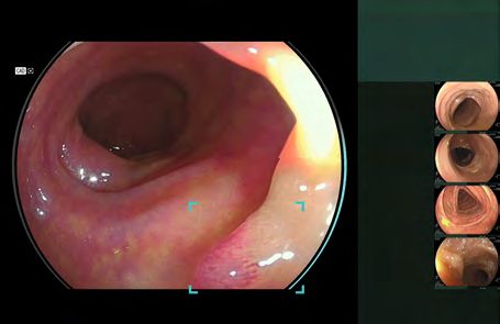
DEEP LEARNING TECHNOLOGY
        CAD EYE has been trained with a powerful supercomputer located in Fujifilm’s global AI technology centre in Tokyo, utilising
        an immense amount of clinical images using Fujifilm endoscopy systems. As a result, CAD EYE is a customised detection and
        characterisation support compatible with the ELUXEO™ system.

                                                                                                                                                   Detection & marking of polyps by CAD EYE

        Collected training data                                        Deep learning

                                                                             Medical AI Technology

                                                                                                                                                       Characterisation of polyps by CAD EYE

                                                    2018                                             Launched AI algorithms
                                                                                                     for bridge crack detection
                                                    Launched the REiLI artificial                    to support infrastructure
                                                                                                     improvements
                                                    intelligence platform and
                                                    deep learning engines                            Entered joint research
                                                                                                     agreement with Indiana
                                                                                                     University School of
                                                                                                     Medicine for AI medical
                                                                                                     imaging development
                       2014                                                                          Announced
                       Launched Virtual Grid™       Brain(s) creative AI center                      joint collaboration with
                       processing, which enhances   installed NVIDIA's DGX-2 for                     Lunit Inc. and Salud
                                                                                                     Digna to help radiologists
                       image contrast and clarity   AI development
                                                                                                                                                       MEETS
                                                                                                     evaluate AI technologies
                                                                                                     for diagnostic imaging
             2014

    Deep learning

Diagnostic support
                                                                                                                                  reili.fujifilm.com

                                                                                                                                                                                               3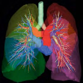
GASTROENTEROLOGY

               REAL-TIME DETECTION
    CAD EYE is aimed to improve the real time polyp detection rate to expert level, helping to recognise flat lesions, multiple
    polyps simultaneously, as well as any lesions at the corner of the image. CAD EYE Detection is possible with White Light and LCI
    (Linked Color Imaging) mode.

    White Light Mode                                                  LCI Mode

    USER-FRIENDLY INTERFACE
    The development of the interface has been designed to enable comfortable procedures. It does not interfere with clinical images
    and minimises required eye movement. Its display is designed to be simple and intuitive for excellent support during long
    hours in the examination room.

                                                                       1            DETECTION BOX
                                                                                    Displays the area where the suspicious
                                                                                    polyp is detected. Different sizes of the
                                                                                    Detection Box are available.

                                                                       2            VISUAL ASSIST CIRCLE
                                                                                    Lights up in the direction where the
                                               2                                    suspicious polyp is detected.

                              1
                                                                                    DETECTION SOUND
                                                                                    Sound signal when a suspicious polyp is
                                                                                    detected. Volume can be defined for each
                                                                                    user.

    REAL-TIME DIAGNOSTIC SUPPORT                                                                                       Detection
                                                                                                                       Mode
    The detection and characterisation results are displayed by processing
    up to 60 frames per second. Without freezing the image, CAD EYE
    supports real-time diagnosis during standard and magnified observation.

4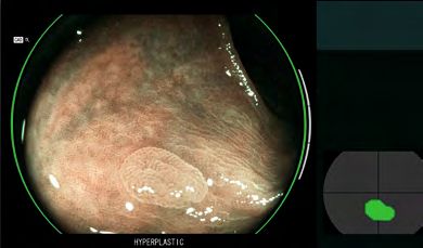
CHARACTERISATION SUPPORT
Once a suspected polyp is detected by CAD EYE Detection (WLI or LCI), CAD EYE Characterisation – in combination with BLI –
can support endoscopists in the diagnosis of the polyp. This function analyses in real-time and without freezing or zooming if a polyp
is hyperplastic or neoplastic, which is visually indicated by the use of different colour codes in the Position Map. CAD EYE Characterisation
is aimed to make procedures more efficient by increasing the accuracy of diagnosis to expert-level.*

BLI Mode – Neoplastic                                                   BLI Mode – Hyperplastic

CAD EYE received the prestigious Good Design Award for its interface.

                                                                         1                STATUS BAR
                                                                                          Indicates the status of characterisation analysis
                                                                                          regarding the suspicious area.

                                                                         2                VISUAL ASSIST CIRCLE
                                                  1                                       GREEN: Characterisation HYPERPLASTIC
                                                                                          YELLOW: Characterisation NEOPLASTIC
                                              2
                                                         3               3                POSITION MAP
                                                                                          Indicates the position of the suspicious area, this
                                                                                          software is characterising.
                                4
                                                                         4                CHARACTERISATION RESULT
                                                                                          HYPERPLASTIC: hyperplastic polyps & SSL
                                                                                          NEOPLASTIC: adenoma and cancer

                                      Characterisation
                                      Mode

                                                                                          *According to the validation study, the accuracy of non experts
                                                                                           with the assistance of CAD EYE Characterisation was equivalent
                                                                                           to that of an expert.

                                                                                                                                                            5
GASTROENTEROLOGY

    FOR YOUR DAILY EXAMINATION
    CAD EYE can be activated and deactivated simply by a push on the endoscope button or directly at the
    processor.

    SCOPE SWITCH 3                                                          SCOPE SWITCH 2

                                                  ON                  OFF                                      ON

    The function of each switch can be defined individually.                         CAD EYE Detection with White Light

    MOVIE AND STILL IMAGE RECORDING FUNCTION*
    Full HD movies and still images can be recorded and stored at
    the expansion unit EX-1. It can be controlled via the scope switch                   CAD EYE Detection with LCI
    or directly at the ELUXEO processor.
                                      ™

                                                                                      CAD EYE Characterisation with BLI

    * Video and still image files should not be used for diagnoses.

6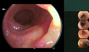
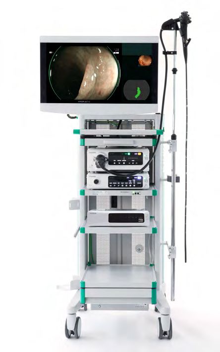
SPECIFICATIONS
CAD EYE works with the expansion unit EX-1 and the CAD EYE software EW10-EC02. With software
EW10-SC01 up to 30 hours of movie and still image material can be stored in the internal memory of EX-1.
It can easily be controlled with the scope switch or directly at the processor.

Expansion Unit EX-1
 Compatible processors         VP-7000, EP-6000
 Compatible scopes             700 series colonoscopes*
 Output                        DVI-I x1, DVI-D x1
 Input                         DVI-I x1
                               30 hours of video (Full HD, MP4) and still image
 Memory
                               material (Full HD or SXGA selectable, TIFF, JPEG)**
 Power rating                  100 – 240 VAC +/- 10%, 50/60 Hz, 1.25 to 0.60 A
 Dimensions (W x H x D)        370.0 x 99.0 x 465.6 mm
 Weight                        7.1 kg

 * Movie and still image recording function is compatible with
   700/600 / 500 scopes excluding EUS scopes
** In combination with EW10-SC01 software

Software EW10-EC02
                               USB flash drive for CAD EYE installment for
 Package Content
                               colon polyp detection and characterisation support

Software EW10-SC01
                                DICOM

                               USB flash drive for basic functions:
                               - Movie and still image recording with
                                 CAD EYE overlay
 Package Content
                               - Network function: Still image transfer via
                                 FTP/FTPS/Dicom storage and for video recording
                                 transfer via SAMBA

                                        FOR COLONIC POLYP
                                        DETECTION & CHARACTERISATION

                                        MOVIE / STILL IMAGE RECORDING
                                        & NETWORK FUNCTION

                                        FUTURE CAD APPLICATIONS                                           Expansion Unit EX-1

                                        CAN BE INSTALLED

                   HD TECHNOLOGY                                 DICOM TECHNOLOGY
                   Combine equipment displaying        DICOM     The goal of the DICOM Standard is
                   this logo to ensure that you view             to achieve compatibility and improve
                   HDTV images on your monitor.                  workflow efficiency between imaging
                                                                 systems and other information systems.

For further information visit www.cadeye.eu

                                                                                                                                7
are trademarks of their respective owners. All rights reserved. SAP70170009710. 01/2021.
FUJIFILM logo are trademarks of FUJIFILM Corporation. All other trademarks shown
Specifications are subject to change without notice. The name FUJIFILM and the
Treier Endoscopie AG
Chrüzmatt 1, 6215 Beromünster
Tel. +41 (0)41 510 07 00
info@treier.com • www.treier.com
FUJIFILM Europe GmbH
Heesenstr. 31, 40549 Düsseldorf, Germany
Tel.: +49 211-50 89 0, Fax: +49 211-50 89 8700
www.fujifilm-endoscopy.com, www.eluxeo-ultra.comYou can also read