Meta-analysis uncovers genome-wide significant variants for rapid kidney function decline
←
→
Page content transcription
If your browser does not render page correctly, please read the page content below
www.kidney-international.org                                                                                   clinical investigation

Meta-analysis uncovers genome-wide significant
variants for rapid kidney function decline                                                                                              OPEN

Mathias Gorski1,2,120, Bettina Jung2,120, Yong Li3,120, Pamela R. Matias-Garcia4,5,6,120, Matthias Wuttke3,7,
Stefan Coassin8, Chris H.L. Thio9, Marcus E. Kleber10, Thomas W. Winkler1, Veronika Wanner1,
Jin-Fang Chai11, Audrey Y. Chu12, Massimiliano Cocca13, Mary F. Feitosa14, Sahar Ghasemi15,16,
Anselm Hoppmann3, Katrin Horn17,18, Man Li19, Teresa Nutile20, Markus Scholz17,18, Karsten B. Sieber21,
Alexander Teumer15,16, Adrienne Tin22,23, Judy Wang14, Bamidele O. Tayo24, Tarunveer S. Ahluwalia25,
Peter Almgren26, Stephan J.L. Bakker27, Bernhard Banas2, Nisha Bansal28,29, Mary L. Biggs30,31,
Eric Boerwinkle32, Erwin P. Bottinger33,34, Hermann Brenner35,36, Robert J. Carroll37, John Chalmers38,39,40,
Miao-Li Chee41, Miao-Ling Chee41, Ching-Yu Cheng41,42,43, Josef Coresh40, Martin H. de Borst27,
Frauke Degenhardt44, Kai-Uwe Eckardt45,46, Karlhans Endlich16,47, Andre Franke44, Sandra Freitag-Wolf48,
Piyush Gampawar49, Ron T. Gansevoort27, Mohsen Ghanbari50,51, Christian Gieger4,5,52,
Pavel Hamet53,54,55, Kevin Ho56,57, Edith Hofer58,59, Bernd Holleczek35, Valencia Hui Xian Foo41,
Nina Hutri-Kähönen60,61, Shih-Jen Hwang62,63, M. Arfan Ikram50, Navya Shilpa Josyula64,
Mika Kähönen65,66, Chiea-Chuen Khor41,67, Wolfgang Koenig68,69,70, Holly Kramer24,71,
Bernhard K. Krämer72, Brigitte Kühnel4, Leslie A. Lange73, Terho Lehtimäki74,75, Wolfgang Lieb76; Lifelines
Cohort Study77, Regeneron Genetics Center77: Ruth J.F. Loos33,78, Mary Ann Lukas79,
Leo-Pekka Lyytikäinen74,75, Christa Meisinger80,81, Thomas Meitinger69,82,83, Olle Melander84,
Yuri Milaneschi85, Pashupati P. Mishra74,75, Nina Mononen74,75, Josyf C. Mychaleckyj86,
Girish N. Nadkarni33,87, Matthias Nauck16,88, Kjell Nikus89,90, Boting Ning91, Ilja M. Nolte9,
Michelle L. O’Donoghue92,93, Marju Orho-Melander26, Sarah A. Pendergrass94, Brenda W.J.H. Penninx85,
Michael H. Preuss33, Bruce M. Psaty95,96, Laura M. Raffield97, Olli T. Raitakari98,99,100, Rainer Rettig101,
Myriam Rheinberger2,102, Kenneth M. Rice31, Alexander R. Rosenkranz103, Peter Rossing25,
Jerome I. Rotter104, Charumathi Sabanayagam41,42, Helena Schmidt49, Reinhold Schmidt58,
Ben Schöttker35,36, Christina-Alexandra Schulz26, Sanaz Sedaghat50,105, Christian M. Shaffer37,
Konstantin Strauch106,107, Silke Szymczak48, Kent D. Taylor104, Johanne Tremblay53,55,54,
Layal Chaker50,108, Pim van der Harst109,110,111, Peter J. van der Most9, Niek Verweij109, Uwe Völker16,112,
Melanie Waldenberger4,5,69, Lars Wallentin113,114, Dawn M. Waterworth21, Harvey D. White115,
James G. Wilson116, Tien-Yin Wong41,42, Mark Woodward38,39,40, Qiong Yang91, Masayuki Yasuda41,117,
Laura M. Yerges-Armstrong21, Yan Zhang35, Harold Snieder9, Christoph Wanner118,
Carsten A. Böger2,102,121, Anna Köttgen3,40,121, Florian Kronenberg8,121, Cristian Pattaro119,121 and
Iris M. Heid1,121
1
 Department of Genetic Epidemiology, University of Regensburg, Regensburg, Germany; 2Department of Nephrology, University Hospital
Regensburg, Regensburg, Germany; 3Institute of Genetic Epidemiology, Department of Biometry, Epidemiology and Medical
Bioinformatics, Faculty of Medicine and Medical Center—University of Freiburg, Freiburg, Germany; 4Research Unit of Molecular
Epidemiology, Helmholtz Zentrum München—German Research Center for Environmental Health, Neuherberg, Germany; 5Institute of
Epidemiology, Helmholtz Zentrum München—German Research Center for Environmental Health, Neuherberg, Germany; 6TUM School of
Medicine, Technical University of Munich, Munich, Germany; 7Renal Division, Department of Medicine IV, Faculty of Medicine and Medical
Center—University of Freiburg, Freiburg, Germany; 8Department of Genetics and Pharmacology, Institute of Genetic Epidemiology,
Medical University of Innsbruck, Innsbruck, Austria; 9Department of Epidemiology, University of Groningen, University Medical Center
Groningen, Groningen, the Netherlands; 10Vth Department of Medicine (Nephrology, Hypertensiology, Rheumatology, Endocrinology,
Diabetology), Medical Faculty Mannheim, University of Heidelberg, Mannheim, Germany; 11Saw Swee Hock School of Public Health,
National University of Singapore and National University Health System, Singapore, Singapore; 12Genetics, Merck & Co., Inc., Kenilworth,

                                                                             120
Correspondence: Iris M. Heid or Mathias Gorski, Department of Genetic          These authors contributed equally.
Epidemiology, University of Regensburg, Franz-Josef-Strauß-Allee 11, 93053   121
                                                                               These authors jointly supervised this work.
Regensburg, Germany. E-mail: mathias.gorski@klinik.uni-regensburg.de or
iris.heid@klinik.uni-regensburg.de                                           Received 4 June 2020; revised 21 August 2020; accepted 17 September
77                                                                           2020; published online 31 October 2020
  Members of the Lifelines Cohort Study and Regeneron Genetics Center
are listed in the Appendix.

Kidney International (2021) -, -–-                                                                                                            1
clinical investigation                                                                            M Gorski et al.: Rapid kidney function decline

New Jersey, USA; 13Institute for Maternal and Child Health, IRCCS “Burlo Garofolo,” Trieste, Italy; 14Division of Statistical Genomics,
Department of Genetics, Washington University School of Medicine, St. Louis, Missouri, USA; 15Institute for Community Medicine,
University Medicine Greifswald, Greifswald, Germany; 16DZHK (German Center for Cardiovascular Research), Partner Site Greifswald,
Greifswald, Germany; 17Institute for Medical Informatics, Statistics and Epidemiology, University of Leipzig, Leipzig, Germany; 18LIFE
Research Center for Civilization Diseases, University of Leipzig, Leipzig, Germany; 19Division of Nephrology and Hypertension, Department
of Medicine, University of Utah, Salt Lake City, Utah, USA; 20Institute of Genetics and Biophysics “Adriano Buzzati-Traverso”—CNR, Naples,
Italy; 21Human Genetics, GlaxoSmithKline, Collegeville, Pennsylvania, USA; 22Memory Impairment and Neurodegenerative Dementia
(MIND) Center, University of Mississippi Medical Center, Jackson, Mississippi, USA; 23Division of Nephrology, Department of Medicine,
University of Mississippi Medical Center, Jackson, Mississippi, USA; 24Department of Public Health Sciences, Loyola University Chicago,
Maywood, Illinois, USA; 25Steno Diabetes Center Copenhagen, Gentofte, Denmark; 26Diabetes and Cardiovascular Disease—Genetic
Epidemiology, Department of Clinical Sciences in Malmö, Lund University, Malmö, Sweden; 27Division of Nephrology, Department of
Internal Medicine, University of Groningen, University Medical Center Groningen, Groningen, the Netherlands; 28Division of Nephrology,
University of Washington, Seattle, Washington, USA; 29Kidney Research Institute, University of Washington, Seattle, Washington, USA;
30
   Cardiovascular Health Research Unit, Department of Medicine, University of Washington, Seattle, Washington, USA; 31Department of
Biostatistics, University of Washington, Seattle, Washington, USA; 32Human Genetics Center, University of Texas Health Science Center,
Houston, Texas, USA; 33Charles Bronfman Institute for Personalized Medicine, Icahn School of Medicine at Mount Sinai, New York, New
York, USA; 34Digital Health Center, Hasso Plattner Institute and University of Potsdam, Potsdam, Germany; 35Division of Clinical
Epidemiology and Aging Research, German Cancer Research Center (DKFZ), Heidelberg, Germany; 36Network Aging Research, University
of Heidelberg, Heidelberg, Germany; 37Department of Biomedical Informatics, Vanderbilt University Medical Center, Nashville, Tennessee,
USA; 38The George Institute for Global Health, University of New South Wales, Sydney, Australia; 39The George Institute for Global Health,
University of Oxford, Oxford, UK; 40Department of Epidemiology, Johns Hopkins Bloomberg School of Public Health, Baltimore, Maryland,
USA; 41Singapore Eye Research Institute, Singapore National Eye Center, Singapore, Singapore; 42Ophthalmology and Visual Sciences
Academic Clinical Program (Eye ACP), Duke—NUS Medical School, Singapore, Singapore; 43Department of Ophthalmology, Yong Loo Lin
School of Medicine, National University of Singapore and National University Health System, Singapore, Singapore; 44Institute of Clinical
Molecular Biology, Christian-AlbrechtsUniversity of Kiel, Kiel, Germany; 45Department of Nephrology and Medical Intensive Care,
Charité—Universitätsmedizin Berlin, Berlin, Germany; 46Department of Nephrology and Hypertension, Friedrich Alexander University
Erlangen-Nürnberg (FAU), Erlangen, Germany; 47Department of Anatomy and Cell Biology, University Medicine Greifswald, Greifswald,
Germany; 48Institute of Medical Informatics and Statistics, Kiel University, University Hospital Schleswig-Holstein, Kiel, Germany; 49Institute
of Molecular Biology and Biochemistry, Center for Molecular Medicine, Medical University of Graz, Graz, Austria; 50Department of
Epidemiology, Erasmus MC, University Medical Center Rotterdam, Rotterdam, the Netherlands; 51Department of Genetics, School of
Medicine, Mashhad University of Medical Sciences, Mashhad, Iran; 52German Center for Diabetes Research (DZD), Neuherberg, Germany;
53
   Montreal University Hospital Research Center, CHUM, Montreal, Quebec, Canada; 54Medpharmgene, Montreal, Quebec, Canada;
55
   CRCHUM, Montreal, Canada; 56Kidney Health Research Institute (KHRI), Geisinger, Danville, Pennsylvania, USA; 57Department of
Nephrology, Geisinger, Danville, Pennsylvania, USA; 58Clinical Division of Neurogeriatrics, Department of Neurology, Medical University of
Graz, Graz, Austria; 59Institute for Medical Informatics, Statistics and Documentation, Medical University of Graz, Graz, Austria;
60
   Department of Pediatrics, Tampere University Hospital, Tampere, Finland; 61Department of Pediatrics, Faculty of Medicine and Health
Technology, Tampere University, Tampere, Finland; 62NHLBI’s Framingham Heart Study, Framingham, Massachusetts, USA; 63The Center
for Population Studies, NHLBI, Framingham, Massachusetts, USA; 64Geisinger Research, Biomedical and Translational Informatics
Institute, Rockville, Maryland, USA; 65Department of Clinical Physiology, Tampere University Hospital, Tampere, Finland; 66Department of
Clinical Physiology, Finnish Cardiovascular Research Center—Tampere, Faculty of Medicine and Health Technology, Tampere University,
Tampere, Finland; 67Genome Institute of Singapore, Agency for Science Technology and Research, Singapore, Singapore; 68Deutsches
Herzzentrum München, Technische Universität München, Munich, Germany; 69DZHK (German Center for Cardiovascular Research),
Partner Site Munich Heart Alliance, Munich, Germany; 70Institute of Epidemiology and Medical Biometry, University of Ulm, Ulm,
Germany; 71Division of Nephrology and Hypertension, Loyola University Chicago, Chicago, Illinois, USA; 72Department of Medicine
(Nephrology, Hypertensiology, Rheumatology, Endocrinology, Diabetology), Medical Faculty Mannheim, University of Heidelberg,
Mannheim, Germany; 73Division of Biomedical Informatics and Personalized Medicine, School of Medicine, University of Colorado
Denver—Anschutz Medical Campus, Aurora, Colorado, USA; 74Department of Clinical Chemistry, Fimlab Laboratories, Tampere, Finland;
75
   Department of Clinical Chemistry, Finnish Cardiovascular Research Center—Tampere, Faculty of Medicine and Health Technology,
Tampere University, Tampere, Finland; 76Institute of Epidemiology and Biobank Popgen, Kiel University, Kiel, Germany; 78The Mindich
Child Health and Development Institute, Icahn School of Medicine at Mount Sinai, New York, New York, USA; 79Target Sciences—
Genetics, GlaxoSmithKline, Albuquerque, New Mexico, USA; 80Independent Research Group Clinical Epidemiology, Helmholtz Zentrum
München, German Research Center for Environmental Health, Neuherberg, Germany; 81Chair of Epidemiology, Ludwig-Maximilians-
Universität München at UNIKA-T Augsburg, Augsburg, Germany; 82Institute of Human Genetics, Helmholtz Zentrum München,
Neuherberg, Germany; 83Institute of Human Genetics, Technische Universität München, Munich, Germany; 84Hypertension and
Cardiovascular Disease, Department of Clinical Sciences Malmö, Lund University, Malmö, Sweden; 85Department of Psychiatry,
Amsterdam Public Health and Amsterdam Neuroscience, Amsterdam UMC/Vrije Universiteit and GGZ inGeest, Amsterdam, the
Netherlands; 86Center for Public Health Genomics, University of Virginia, Charlottesville, Charlottesville, Virginia, USA; 87Division of
Nephrology, Department of Medicine, Icahn School of Medicine at Mount Sinai, New York, New York, USA; 88Institute of Clinical Chemistry
and Laboratory Medicine, University Medicine Greifswald, Greifswald, Germany; 89Department of Cardiology, Heart Center, Tampere

2                                                                                                                Kidney International (2021) -, -–-
M Gorski et al.: Rapid kidney function decline                                                             clinical investigation

University Hospital, Tampere, Finland; 90Department of Cardiology, Finnish Cardiovascular Research Center—Tampere, Faculty of
Medicine and Health Technology, Tampere University, Tampere, Finland; 91Department of Biostatistics, Boston University School of Public
Health, Boston, Massachusetts, USA; 92Cardiovascular Division, Brigham and Women’s Hospital, Boston, Massachusetts, USA; 93TIMI
Study Group, Boston, Massachusetts, USA; 94Geisinger Research, Biomedical and Translational Informatics Institute, Danville,
Pennsylvania, USA; 95Cardiovascular Health Research Unit, Department of Medicine, Department of Epidemiology, Department of Health
Services, University of Washington, Seattle, Washington, USA; 96Kaiser Permanente Washington Health Research Institute, Seattle,
Washington, USA; 97Department of Genetics, University of North Carolina, Chapel Hill, North Carolina, USA; 98Department of Clinical
Physiology and Nuclear Medicine, Turku University Hospital, Turku, Finland; 99Research Center of Applied and Preventive Cardiovascular
Medicine, University of Turku, Turku, Finland; 100Centre for Population Health Research, University of Turku and Turku University Hospital,
Turku, Finland; 101Institute of Physiology, University Medicine Greifswald, Karlsburg, Germany; 102Department of Nephrology and
Rheumatology, Kliniken Südostbayern, Regensburg, Germany; 103Department of Internal Medicine, Division of Nephrology, Medical
University Graz, Graz, Austria; 104The Institute for Translational Genomics and Population Sciences, Department of Pediatrics, The
Lundquist Institute for Biomedical Innovation at Harbor-UCLA Medical Center, Torrance, California, USA; 105Department of Preventive
Medicine, Northwestern University, Feinberg School of Medicine, Chicago, Illinois, USA; 106Institute of Genetic Epidemiology, Helmholtz
Zentrum München—German Research Center for Environmental Health, Neuherberg, Germany; 107Chair of Genetic Epidemiology, IBE,
Faculty of Medicine, Ludwig-Maximilians-Universität München, München, Germany; 108Department of Internal Medicine, Erasmus MC,
University Medical Center Rotterdam, Rotterdam, the Netherlands; 109Department of Cardiology, University of Groningen, University
Medical Center Groningen, Groningen, the Netherlands; 110Department of Genetics, University of Groningen, University Medical Center
Groningen, Groningen, the Netherlands; 111Durrer Center for Cardiovascular Research, The Netherlands Heart Institute, Utrecht, the
Netherlands; 112Interfaculty Institute for Genetics and Functional Genomics, University Medicine Greifswald, Greifswald, Germany;
113
   Cardiology, Department of Medical Sciences, Uppsala University, Uppsala, Sweden; 114Uppsala Clinical Research Center, Uppsala
University, Uppsala, Sweden; 115Green Lane Cardiovascular Service, Auckland City Hospital and University of Auckland, Auckland, New
Zealand; 116Department of Physiology and Biophysics, University of Mississippi Medical Center, Jackson, Mississippi, USA; 117Department
of Ophthalmology, Tohoku University Graduate School of Medicine, Miyagi, Japan; 118Division of Nephrology, University Clinic, University
of Würzburg, Würzburg, Germany; and 119Eurac Research, Institute for Biomedicine (affiliated with the University of Lübeck), Bolzano,
Italy

Rapid decline of glomerular filtration rate estimated from              interval 1.08-1.33). Thus, our identified loci for rapid kidney
creatinine (eGFRcrea) is associated with severe clinical               function decline may help prioritize therapeutic targets and
endpoints. In contrast to cross-sectionally assessed                   identify mechanisms and individuals at risk for sustained
eGFRcrea, the genetic basis for rapid eGFRcrea decline is              deterioration of kidney function.
largely unknown. To help define this, we meta-analyzed 42               Kidney International (2021)   -, -–-;   https://doi.org/10.1016/
genome-wide association studies from the Chronic Kidney                j.kint.2020.09.030
Diseases Genetics Consortium and United Kingdom                        KEYWORDS: acute kidney injury; end-stage kidney disease; genome-wide
Biobank to identify genetic loci for rapid eGFRcrea decline.           association study; rapid eGFRcrea decline
Two definitions of eGFRcrea decline were used: 3 mL/min/                Copyright ª 2020, International Society of Nephrology. Published by
                                                                       Elsevier Inc. This is an open access article under the CC BY-NC-ND license
1.73m2/year or more (“Rapid3”; encompassing 34,874
                                                                       (http://creativecommons.org/licenses/by-nc-nd/4.0/).
cases, 107,090 controls) and eGFRcrea decline 25% or more
and eGFRcrea under 60 mL/min/1.73m2 at follow-up

                                                                      R
among those with eGFRcrea 60 mL/min/1.73m2 or more at                          apid kidney function decline is an important risk factor
baseline (“CKDi25”; encompassing 19,901 cases, 175,244                         for end-stage kidney disease (ESKD), cardiovascular
controls). Seven independent variants were identified                           events, and early mortality.1,2 ESKD is a life-
across six loci for Rapid3 and/or CKDi25: consisting of five            threatening condition with substantial individual and public
variants at four loci with genome-wide significance (near               health burden3–5 and a major endpoint in clinical nephrology
UMOD-PDILT (2), PRKAG2, WDR72, OR2S2) and two variants                 trials. However, identifying and monitoring individuals at risk
among 265 known eGFRcrea variants (near GATM, LARP4B).                 for ESKD is challenging. Two definitions of rapid decline in
All these loci were novel for Rapid3 and/or CKDi25 and our             creatinine-based eGFR (eGFRcrea) are reported to increase
bioinformatic follow-up prioritized variants and genes                 ESKD risk 5- and 12-fold,6,7 respectively, and thus recom-
underneath these loci. The OR2S2 locus is novel for any                mended for clinical use: (i) rapid eGFRcrea decline of >5
eGFRcrea trait including interesting candidates. For the five           ml/min per 1.73 m2 per year and (ii) a $25% decline of
genome-wide significant lead variants, we found                         eGFRcrea along with movement into a lower category of
supporting effects for annual change in blood urea                     chronic kidney disease.7 Other surrogate endpoints of ESKD
nitrogen or cystatin-based eGFR, but not for GATM or                   were implemented by interventional trials with a follow-up
LARP4B. Individuals at high compared to those at low                   duration of
clinical investigation M Gorski et al.: Rapid kidney function decline
Figure 1 | Illustration of the case-control definitions of Rapid3 and CKDi25. Rapid3 defines cases as individuals with an glomerular
filtration rate estimated from creatinine (eGFRcrea) decline >3 ml/min per 1.73 m2 per year and controls with an eGFRcrea decline between 1
and þ1 ml/min per 1.73 m2 per year. CKDi25 defines cases as a $25% drop from baseline eGFRcrea $60 ml/min per 1.73 m2 into
eGFRcrea 270,000 individuals with 2 eGFRcrea
ulopathies such as immunosuppressive agents11 or tolvaptan measurements across a time period of 1–15 years of follow-
in polycystic kidney disease,12 therapeutic options to slow up. We implemented 2 definitions of rapid eGFRcrea
down kidney function decline are largely limited to glycemic decline that were feasible in population-based studies while
and blood pressure control as well as lipid-lowering drugs. preserving similarity to recommended surrogate clinical
Before the recent advent of SGLT2 inhibitors in large clinical endpoints: (i) “Rapid3” cases defined as eGFRcrea decline of
trials,13 these therapies had shown only a moderate, if any, >3 ml/min per 1.73 m2 per year compared with “no decline”
effect on clinically relevant renal endpoints.14 Selecting (“Rapid3” controls, 1 to þ1 ml/min per 1.73 m2 per year);
genetically supported drug targets was estimated to double and (ii) “CKDi25” cases defined as $25% eGFRcrea decline
success rate in drug discovery,15 in particular when the causal during follow-up together with a movement from
gene was suggested by Mendelian diseases or from genome- eGFRcrea $60 ml/min per 1.73 m2 at baseline to
wide associations driven by coding variants.16 This moti- eGFRcrea 1,000,000 in- RESULTS
dividuals identified 264 loci associated with eGFRcrea based Rapid eGFRcrea decline in 42 longitudinal studies
on 1 creatinine measurement (“cross-sectional eGFRcrea”).17 We collected phenotype summary statistics for Rapid3 and
However, little is known about whether these or additional CKDi25 from 42 studies with genetic data and at least 2
genetic factors are associated with rapid kidney function measurements of creatinine (study-specific mean age of par-
decline (“longitudinal kidney function traits”). Given the ticipants 33–68 years, study-specific median follow-up time
substantial organizational and temporal requirements of 1–15 years; Methods, Supplementary Table S1). Most studies
longitudinal studies, sample sizes for these studies are still were from European ancestry and population (32 European
limited compared with cross-sectional studies. Our previous ancestry–based, 34 population-based).
longitudinal GWAS based on 61,078 individuals and Several interesting aspects emerged: (i) as expected for
approximately 3 million genetic variants did not identify any studies covering general populations as well as elderly and
locus for rapid eGFRcrea decline.18 New studies with longi- patient populations, study-specific median baseline eGFRcrea
tudinal eGFRcrea measurements and new genomic reference ranged from 46.4 to 115.0 ml/min per 1.73 m2 (overall
panels enabling a denser and more precise genetic variant median ¼ 87.3 ml/min per 1.73 m2); (ii) case proportions
imputation now allow for a more powerful investigation. ranged from 11% to 72% for Rapid3 and from 3% to 52% for
We thus performed a GWAS meta-analysis across 42 lon- CKDi25 (median ¼ 30% or 11%, respectively); (iii) there was
gitudinal studies, consisting of 41 studies from the Chronic no association of study-specific median age of participants or
Kidney Disease Genetics (CKDGen) Consortium and UK median follow-up time with Rapid3 or CKDi25
4 Kidney International (2021) -, -–-M Gorski et al.: Rapid kidney function decline clinical investigation
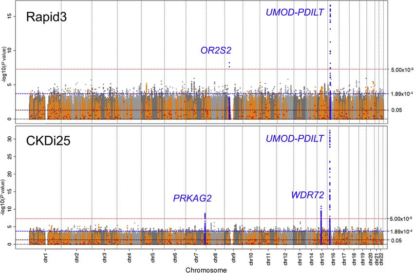
Figure 2 | Four loci identified with genome-wide significance for Rapid3 or CKDi25. Shown are association P values versus genomic
position for Rapid3 (34,874 cases; 107,090 controls) and CKDi25 (19,901 cases; 175,244 controls). Horizontal dashed lines indicate genome-wide
(5.00 108), Bonferroni-corrected (0.05/265 z 1.89 104), and nominal (0.05) significance thresholds. The 4 identified genome-wide
significant loci are annotated by the nearest genes (blue). The 264 loci reported previously for cross-sectional eGFRcrea17 are marked in orange
and respective lead variants as red dots. eGFRcrea, glomerular filtration rate estimated from creatinine.
(Supplementary Figure S1); (iv) most CKDi25 cases were a CKDi25 and showed a second independent signal for CKDi25
subgroup of Rapid3 cases in 3 example studies with different (rs77924615; P-adjusted ¼ 2.98 1010). For CKDi25, the
lengths of follow-up (Supplementary Table S2). independent odds ratios (ORs) for the 2 UMOD-PDILT lead
variants (rs12922822, rs77924615) were 1.06 per adverse
Four new genome-wide significant loci for rapid eGFRcrea allele per variant in a model containing both variants. (ii) One
decline variant in each of the WDR72 and PRKAG2 loci was identified
In each of the 42 studies, the >8 million genetic variants for CKDi25. (iii) A variant near OR2S2 was associated with
imputed via 1000 Genomes19 or Haplotype Reference Con- Rapid3.
sortium20 reference panels were tested for association with For all variants and both outcomes, we observed no to
Rapid3 and CKDi25 using logistic regression adjusting for moderate heterogeneity across studies (I2 ¼ 0%–43%). A
age, sex, and baseline eGFRcrea (Supplementary Table S3, sensitivity analysis restricted to European ancestry (31,101
Methods). We meta-analyzed study-specific summary statis- cases, 102,485 controls for Rapid3; 19,419 cases, 169,087
tics by outcome (34,874 cases, 107,090 controls for Rapid3; controls for CKDi25) identified the same loci with the same
19,901 cases, 175,244 controls for CKDi25; Methods). or highly correlated lead variants (r2 > 0.84, Supplementary
In our genome-wide approach, we selected genome-wide Table S4A). We also conducted a meta-analysis restricting to
significant loci (i.e., $1 variant with a P value of6
clinical investigation
Table 1 | Six loci from the genome-wide and candidate-based search for association with Rapid3 or CKDi25
Rapid3 CKDi25
RSID Chr:Position Identifying analysis Locus name EA/OA EAF OR P OR P Locus/signal no. Reference variant (R2)
Genome-wide search (genome-wide significance, P valueM Gorski et al.: Rapid kidney function decline clinical investigation
Biobank (n up to 364,819 and 358,791) as well as previously reported BUN results from CKDGen17 (n ¼ 416,076), where 1-sided P values test the eGFRcrea-lowering allele into the direction of decreased eGFRcys and increased BUN
(1.62 1022)
1.09 1018 (8.79 1021)
2.38 1026 (2.57 1042)
8.73 1080 (1.69 1041)
Annual change of eGFRcys and BUN was calculated as the baseline value minus the follow-up value divided by the years between baseline and follow-up. The age, sex, and baseline eGFRcys/BUN-adjusted residuals were regressed
Association results for annual change in eGFRcys and BUN in UK Biobank (n up to 15,746 or 15,277, respectively). One-sided P values are provided testing the allele that increased the risk of rapid eGFRcrea decline (usually the
eGFRcrea-lowering allele, except for the OR2S2 lead variant) into the direction of annual eGFRcys decline and annual BUN increase. For completeness, also shown are association results for cross-sectional eGFRcys and BUN from UK
BUN, blood urea nitrogen; effect, genetic effect; eGFRcys, estimated glomerular filtration rate based on cystatin C; locus/signal no. [name], locus number and signal number [locus name]; P, one-sided association P value; RSID,
Two additional loci for rapid eGFRcrea decline from a
1.59 109 (8.46 1017)
2.49 1011 (4.90 107)
candidate-based search
Genetic variants with established association for cross-
0.70 (0.89)
0.95 (0.46)
sectional eGFRcrea are candidates for association with rapid
P
eGFRcrea decline. For our candidate-based approach, we
18
BUNb UKBB (CKDGen)
selected the 264 lead variants and the second signal lead
1.08 10
variant in the UMOD-PDILT locus reported previously for
eGFRcrea17 and tested these for association with Rapid3 and
CKDi25 (judged at Bonferroni-corrected significance; 0.05/
265 ¼ 1.89 104). Among these, we found 6 variants in 5
0.00345 (0.0018)
0.0004 (clinical investigation M Gorski et al.: Rapid kidney function decline
Table 3 | Size of 99% credible sets of variants for the 7 identified signals for Rapid3 or CKDi25
No. of variants in No. of variants in
99% credible set 99% credible set
(overlap with (overlap with
Locus regionc eGFRcrea sets) CKDi25 sets)
Locus/ No. of
a b
signal no. Locus name Identifying trait Chr Start Stop genes Rapid3d CKDi25d eGFRcread
1.1 [UMOD-PDILT] Rapid3, CKDi25 16 19,866,507 20,867,645 13 14 (10) 13 (11) 16 (10)
1.2 [UMOD-PDILT] CKDi25 2nd 16 19,866,507 20,867,645 s.a. 1059 1 (1) 1 (1)
2 [WDR72] CKDi25 15 53,502,606 54,502,606 1 2931 37 (0) 41 (0)
3 [PRKAG2] CKDi25 7 150,906,788 151,906,788 14 2671 16 (6) 6 (6)
4 [OR2S2] Rapid3 9 35,437,931 36,437,931 36 2 2573 NA
5 [LARP4B] CKDi25 10 399,071 1,399,071 10 2955 2806 1e
6 [GATM] Rapid3 15 45,183,795 46,183,795 17 1438 2493 1e
Chr, chromosome of the locus region; s.a., see above; start/stop, start and stop of the locus region on GRCh37.
a
Nearest gene(s), stated in brackets to distinguish from gene and protein names.
b
Indicates the trait for which the variant was identified with significant association (“CKDi25 2nd” indicating that this is the second independent signal for the CKDi25 trait
analysis).
c
Locus region defined as the region of the 2 lead variants identified for Rapid3 and CKDi25 in [UMOD-PDILT] or for the single lead variant identified for Rapid3 or CKDi25 in the
other loci 500 kB. The CKDi25 2nd signal (signal no. 1.2) is mapped to the [UMOD-PDILT] locus region from signal no. 1.1.
d
Bold values indicate the credible set of variants for the analysis that identified the locus/signal.
e
For the candidate-based identified loci [LARP4B] and [GATM], the statistics for the credible sets were instable due to the lack of genome-wide significance and yielded
extremely wide credible set intervals. Because the CKDi25 or Rapid3 signal was very similar to the signal for cross-sectional eGFRcrea (Supplementary Figure S4E and F), we
conducted the bioinformatic follow-up for the credible set variant derived from eGFRcrea previously.
Number of genes overlapping each of the 6 locus regions (lead variant 500 kB) and the number of variants in the 99% credible set for each of the 7 signals. The credible sets
of variants were computed (i) for the 2 rapid eGFRcrea decline traits (Rapid3 and CKDi25) highlighting the set for the analysis that identified the locus/signal (signals 1.1–4
from the genome-wide approach, signals 5 and 6 from the candidate-based approach) and (ii) for cross-sectional eGFRcrea from CKDGen data as reported previously.17
eGFRcys and BUN (n ¼ 364,819 and 358,791). These analyses in the OR2S2 locus for Rapid3 formed the credible set
with alternative renal biomarkers supported UMOD-PDILT, (posterior probability 77% and 23%, respectively); (iv) the
WDR72, PRKAG2, and OR2S2, but not LARP4B or GATM credible sets for the 2 candidate-approach–derived loci,
loci (Table 2, Supplementary Note S3). LARP4B and GATM, included 1438–2955 variants for
Rapid3 and CKDi25, which was due insufficiently strong
From lead variants to the statistical signals associations resulting from the lack of genome-wide signif-
Each lead variant represents a signal consisting of correlated icance. We thus considered these credible sets unsuitable for
variants. Regional association plots (Supplementary in silico follow-up and focused on further evaluation on the
Figure S4) illustrate that the 7 rapid eGFRcrea decline sig- 5 genome-wide significant signals.
nals mostly coincided with the cross-sectional eGFRcrea
signal, except for a weaker signal in the WDR72 locus and no From statistical evidence to biology
corresponding OR2S2 signal for cross-sectional eGFRcrea. One of the key challenges in translating GWAS associations
Between the 2 traits, Rapid3 and CKDi25, the signals were into an understanding of the underlying biology is the
mostly comparable, except for LARP4B and OR2S2. identification of variants and genes causing the statistical
To prioritize variants at identified signals, we ranked each signal. It is unclear exactly what evidence to weigh in and how
signal variant by its posterior probability of driving the expansive the search for causal genes should be; 500 kB
observed association and added them to the “99% credible around the lead variant is often used (“locus region”). A
set of variants” until the cumulative posterior probability variant is often considered more likely causal when it is in a
was >99% (Methods). Such a credible set is thus a parsi- credible set and predicted to have a relevant function, such as
monious set of variants that most likely include the causal protein-altering (e.g., changing the peptide sequence, trun-
variant, assuming that there is exactly 1 causal variant per cating, affecting RNA splicing) or modulating a gene’s
signal and that this variant was analyzed.23 When deriving expression24 (expression quantitative trait locus [eQTL]). A
the 99% credible sets of variants for each of the 7 identified gene is often considered more likely causal when it (i) con-
signals for Rapid3 and CKDi25 (Methods) and comparing tains a protein-altering credible set variant, (ii) is a target of
them with cross-sectional eGFRcrea credible sets,17 we an eQTL variant, or (iii) has a kidney-related phenotype re-
found the following (Table 3): (i) for most GWAS-derived ported from animal models or monogenic disease. We an-
signals, the credible sets coincided with those for cross- notated the credible set variants and the 64 genes across the 5
sectional eGFRcrea, except for the WDR72 locus; (ii) the genome-wide significant signals accordingly (Methods,
credible set of the second UMOD-PDILT signal for CKDi25 Supplementary Tables S6A and B and S7A and B). We sum-
consisted of precisely 1 variant, rs77924615, which was marized the evidence per gene in a Gene PrioritiSation table
exactly the 1 credible set variant for eGFRcrea supporting and implemented a customizable score, where each category’s
this as the most likely causal variant for this association weight can be modified according to personal interest or
signal; (iii) the 2 correlated genome-wide significant variants preference (Supplementary Table S8).
8 Kidney International (2021) -, -–-M Gorski et al.: Rapid kidney function decline clinical investigation
eQTL-modulated
Any credible expression by Evidenced
set variants any credible set kidney
in gene variant phenotype
Weight 1 1 1 1 1 1 1 1 1
# Credible set variants in gene
Distance to 1st signal variant
NephQTL tubulointerstitium
GTEx v8 any other tissue
GTEx v8 kidney tissue
NephQTL glomerulus
Gene Priority Score
In human (OMIM)
Altered splicing
Chromosome
In mice (MGI)
Locus name
Locus no.
Missense
Gene
NMD
[UMOD-PDILT] 1 UMOD 16 0 10 2 0 0 0 0 0 0 0 1 1
[UMOD-PDILT] 1 PDILT 16 2,846 1 1 0 0 0 0 0 0 1 0 0
[WDR72] 2 WDR72 15 0 37 2 1 0 0 0 0 0 1 0 0
[PRKAG2] 3 PRKAG2 7 0 16 2 0 1 0 0 0 0 0 0 1
[PRKAG2] 3 GALNTL5 7 246,675 0 1 0 0 0 0 1 0 0 0 0
[OR2S2] 4 OR2S1P 9 75,251 0 1 0 0 0 0 0 0 1 0 0
[OR2S2] 4 GNE 9 276,506 0 1 0 0 0 0 0 0 0 1 0
[OR2S2] 4 CD72 9 -319,507 0 1 0 0 0 0 0 0 0 1 0
Figure 3 | Gene PrioritiSation (GPS) for the genes across the 4 loci identified with genome-wide significance. Shown are genes across
the 4 loci, for which we found any relevant evidence: (i) blue: gene contains at least 1 credible set variant that was protein-altering (missense,
nonmediated decay, NMD, or altered splicing; Supplementary Table S6A, information obtained from VEP25); (ii) orange: the gene’s expression
shows a modulation by any of the signal’s credible set variant (expression quantitative trait loci, eQTL, in NephQTL26 or GTEx v8;27
Supplementary Table S6B), (iii) gene shows a kidney phenotype in mouse or human (MGI,28 OMIM;29 Supplementary Tables S7A and B).
The full GPS shows all genes overlapping the 4 loci (Supplementary Table S8) and the online version is searchable and customizable (i.e.,
the weights per column can be altered) to re-sort the table reflecting other preferences (www.genepi-regensburg.de/rapiddecline). Locus
name ¼ nearest gene(s), stated in brackets to distinguish from gene or protein names; #credible set variants in gene region ¼ no. of variants
in the 99% credible set overlapping the gene’s region; Gene Priority Score ¼ cumulative score (here, weighing all categories equally; see
Supplementary Table S8 for all genes in locus regions and online version for customization of weights). Blue section: gene contains $1
credible set variant overlapping the gene with relevant function (yes, blue; no, white); orange section: locus/signal contains $1 credible set
variant that modulates gene expression (yes, orange; no, white) in NephQTL glomerulus, NephQTL tubulointerstitium, GTEx v8 kidney tissue,
or GTEx v8 any tissue; green section: gene shows a kidney-related phenotype (yes, green; no, white) in MGI Mouse kidney phenotype or
OMIM Human kidney phenotype.
By this, we identified 8 genes with functional evidence computed the GRS across the 7 variants in 4 studies for
(score $1; Figure 3, customizable version of the Figure als.xls Rapid3 and CKDi25 (overall 3683 cases vs. 8579 controls for
at www.genepi-regensburg.de/rapiddecline): 2 genes with Rapid3; 895 cases vs. 21,472 controls for CKDi25) and
protein-altering variant (WDR72, PRKAG2), 4 genes as a defined genetic high-risk and low-risk groups (individuals
target of a significant eQTL variant (PDILT, WDR72, with 8–14 adverse alleles, approximately 30% in UK Biobank;
GALNTL5, and OR2S1P), and 4 genes with a phenotype in 0–5 alleles, approximately 20%, respectively; Methods). In the
mice and/or human (UMOD, PRKAG2, GNE, and CD72). meta-analysis of study-specific ORs, we found a 1.11-fold
Particularly interesting were the 36 genes in the OR2S2 locus increased risk for Rapid3 (95% confidence interval ¼ 0.99–
(Supplementary Table S9) and the findings from in silico 1.24, P value ¼ 0.07) and a 1.29-fold increased risk for
follow-up in 3 of these genes: OR2S1P as an eQTL target of CKDi25 (1.06–1.57, P value ¼ 0.01, Table 4). The lower risk
the lead variant rs141809766 in lung tissue with a particularly for Rapid3 compared withCKDi25 can be explained by the
high effect estimate also for kidney tissue (Supplementary less pronounced effect sizes for Rapid3 for most variants in
Figure S5; no data available in NephQTL) and GNE as well the GRS and by the fact that the only variant with a high effect
as CD72 with abnormal morphology of podocytes or renal for Rapid3 (near OR2S2) was rare and thus with little impact
glomerulus in mice providing candidates for a potential on the distribution of the GRS.
kidney function biology. Because rapid eGFRcrea decline is known to be associated
with high ESKD risk, we were interested to see whether the
The cumulative genetic effect genetic risk carried forward also to the severe renal endpoint
A genetic risk score (GRS) is an approach to summarize the further down the road. We gathered data on individuals with
genetic profile of a person across the identified variants. We ESKD from 3 different sources (International Classification of
Kidney International (2021) -, -–- 9clinical investigation M Gorski et al.: Rapid kidney function decline
Table 4 | GRS analyses of Rapid3, CKDi25, ESKD, and AKI
High- versus low-risk group: 8–14 adverse alleles versus 0–5
High-risk group Low-risk group
Number Number Number of Number of Number of Number of
Study of cases of controls OR L95 U95 P cases controls cases controls
Rapid3
UK Biobank 2416 5828 1.05 0.92 1.20 0.49 488 1205 721 1840
DIACORE 705 532 0.95 0.70 1.31 0.77 169 136 189 147
KORA-F3 321 851 1.85 1.26 2.72 0.00 85 184 69 250
KORA-F4 241 1368 1.34 0.88 2.03 0.17 52 314 61 388
Meta-analysis 3683 8579 1.11 0.99 1.24 0.07 794 1839 1040 2625
CKDi25
UK Biobank 518 14,518 1.19 0.92 1.53 0.18 113 2972 142 4514
DIACORE 124 1584 1.22 0.72 2.05 0.46 34 359 32 449
KORA-F3 168 2651 1.68 1.03 2.74 0.04 49 592 32 735
KORA-F4 85 2719 1.50 0.79 2.83 0.21 25 598 21 773
Meta-analysis 895 21,472 1.29 1.06 1.57 0.01 221 4521 227 6471
ESKDa
4D_KORA-F3 1100 1601 0.91 0.73 1.14 0.43 227 363 298 438
GENDIAN_KORA- 470 1545 1.11 0.82 1.50 0.50 103 345 124 455
F4
UKBBCaCo 528 1584 1.09 0.82 1.45 0.56 108 329 153 504
Meta-analysis 2098 4730 1.01 0.87 1.18 0.91 438 1037 575 1397
AKIb
UKBBCaCo 4123 12,369 1.20 1.08 1.33 4.45 104 889 2398 1243 3956
GRS, Genetic Risk Score; L95/ U95, lower and upper 95% confidence intervals; OR, odds ratio; study, study name; UKBBCaCo, cases and controls from UK Biobank.
a
ESKD, end-stage kidney disease, cases: ICD10 code N18.0 or N18.5; controls: no ICD10 code N18, eGFRcrea > 60 ml/min per 1.73 m2, frequency-matched by age group and
sex.
b
AKI, acute kidney injury, cases: ICD 10 code N17; controls: no ICD10 code N17, frequency-matched by age group and sex.
The results of the unweighted GRS across the 7 variants identified for Rapid3 and/or CKDi25 counting Rapid3- or CKDi25-risk increasing alleles and its association with Rapid3,
CKDi25, ESKD, and AKI. We show ORs for the comparison of genetic high-risk versus low-risk individuals (GRS $ 7.5 vs. GRS # 5.5). Associations are adjusted for age, sex, and
baseline eGFRcrea for Rapid3 and CKDi25 and adjusted for matching variables age group and sex as well as quantitative age for ESKD and AKI.
Diseases, 10th Revision codes N18.5 and N18.6; UK Biobank, of up to 15 years, we provide—to our knowledge—the first
GENDIAN30 and 4D,31 together 2098 cases) and compared record of genome-wide significant variants for these traits.
them with “healthy” individuals frequency-matched by age Although there are several genetic studies for cross-sectional
groups and sex per case source (eGFRcrea >60 ml/min per eGFRcrea (e.g., papers by Wuttke et al.17 and Hellwege
1.73 m2, no health record for chronic kidney impairment; UK et al.,32 summarized in a review33) and some on annual
Biobank, KORA-F3, KORA-F4, together 4730 controls). eGFRcrea decline,18,34,35 we adopted this extreme phenotype
When comparing the same GRS high-risk versus low-risk approach and focused on 2 binary traits for rapid eGFRcrea
group as defined above, we found no association with decline reported for increased ESKD risk.6 Our work is
ESKD risk (OR ¼ 1.01, 95% confidence interval ¼ 0.87–1.18, unique in its large sample size for these 2 case-control defi-
P value ¼ 0.91; Table 4). nitions with approximately 35,000 Rapid3 cases and
When comparing the same GRS high-risk versus low-risk approximately 20,000 CKDi25 cases versus >100,000 con-
group for acute kidney injury (AKI) risk in UK Biobank trols. These trait definitions were based on precisely 2 creat-
(International Classification of Diseases, 10th Revision codes inine measurements over time, which does not allow for a
N17.0–N17.9, 4123 cases; 12,369 controls frequency-matched characterization of the slope, but for differentiating persons
on age group and sex, eGFRcrea >60 ml/min per 1.73 m2, no with rapid decline yes/no. Besides the fact that these traits
record of AKI), we found a 1.20-fold statistically significant require longitudinal data with all known challenges to
increased risk (95% confidence interval ¼ 1.08–1.33, P maintain sample size, another challenge is the stringent case-
value ¼ 4.45 104; Table 4). Thus, the derived GRS across control definitions as they exclude individuals with moderate
the 7 identified variants was associated with increased risk of decline or baseline eGFRcrea 270,000 individuals with at least 2
DISCUSSION assessments of kidney function from population-based
Overall, we identified 7 independent genetic variants across 6 studies, exceeding previous work18 by >4-fold. Despite the
loci that were significantly associated with 2 binary traits of relatively large sample size, we cannot exclude that the lack of
rapid eGFRcrea decline, Rapid3 and/or CKDi25. In this association of an identified variant for one trait or the other as
GWAS meta-analysis of >40 studies with the follow-up time well as differences in effect sizes between traits might result
10 Kidney International (2021) -, -–-M Gorski et al.: Rapid kidney function decline clinical investigation from chance. We expect that the analysis of even larger that overlapping single-variant credible sets between cross- samples in the future might increase the overlap of findings sectional and longitudinal traits may be indicative of the between the 2 traits and allow for a more formal comparison causal variant. of effect sizes. Particularly interesting is the OR2S2 locus, which was not It might be considered a limitation that these binary traits identified by the previous GWAS of cross-sectional eGFR- were only similar, but not identical to KDIGO- crea17 and showed no association with cross-sectional recommended surrogate endpoints for ESKD. However, eGFRcys or BUN here. In this locus, the genes OR2S1P, those endpoints would have limited the GWAS sample size GNE, and CD72 were supported by our Gene PrioritiSation: even more. Our sample size is still much smaller than GWAS CD72 and GNE with evidence of abnormal morphology of sample sizes for cross-sectional eGFRcrea, which might podocytes or renal glomerulus, respectively, and by a link of explain the relatively few identified loci for rapid decline, CD72 molecules to patients with systemic lupus erythema- even with the candidate approach allowing for a less strin- tosus with renal involvement39 or GNE mutation in mice as a gent threshold of significance, compared with the vast model for human glomerulopathy.40 There is little published number of loci identified for cross-sectional eGFRcrea.17 For evidence on OR2S1P, but we find OR2S1P as a target of an example, our sample size for Rapid3 enabled a power of eQTL variant that is a credible set variant and thus a likely >80% to detect a variant with MAF ¼ 30% (2%) with 1.13- variant to drive the association signal. We provide no inde- fold (1.28-fold) increased Rapid3 risk with genome-wide pendent replication for this locus association due to the lack significance. There might be genetic variants with smaller of available comparable data for the low-frequency (MAF MAF or smaller risk that have been missed. The sample size approximately 2%) driver variants, but our sensitivity ana- in non–European ancestry individuals was too small for lyses supported the signal as genuine. separate evaluation. There are current efforts to substantially The genuineness of the OR2S2 locus for rapid kidney enhance longitudinal studies and their molecular content,36– function decline was supported by consistent association with 38 also with non–European ancestry, which will foster more annual change in eGFRcys and BUN. These alternative GWAS on clinical endpoints in the future. Among the 6 biomarker results also supported 5 of the 7 identified variants identified loci for Rapid3 and/or CKDi25, 4 were identified to be associated with kidney function (UMOD-PDILT [2 with genome-wide significance (near UMOD-PDILT [2 variants], WDR72, PRKAG2, OR2S2), but not the loci near signals], PRKAG2, WDR72, and OR2S2) and 2 among pre- GATM and LARP4B. viously reported loci for cross-sectional eGFRcrea17 A challenge in clinical practice is the identification of in- (LARP4B and GATM). Our in silico follow-up highlighted dividuals at increased risk of ESKD and little evidence on the relevance of genome-wide significant associations for genetic factors for ESKD. Some GWAS including 500–4000 fine-mapping: credible sets identified via candidate-based ESKD cases reported genome-wide significant loci, but none approach contained >1000 variants, rendering the Gene of these overlap with the loci identified here.34,41–49 Two ge- PrioritiSation unfeasible. For the 4 loci with genome-wide netic variants were identified in approximately 4000 ESKD significance, the credible sets contained 1–40 variants, cases and equal number of controls41 testing 16 variants providing a more practical number of targets to turn the known for cross-sectional eGFRcrea. One variant, statistical signals into potentially relevant biological findings. rs12918807, is highly correlated with our UMOD-PDILT lead For the 4 loci with genome-wide significance, our Gene variant rs12922822 (R2 ¼ 1.00), but the other variant PrioritiSation helps prioritize genes for functional follow-up rs1260326, near GCKR, was not associated with rapid and provides the opportunity to customize the weighing of eGFRcrea decline (OR ¼ 1.01 and 1.00, P value ¼ 0.396 and each piece of bioinformatic evidence. Although some of the 0.757). Previous GWAS on ESKD may have been hampered findings overlap with previous reports17 including func- by sample size: to detect a variant with MAF 30% (10%) and tionally interesting variants’ mapping to the PRKAG2 and 1.1-fold increased disease risk at genome-wide significance GALNTL5 genes both residing in the PRKAG2 locus, the with 80% power, the required sample size sizes is 13,500 WDR72 gene is supported with a missense variant that was (31,000) cases and a similar number of controls; to detect not among credible set variants for cross-sectional eGFR- such a variant with nominal significance, 2700 (6100) cases crea. Our data also highlight the 2 independent variants in are needed. Therefore, ESKD case-control data with thou- the UMOD-PDILT locus known for large effects on eGFR- sands of cases might work for candidate-based approaches crea17 as the 2 strongest genetic risk factors for rapid but will be underpowered for GWAS. Although the genetic eGFRcrea decline with each of the 4 adverse alleles variants identified for rapid kidney function decline might be increasing CKDi25 risk by 1.06-fold. One variant captures effective candidates, we did not find increased ESKD risk the signal in UMOD with unclear function and the other is comparing the high versus low genetic profile in >2100 pa- the PDILT-residing variant rs77924615. The rs77924615 was tients with ESKD and health controls. This could be due to reported as likely causal, modulating UMOD expression and insufficient power or survival bias on the adverse alleles,50 but urinary uromodulin concentrations.17 The fact that this the data would also be in line with a lack of effect. variant is the sole variant in the credible set for CKDi25 and We did find a 1.20-fold increased risk for AKI comparing for cross-sectional eGFRcrea17 provides a proof-of-concept the genetic high-risk versus low-risk group in UK Biobank Kidney International (2021) -, -–- 11
clinical investigation                                                                             M Gorski et al.: Rapid kidney function decline

including 4000 individuals recorded for AKI. Although AKI is           JW, TSA, PA, RJC, FD, AF, PG, PH, EH, NSJ, CK, LLy, YM, PPM, SAP,
defined as an acute event, AKI and particularly repeated ep-            MHP, CSc, SSe, CMS, SSz, JTr, PJvdM, LMY, CW, CAB, and IMH
                                                                       performed bioinformatics. MG, BJ, YL, MWu, ST, TW, VW, MF, SG, KH,
isodes of AKI are known to deteriorate patients’ kidney                ML, MS, KBS, AT, BOT, TSA, JChal, KEn, MGh, CG, PH, KeH, SH, WKo,
function also chronically, at least for a subgroup.51 Because of       SAP, MR, SSe, JTr, LC, PvdH, NV, LWal, HDW, MW, MY, LMY, CAB, AK,
the nature of population-based studies in contrast to hospital-        CP, and IMH interpreted results. MK, MF, AT, EB, CC, AF, RTG, PH,
based studies, it is conceivable that some of the individuals in       MKä, CK, WKo, LAL, TL, LLy, TM, OM, YM, NM, JcM, MO, BWJHP, MHP,
the GWAS studies had AKI between baseline and follow-up                OTR, JIR, KDT, JTr, PvdH, UV, MWa, JGW, CW, CAB, and FK performed
                                                                       genotyping. MG, BJ, YL, PRM-G, MWu, ST, TW, VW, AC, MF, SG, AH,
and that those with chronically rather than transiently                ML, TN, MS, KBS, AT, ATi, BOT, TSA, PA, SJLB, NB, MLBig, EPB, HB,
reduced kidney function could have become cases for rapid              JChal, JCo, MHdB, KEc, KEn, AF, PG, MGh, CG, PH, KeH, BH, NH, SH,
decline. We assume it unlikely that persons in the acute phase         MKä, WKo, HKr, BKK, BK, LAL, TL, WL, RJFL, LLy, CM, TM, OM, GNN,
of AKI come to the study center for a follow-up visit.                 MN, KN, BN, IMN, MLO, MO, SAP, BWJHP, MHP, BMP, LMR, OTR, RRe,
Although not each patient with an AKI episode will experi-             MR, KMR, AR, PR, CSa, BS, CSc, SSe, KStr, JTr, LC, PvdH, NV, UV, MWa,
                                                                       LWal, DMW, HDW, JGW, MW, QY, YZ, HSn, CAB, AK, FK, CP, and IMH
ence long-term and rapid deterioration of kidney function,             critically reviewed the manuscript. BJ, EPB, HB, JChal, MLingC, CC,
individuals in the genetic high-risk group might include in-           JCo, KEc, RTG, PH, VHXF, NH, MKä, TL, WL, CM, KN, MLO, SAP, BWJHP,
dividuals at a higher risk of sustained deterioration of kidney        OTR, MR, AR, PR, RS, LWal, HDW, JGW, TWo, MW, CAB, AK, and CP
function after AKI. Therefore, the genetic variants identified          recruited subjects.
for rapid kidney function decline might capture mechanisms
and individuals at increased risk for sustained kidney function        APPENDIX
deterioration after AKI.                                               LifeLines Cohort Study authors (LifeLines group author genetics)
                                                                       Behrooz Z. Alizadeh (Department of Epidemiology, University of
                                                                       Groningen, University Medical Center Groningen, The Netherlands), H.
METHODS                                                                Marike Boezen (Department of Epidemiology, University of Groningen,
Overall, 42 studies contributed GWAS results estimated via lo-         University Medical Center Groningen, The Netherlands), Lude Franke
gistic regression on Rapid3 and CKDi25 with 1000 Genomes               (Department of Genetics, University of Groningen, University Medical
phase 3 v5 ALL52 or Haplotype Reference Consortium v.1.153             Center Groningen, The Netherlands), Pim van der Harst (Department of
reference variants. After an inverse-variance weighted meta-           Cardiology, University of Groningen, University Medical Center Groningen,
                                                                       The Netherlands), Gerjan Navis (Department of Internal Medicine, Division
analysis, genome-wide significantly associated loci including pri-      of Nephrology, University of Groningen, University Medical Center
mary and secondary lead variants were identified. In addition, we       Groningen, The Netherlands), Marianne Rots (Department of Pathology and
identified loci among known loci for cross-sectional eGFRcrea.17        Medical Biology, University of Groningen, University Medical Center
We validated identified effects by alternative cross-sectional and      Groningen, The Netherlands), Harold Snieder (Department of Epidemiology,
                                                                       University of Groningen, University Medical Center Groningen, The
longitudinal renal markers eGFRcys and BUN. We derived cred-           Netherlands), Morris Swertz (Department of Genetics, University of
ible sets of variants for each identified signal and conducted a        Groningen, University Medical Center Groningen, The Netherlands), Bruce
comprehensive in silico follow-up for all genes underneath iden-       H.R. Wolffenbuttel (Department of Endocrinology, University of Groningen,
tified loci. Finally, we estimated the cumulative genetic effect of     University Medical Center Groningen, The Netherlands), and Cisca
                                                                       Wijmenga (Department of Genetics, University of Groningen, University
the identified lead variants on rapid kidney function decline,
                                                                       Medical Center Groningen, The Netherlands).
ESKD, and AKI. A detailed description of the methods can be
found in the Supplementary Methods.
                                                                       Regeneron Genetics Center authors (Regeneron Genetics Center
                                                                       banner author list and contribution statements)
ACKNOWLEDGMENTS                                                        All authors/contributors are listed in alphabetical order.
We thank Daniele Di Domizio (Eurac Research) and Randy Rückner             RGC management and leadership team. Goncalo Abecasis, Aris
(University of Regensburg) for IT assistance. The University of        Baras, Michael Cantor, Giovanni Coppola, Aris Economides, Luca A. Lotta,
Regensburg provided computing resources for the meta-analysis. We      John D. Overton, Jeffrey G. Reid, and Alan Shuldiner.
conducted this research using the UK Biobank resource under the            All authors contributed to securing funding, study design, and oversight.
application number 20272. General and study-specific                    All authors reviewed the final version of the manuscript.
acknowledgements and funding sources are provided in the                   Sequencing and lab operations. Christina Beechert, Caitlin
Supplementary Material.                                                Forsythe, Erin D. Fuller, Zhenhua Gu, Michael Lattari, Alexander Lopez, John
                                                                       D. Overton, Thomas D. Schleicher, Maria Sotiropoulos Padilla, Karina Toledo,
                                                                       Louis Widom, Sarah E. Wolf, Manasi Pradhan, Kia Manoochehri, and Ricardo H.
AUTHOR CONTRIBUTIONS                                                   Ulloa.
MG, BJ, PRM-G, CAB, AK, FK, CP, and IMH wrote the manuscript. MG,          CB, CF, KT, AL, and JDO performed and are responsible for sample
MWu, AT, CAB, AK, and CP designed the study. BJ, MS, BOT, TSA,         genotyping. CB, CF, EDF, ML, MSP, KT, LW, SEW, AL, and JDO performed and are
SJLB, BB, EB, HB, RJC, JChal, CC, JCo, MHdB, KEc, RTG, CG, PH, KeH,    responsible for exome sequencing. TDS, ZG, AL, and JDO conceived and are
BH, MAI, MKä, CK, WKo, HKr, BKK, TL, RJFL, MAL, OM, YM, GNN, MLO,      responsible for laboratory automation. MP, KM, RU, and JDO are responsible for
                                                                       sample tracking and the library information management system.
MO, SAP, BWJHP, BMP, OTR, RRe, MR, PR, CSa, HSc, RS, BS, KStr, PvdH,
UV, LWal, DMW, HDW, JGW, TWo, MW, QY, MY, YZ, HSn, CAB, AK, FK,            Genome informatics. Xiaodong Bai, Suganthi Balasubramanian,
and CP managed an individual contributing study. MG, BJ, YL, MWu,      Leland Barnard, Andrew Blumenfeld, Gisu Eom, Lukas Habegger, Alicia
                                                                       Hawes, Shareef Khalid, Jeffrey G. Reid, Evan K. Maxwell, William Salerno, and
CHLT, TW, VW, JChai, AC, MC, MF, SG, AH, KH, ML, TN, MS, KBS, AT,
                                                                       Jeffrey C. Staples.
ATi, JW, BOT, TSA, PA, MLBig, RJC, JChal, MLiC, SF, MGh, PH, EH, SH,       XB, AH, WS, and JGR performed and are responsible for analysis needed to
NSJ, CK, HKr, BK, LAL, LLy, PPM, NM, MN, BN, IMN, SAP, MHP, LMR,       produce exome and genotype data. GE and JGR provided compute
MR, KMR, CSc, SSe, SSz, JTr, PvdH, PJvdM, NV, MW, QY, LMY, CW,         infrastructure development and operational support. SK, SB, and JGR provided
CAB, AK, CP, and IMH performed statistical methods and analysis.       variant and gene annotations and their functional interpretation of variants.
MG, YL, MWu, ST, MK, TW, VW, AC, MC, SG, AH, KH, ML, TN, MS, KBS,      EM, LB, JS, AB, LH, and JGR conceived and are responsible for creating,

12                                                                                                                   Kidney International (2021) -, -–-
You can also read