Molecular Characterization and Phylogenetic Analyses of Oestrus ovis Larvae Causing Human Naso-pharyngeal Myiasis Based on CO1 Barcode Sequences
←
→
Page content transcription
If your browser does not render page correctly, please read the page content below
43   Original Investigation
                              Özgün Araştırma
                                                                                                                                            Turkiye Parazitol Derg 2020;44(1):43-7
                                                                                                                                              DOI: 10.4274/tpd.galenos.2020.6852

                              Molecular Characterization and Phylogenetic
                              Analyses of Oestrus ovis Larvae Causing Human
                              Naso-pharyngeal Myiasis Based on CO1 Barcode
                              Sequences
                              İnsan Naso-pharyngeal Myiasis’ine Neden Olan Oestrus ovis Larvalarının
                              CO1 Barkod Sekanslarına Göre Moleküler Karakterizasyonu ve
                              Filogenetik Analizi
                                 Gupse Kübra Karademir,                    Sadullah Usluğ,             Mübeccel Okur,              Abdullah İnci,          Alparslan Yıldırım
                              Erciyes University Faculty of Veterinary Medicine, Parasitology Department, Kayseri, Turkey

                                Cite this article as: Karademir GK, Usluğ S, Okur M, İnci A, Yıldırım A. Molecular Characterization and Phylogenetic Analyses
                                of Oestrus ovis Larvae Causing Human Naso-pharyngeal Myiasis Based on CO1 Barcode Sequences. Turkiye Parazitol Derg
                                2020;44(1):43-7.

                              ABSTRACT
                              Objective: The identification and molecular characterization of the bot fly larvae from an infected human with naso-pharyngeal
                              myiasis in Turkey were aimed in this study.
                              Methods: A total of 8 bot fly larvae from a 49-year-old woman with naso-pharyngeal infection in Adana province constituted the
                              materials of this study. Morphological identification was performed on the larvae according to described keys. The barcode region
                              of the CO1 gene from the genomic DNA extracts of the larvae was amplified and sequence analyses were utilized. Haplotype and
                              genetic distance analyses were performed in CO1 sequences and a phylogenetic tree was built revealing phylogenetic relationships.
                              Results: All bot fly larvae were identified as second stage larvae of Oestrus ovis in terms of morphologic characteristics. There
                              was no polymorphism among the CO1 sequences of all isolates leading to detection of a single novel haplotype. The newly
                              characterized haplotype in this study clustered with the O. ovis haplotypes from Bosnia and Herzegovina, Croatia, Brazil, and Iran
                              in a monophyletic clade with an overall identity of 99.5%. Interspecific genetic differences among the subfamilies of Oestridae
                              were in the range of 19.8% to 30.8%.
                              Conclusion: This study has provided the first molecular characterization data on O. ovis larvae from an accidental human host
                              in Turkey based on CO1 barcode sequences.
                              Keywords: Oestrus ovis, human naso-pharyngeal myiasis, DNA barcoding, molecular characterization, Turkey

                              ÖZ
                              Amaç: Bu çalışmada, naso-pharyngeal myiasis ile enfekte bir insandan elde edilmiş bot sineği larvalarının identifikasyonu ve
                gisisi

                              moleküler karakterizasyonu amaçlanmıştır.
             Dergi

                              Yöntemler: Adana bölgesinde naso-pharyngeal myiasis enfeksiyonu olan 49 yaşındaki bir kadından elde edilen sekiz adet bot
PARAZITOLOJI Der

                              sineği larvası çalışmanın materyalini oluşturmuştur. Larvaların teşhis anahtarlarına göre morfolojik teşhisleri yapılmıştır. Larva
                              örneklerinin genomik DNA ekstraktlarında barkod CO1 geni amplifiye edilmiştir. CO1 sekanslarında haplotip ve genetik farklılık
                              analizleri yapılmış ve ilişkileri göstermek için filogenetik ağaç oluşturulmuştur.
                              Bulgular: Morfolojik özelliklerine göre tüm bot sineği larvaları O. ovis’in ikinci dönem larvası olarak teşhis edilmiştir. İzolatların
                              CO1 sekansları arasında polimorfizm belirlenmemiş olup bu sonuç O. ovis için yeni bir haplotipin varlığını ortaya çıkarmıştır.
                              Çalışmada yeni karakterize edilen haplotip Bosna-Hersek, Hırvatistan, Brezilya ve İran’dan rapor edilmiş O. ovis haplotipleriyle
                              ortalama %99,5 identiklik göstererek kümelenme göstermiştir. Oestridae alt aileleri arasındaki interspesifik genetik farklılıklar
                              %19,8-%30,8 olarak belirlenmiştir.
                              Sonuç: Bu çalışma Türkiye’de rastlantısal bir insan konakta belirlenen O. ovis larvaları üzerine CO1 barkod sekansları temelinde
                              ilk moleküler karakterizasyon verilerini sağlamıştır.
                              Anahtar Kelimeler: Oestrus ovis, insan naso-pharyngeal myiasis, DNA barkodlama, moleküler karakterizasyon, Türkiye

                              Received/Geliş Tarihi: 02.03.2020 Accepted/Kabul Tarihi: 05.03.2020
                              Address for Correspondence/Yazar Adresi: Gupse Kübra Karademir MD, Erciyes University Faculty of Veterinary Medicine, Parasitology
                              Department, Kayseri, Turkey
                              Phone/Tel: +90 352 207 66 66 E-mail/E-Posta: gupsekarademir@gmail.com ORCID ID: orcid.org/0000-0002-3594-1770

                              ©Copyright 2020 Turkish Society for Parasitology - Available online at www.turkiyeparazitolderg.org
                              ©Telif hakkı 2020 Türkiye Parazitoloji Derneği - Makale metnine www.turkiyeparazitolderg.org web sayfasından ulaşılabilir.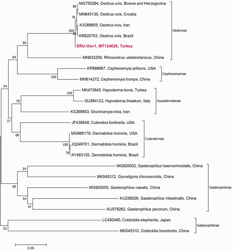
Karademir et al. Molecular Characterization of O. ovis                                                Turkiye Parazitol Derg 2020;44(1):43-7
44
     INTRODUCTION                                                           to parasitology laboratory in the sterile tubes with 70% ethanol
                                                                            by a public veterinarian working in the same province. No need
     The infestations caused by larvae of Diptera in animals and            to take ethical approval for the study according to directions
     humans are known as myiasis. The larvae belong to the species of       of the Erciyes University Local Ethics Committee for Animal
     Calliphoridae, Sarcophagidae and Oestridae families are the most       Experiments with the date 29.01.2016 and no 04 due to the
     common and important causative myiases agents (1). Oestridae is
                                                                            material comprised of insect species. The identification of fly
     a diverse family of Diptera comprising about 180 described species
                                                                            larvae was utilized under a stereo microscope (SZX16, Olympus,
     worldwide and their larvae have a variety of feeding habits, mostly
                                                                            Japan) with the identification keys described by Zumpt (1) and
     saprophages, endoparasites, parasitoids, and predators (2). They
                                                                            the images were recorded.
     cause often severe economic losses by reducing production,
     causing damages to hides and increasing treatment costs. Myiasis       Genomic DNA Extraction and PCR Amplification of
     caused by O. ovis has a global distribution, especially in rural and   CO1 Gene
     underdeveloped regions of tropical and subtropical areas. Domestic
     sheep and goats are thought to be the main host and also reservoir     Genomic DNA (gDNA) was extracted from the tissue sections
     to other hosts especially those in wildlife (3). The transmission      of each individual larvae specimens using GeneJET gDNA
     of O. ovis occurs when females deposit larvae onto the nostrils        Purification Kit (Thermo Fisher Scientific, USA). The tissue
     of sheep and goats. Their movement and development in the              pieces from the larvae were homogenized with the TissueLyser LT
     nasal-sinus cavities can cause pathologic and clinical symptoms        (Qiagen, UK) prior to gDNA extraction. The gDNA concentration
     including rhinitis, frequent sneezing, nasal discharge, breathing      of larvae was measured by Qubit Fluorometric Quantitation
     difficulties, and emaciation. The annoyance of the adult flies can     (Thermo Fisher Scientific, USA) to determine the optimum
     also lead to significantly reduced animal production by effecting      amount of DNA in the PCR mastermix. gDNA isolates were
     meat, wool and milk production (2). The gravid females are not         stored at -20 °C until PCR analyses.
     strictly host-specific and even only a few reports (4), ophthalmic     The universal barcode region of CO1 gene (709 bp) of the
     and naso-pharyngeal myiasis also occurred in humans especially         individual larvae was amplified using the common primers of
     the ones close relationship to livestock in several countries.         Folmer et al. (20). Amplifications were utilized in C1000 Touch™
     Oestrosis caused by O. ovis also common in sheep and goats             Thermal Cycler (Bio-Rad, USA) using described conditions (20) by
     in Turkey and several studies have reported a prevalence rate          adjusting the annealing temperature to 50 °C. The amplicons were
     of 36% to 59% in different regions (5-7). Sporadic cases of            visualized in the Fusion FX Gel Documentation System (Vilber
     human ophthalmic and naso-pharyngeal myiasis have been                 Lourmat, France) by loading 5µL of PCR products on 1.5% w/v
     also documented from some regions in Turkey (8-12). However,           agarose gel.
     there has been no study regarding molecular and phylogenetic
     characterization of O. ovis lineages found in Turkey except a single
     GenBank record of partial mt-CO1 sequence of an isolate from           CO1    Sequence     Analysis             and       Phylogenetic
     Diyarbakır province (GenBank accession: KT761199).                     Relationships of Bot Fly
     Molecular phylogenetic studies on O. ovis populations are very         PCR products were gel purified using a commercial kit (Thermo
     limited compared to the studies based on etiology, taxonomy,           Fisher Scientific, USA) and purified amplicons were sequenced
     biology, immunology, treatment and control approaches.                 in both directions with the amplification primers in the Sanger
     Mitochondrial DNA (mtDNA) is a useful tool for evaluating
                                                                            Sequencing Platform (Macrogen, Netherlands). The primer
     genomic differences and homologies among taxonomically
                                                                            sequences in all reads were trimmed prior to analyses. The final
     related species of both vertebrates and invertebrates. Due to its
                                                                            forward and reverse sequences were assembled in Geneious Prime
     high number of copies, easier isolation, and high phylogenetic
                                                                            2019.2.1 (https://www.geneious.com) and a single consensus
     signal and mutational rates, mtDNA has been widely used for
     taxonomic, population and evolutionary investigations across           sequence was obtained with the De Novo Assemble tool in the
     metazoan taxa (13-15). The 658 bp region of CO1 is widely              related software.
     accepted as a universal and standard marker for all animal taxa        The final sequences were searched in the NCBI database by using
     (14). During the last decade, DNA barcoding using CO1 sequences        the BLASTn algorithm. A CO1 data set was constituted with
     has provided an efficient tool for molecular identification and        totally 22 sequences belong to genetically related sequences from
     phylogenetic characterization of several kinds of insect species       different countries in the GenBank for phylogenetic analyses.
     including Oestridae family (16-19).                                    Multiple alignments of the sequences in CO1 dataset were
     In this study, we aimed to characterize the O. ovis larvae from        utilized using MUSCLE plugin in Geneious Prime (21). DNA
     an infested human with naso-pharyngeal myiasis by using                polymorphism and haplotype analyses were performed with
     CO1 barcode sequences. Phylogenetic relationships among the            DnaSp v.5.1 (22). Intraspecific and interspecific differences were
     identified larvae and the corresponding species in the Oestridae       calculated in MEGA 7 (23) using the Kimura two-parameter
     were also revealed in the study.                                       model (24).
                                                                            For inferring phylogenetic relationships in the CO1 dataset
     METHODS                                                                Maximum Likelihood (ML) estimation was utilized. GTR+G+I
                                                                            was determined as the suitable DNA substitution model for ML
     Bot Fly Larvae from a Human with Naso-pharyngeal                       analyses using the software jModeltest v.0.1.1 (25). The plugin of
     Myiasis                                                                PhyML in Geneious Prime was used for the construction of ML
     A total of 8 bot fly larvae obtained from a 49-year-old woman          tree (26). The reliability of branching of the ML tree was analyzed
     with naso-pharyngeal myiasis living in Adana province were sent        using a bootstrap resampling of 1000 replications.
Turkiye Parazitol Derg 2020;44(1):43-7 Karademir et al. Molecular Characterization of O. ovis
45
RESULTS found among the CO1 sequences of all eight isolates resulting
in the detection of a single haplotype (ERU-Oov1). The CO1
Human Case of Naso-pharyngeal Myiasis sequence of ERU-Oov1 was deposited to the GenBank with
accession MT124626. The base composition of the barcode
A 49-year-old woman living in Adana province was diagnosed with
sequence of the obtained haplotype was highly AT biased with a
naso-pharyngeal myiasis in the hospital. In our conversation, she
complained of about two months of nasal pruritus and sensation mean GC content of 34.2%.
of congestion with sanguinolent discharge from her nostrils. She A total of 303 polymorphic sites, of which 244 were parsimony
also reported having frequent sneezing, coughing, tear in gland informative, were determined within the entire CO1 data
nausea in the related period. After getting non-specific medical set including the sequences of the species belonging to
approaches such as using cortisone or decongestants two bot fly Gasterophilinae, Cuterebrinae, Oestrinae, Cephenemyiinae and
larvae were dropped. Following the detection of these parasites, Hypodermatinae subfamilies of Oestridae. Totally four different
she got antiparasitic treatment and further six larvae were haplotypes were determined among the CO1 sequences of the
dropped from the nostrils. O. ovis and the ERU-Oov1 represented a new haplotype of the
corresponding species. Mean haplotype and nucleotide diversities
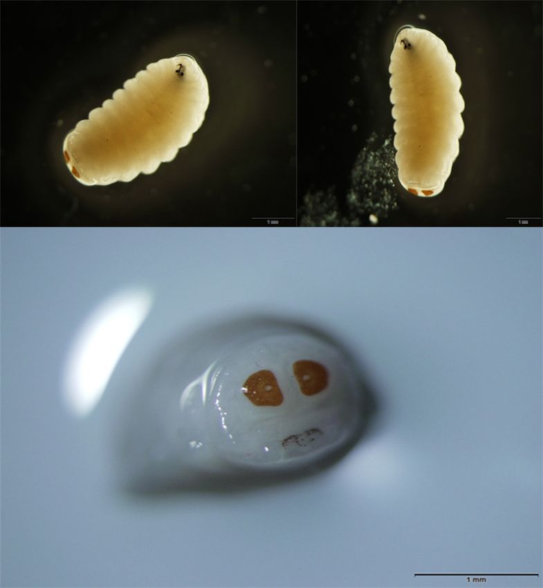
Morphologic Identification of the Bot Fly Larvae for O. ovis were 0.900 and 0.026, respectively. Intraspecific
All the eight larvae were sent to our parasitology laboratory nucleotide differences for O. ovis were determined within range of
and identified as second stage larvae of O. ovis according to the 0.00% to 0.61% and the mean genetic difference was 0.37±0.15%.
morphological features under stereo microscope (Figure 1). The Interspecific differences among the subfamilies of Oestridae
size of larvae specimens was 4-5 mm and they demonstrated in the CO1 data set were presented in Table 1. The species
anterior hooks, dark posterior spiracles with a flat side medially, of Hypodermatinae and Cuterebrinae; Cephenemyiinae and
and respiratory holes arranged radially. Oestrinae were closer to each other as sister taxon. The species of
Gasterophilinae were clustered into two distinct clades and found
Sequence Characterization and Phylogenetic more distant from other subfamilies of Oestridae and the clade
Relationships including Cobboldia species constituted outer taxa (Figure 3).
The barcode region of CO1 mtDNA was successfully amplified for The ML tree is presented in Figure 3 with bootstrap support
all the eight isolates in PCR analyses (Figure 2). The consensus values. All the sequences classified within species-based clades in
sequences covering the 658 bp barcode CO1 region from all reads subfamily taxa with the support of bootstrap values over 69.0%.
were successfully obtained with high-quality chromatogram The novel ERU-Oov1 haplotype clustered in a monophyletic clade
scores. The sequences were validated by translation analyses with with the published haplotypes of O. ovis indicated in Figure 3 with
the absence of insertions, deletions, or stop codons indicating a high bootstrap value (100.0%). The ERU-Oov1 was exhibited
functional mitochondrial products. The BLASTn analysis of the highest identity of 99.5% to the O. ovis isolates reported from
obtained sequences confirmed the morphological identifications Bosnia and Herzegovina (MG755264), Croatia (MN845130) and
as the specimens belonged to O. ovis. No polymorphic sites were Brazil (KR820703) and also showed an identity rate of 99.4% to
the isolate reported from Iran (KX268655).
DISCUSSION
A naso-pharyngeal myiasis case in a woman living in Adana
province was determined and the recovered bot fly larvae
identified as the second stage larvae of O. ovis based on the
morphological characters. The size and the shape of larvae and the
shape of posterior spiracles were consistent with those reported
by Zumpt (1). The clinical signs of the infection in the woman
patient also consistent with the several naso-pharyngeal myiasis
Figure 2. PCR amplicons of the barcode CO1 region of O. ovis
Figure 1. Second stage larvae of O. ovis causing human naso- isolates. M: Marker (100 bp), 1-8: gDNA from O. ovis larvae
pharyngeal myiasis specimensKarademir et al. Molecular Characterization of O. ovis Turkiye Parazitol Derg 2020;44(1):43-7
46
our newly characterized haplotype clustered together with those
reported from Bosnia and Herzegovina (MG755264), Croatia
(MN845130), Brazil (KR820703) and Iran (KX268655) in a
monophyletic clade and intra-specific genetic distance among the
haplotypes were equal or less than 0.6%. This result may indicate
a possible low polymorphism in this species with scarce gene
flow among the populations from different geographical regions.
Low intra-specific distances based on different fragments of CO1
from other bot fly species such as Przhevalskiana silenus (18,37),
Hypoderma bovis, H. lineatum, H. diana, H. tarandi, Gasterophilus
intestinalis, G. haemorrhoidalis, G. nasalis and Cuterebra baeri
(37) in different countries were also emphasized and all these
results might provide an evidence on the absence or lower cryptic
diversity in the species of Oestridae. However, for a better
understanding of the evolution of O. ovis, more comprehensive
set of samples from different regions of Turkey and also the world
is needed to be analyzed by using both mitochondrial and nuclear
markers preferably mitogenomes.
CONCLUSION
This study provides the first molecular characterization data
on O. ovis larvae causing naso-pharyngeal myiasis in a human
in Turkey based on barcode CO1 sequences. Although limited
Figure 3. Phylogenetic analysis of O. ovis and other species of sequences were included in the data set, our findings revealed
Oestridae subfamilies. The characterized haplotype was shown an evidence on the close association within O. ovis from different
in bold red character. Bootstrap values (1000 replicates) was countries. The barcode CO1 sequence analyses were successfully
shown at the nodes. The scale bar represents a 0.05% divergence differentiate the species of the subfamilies in Oestriade and this
result indicates the usefulness of the CO1 as a suitable diagnostic
molecular marker for identification and characterization of bot
Table 1. Interspecific genetic distances (below the diagonal) fly species.
± standard deviations (above the diagonal) for Oestridae
Acknowledgements: The authors thank Nuri Güngör, DVM for
subfamilies
giving kind information on the naso-pharyngeal myiasis human
1 2 3 4 5 case and also providing and sending the larvae specimens to our
1 Oestrinae - 0.021 0.022 0.022 0.022 laboratory.
2 Cephenemyiinae 0.203 - 0.022 0.020 0.024
* Ethics
3 Cuterebrinae 0.221 0.226 - 0.019 0.021
Ethics Committee Approval: As this study conducted on insect
4 Hypodermatinae 0.234 0.212 0.198 - 0.022
species, there was no need to take ethical approval.
5 Gasterophilinae 0.284 0.308 0.277 0.296 -
Informed Consent: Patient approval has not been obtained in
case of using insect species in the study.
cases reported from several countries including Turkey (27-32). Peer-review: Externally and internally peer-reviewed.
Accidental myiasis caused by O. ovis larvae has been also reported * Authorship Contributions
from conjunctival sac (ophthalmomyiasis) (33), the throat (34),
the nose (35), and the ears of humans (36). Concept: G.K.K., A.Y., Design: A.Y., G.K.K., M.O., S.U., A.I., Data
Molecular data are still rather limited for bot flies, and the data Collection or Processing: G.K.K., A.Y., M.O., S.U., Analysis or
Interpretation: A.Y., G.K.K., M.O., S.U., A.I., Literature Search:
currently available for Oestridae include mainly partial sequences
S.U., G.K.K., M.O., Writing: A.Y., G.K.K., M.O., S.U., I.A.
of mitochondrial genes especially CO1. However, most of the
sequences available in GenBank are from different fragments Conflict of Interest: No conflict of interest was declared by the
of CO1 rather than 658 bp barcode region commonly used as a authors.
diagnostic marker (13-15). Therefore, it is not possible to compare Financial Disclosure: The authors declared that this study
all CO1 sequences of bot flies including O. ovis in our study. However, received no financial support.
it was obvious that the barcode region successfully discriminates
the species in all subfamilies of Oestridae with relatively high rate REFERENCES
of barcoding gaps. Our result is also consistent with the findings 1. Zumpt F. Myiasis in Man and Animals in the Old-world Ed. London:
of Otranto et al. (37), who reported significant interspecific Butterworth, 1965.
divergences among the 18 species of Oestridae that cause myiasis 2. Scholl PJ, Colwell DD, Cepeda-Palacios R. Myiasis (Muscoidea, Oestroidea).
by using a different fragment of CO1 as a phylogenetic marker. In: Mullen GR, Durden LA, editors. Medical and Veterinary Entomology.
With only available four O. ovis barcode sequences in GenBank, London: Academic Press; 2019. p.383-419.Turkiye Parazitol Derg 2020;44(1):43-7 Karademir et al. Molecular Characterization of O. ovis
47
3. Colwell DD. Bot flies and warble flies (Order Diptera: Family Oestridae). 21. Edgar RC. MUSCLE: multiple sequence alignment with high accuracy and
In: Samuel WM, Pybus MJ, Kocan AA, editors. Parasitic Diseases of Wild high throughput. Nucleic Acids Res. 2004;32:1792-1797.
Mammals, London: Manson Publishing/The Veterinary Press; 2001.
22. Librado P, Rozas J. DnaSP v5: a software for comprehensive analysis of
p.46-71.
DNA polymorphism data. Bioinformatics. 2009;25:1451-1452.
4. Panadero-Fontán R, Otranto D. Arthropods affecting the human eye. Vet
23. Kumar S, Stecher G, Tamura K. MEGA7: Molecular evolutionary genetics
Parasitol. 2015;208:84-93.
analysis version 7.0 for bigger datasets. Mol Biol Evol. 2016;33:1870-
5. Gokcen A, Sevgili M. Prevalence and larval burden of Oestrus ovis in Awassi 1874.
sheep from Sanliurfa region of Turkey. Indian Vet J .2004;81:1168-1169.
24. Kimura M. A simple method for estimating evolutionary rates of base
6. Uslu U, Dik B. Prevalence and intensity of Oestrus ovis in Akkaraman substitutions through comparative studies of nucleotide sequences. J Mol
sheep in the Konya region of Turkey. Med Vet Entomol. 2006;20:347-349. Evol. 1980;16:111-120.
7. Arslan MO, Kara M, Gicik Y. Epidemiology of Oestrus ovis infestations in 25. Posada D. jModelTest: phylogenetic model averaging. Mol Biol Evol.
sheep in Kars province of north-eastern Turkey. Trop Anim Health Prod. 2008;25:1253-1256.
2009;41:299-305.
26. Guindon S, Gascuel O. A simple, fast, and accurate algorithm to estimate
8. Yar K, Özcan AA, Koltaş İS. External ophthalmomyiasis: case reports. large phylogenies by maximum likelihood. Syst Biol. 2003;52:696-704.
Turkiye Parazitol Derg. 2011;35:224-226.
27. Verstrynge K, Foets B. External ophthalmomyiasis: a case report. Bull Soc
9. Akdemir MO, Ozen S. External ophthalmomyiasis caused by Oestrus ovis Belge Ophtalmol. 2004;67-71.
misdiagnosed as bacterial conjunctivitis. Trop Doct. 2013;43:120-123.
28. Yaghoobi R, Tirgari S, Sina N. Human auricular myiasis caused by Lucilia
10. Calışkan S, Ugurbaş SC, Sağdık M. Ophthalmomyiasis externa: three cases sericata: clinical and parasitological considerations. Acta Med Iran.
caused by Oestrus ovis larvae in Turkey. Trop Doct. 2014;44:230-232. 2005;43:155-157.
11. Istek Ş. Ophthalmomyiasis externa from Hakkari, the south east border 29. Akdemir MO, Ozen S. External ophthalmomyiasis caused by Oestrus ovis
of Turkey. BMJ Case Rep. 2014;bcr2013201226. misdiagnosed as bacterial conjunctivitis. Trop Doct. 2013;43:120-123.
12. Özyol P, Özyol E, Sankur F. External ophthalmomyiasis: a case series and 30. Najjari M, Shafiei R, Fakoorziba MR. Nosocomial myiasis with Lucilia
review of ophthalmomyiasis in Turkey. Int Ophthalmol. 2016;36:887- sericata (Diptera: Calliphoridae) in an ICU patient in Mashhad,
891. Northeastern of Iran. Arch Iran Med. 2014;17:523-525.
13. Hebert PD, Penton EH, Burns JM, Janzen DH, Hallwachs W. Ten species 31. Alizadeh M, Mowlavi G, Kargar F, Nateghpour M, Akbarzadeh K,
in one: DNA barcoding reveals cryptic species in the neotropical skipper Hajenorouzali-Tehrani M. A review of myiasis in Iran and a new
butterfly Astraptes fulgerator. Proc. Natl Acad Sci USA. 2004;41:14812- nosocomial case from Tehran Iran. J Arthropod Borne Dis. 2014;8:124-
14817. 131.
14. Hebert PD, Stoeckle MY, Zemlak TS, Francis CM, Identification of birds 32. Hazratian T, Tagizadeh A, Chaichi M, Abbasi M. Pharyngeal Myiasis
through DNA Barcodes. PLoS Biol. 2004;2:e312. Caused by Sheep Botfly, Oestrus ovis (Diptera: Oestridae) Larva, Tabriz,
15. Hebert PD, Gregory TR. The promise of DNA barcoding for taxonomy. East Azarbaijan Province, Iran: a Case Report. J Arthropod Borne Dis.
Syst Biol. 2005;54:852-859. 2017;11:166-170.
16. Moreno V, Romero-Fernández I, Marchal JA, Beltrán M, Granados JE, 33. Fries FN, Pattmöller M, Seitz B, Berger F, Kampen H, Szentmáry N, et
Habela MA, et al. Molecular characterization of bot flies, Oestrus spp. al. Ophthalmomyiasis externa due to Oestrus ovis in a traveller returning
(Diptera, Oestridae), from domestic and wild Bovidae hosts.Vet Parasitol. from Greece. Travel Med Infect Dis. 2018;23:101-102.
2015;212:473-477. 34. Masoodi M, Hosseini K. The respiratory and allergic manifestations
17. Cavallero S, Pombi M, Perrone V, Milardi GL, D’Amelio S, Giuliani C, et of human myiasis caused by larvae of the sheep bot fly (Oestrus ovis):
al. Gasterophilus intestinalis (Diptera: Oestridae) in the diaphragmatic A report of 33 pharyngeal cases from southern Iran. Ann Trop Med
muscle: An unusual finding. Vet Parasitol. 2017;237:117-121. Parasitol. 2003;97:75-81.
18. Rakhshandehroo E, Razavi SM, Farzaneh R, Esmailnejad A, Asadpour M, 35. Hoyer P, Williams RR, Lopez M, Cabada MM. Human nasal myiasis caused
Shams S. Phylogenetic analysis of goat warble fly (Przhevalskiana silenus) by Oestrus ovis in the highlands of Cusco, Peru: report of a case and review
based on mitochondrial COI gene. J Parasit Dis. 2019;43:304-307. of the literature. Case Rep Infect Dis. 2016;2456735.
19. Li XY, Pape T, Zhang D. Gasterophilus flavipes (Oestridae: Gasterophilinae): 36. White ZL, Chu MW, Hood RJ. Nasal myiasis: A case report. Ear Nose
A horse stomach bot fly brought back from oblivion with morphological Throat J. 2015;94:E24-25.
and molecular evidence. PLoS One. 2019;14:e0220820. 37. Otranto D, Traversa D, Guida B, Tarsitano E, Fiorente P, Stevens JR.
20. Folmer O, Black M, Hoeh W, Lutz R, Vrijenhoek R. DNA primers for Molecular characterization of the mitochondrial cytochrome oxidase
amplification of mitochondrial cytochrome c oxidase subunit I from I (COI) gene of Oestridae larvae causing obligate myiasis. Med Vet
diverse metazoan invertebrates. Mar Biotechnol (NY). 1994;3:294-299. Entomol. 2003;17:307-315.You can also read