First observation of larval oarfish, Regalecus russelii, from fertilized eggs through hatching, following artificial insemination in captivity ...
←
→
Page content transcription
If your browser does not render page correctly, please read the page content below
Oka et al. Zoological Letters    (2020) 6:4
https://doi.org/10.1186/s40851-020-00156-6

 RESEARCH ARTICLE                                                                                                                                Open Access

First observation of larval oarfish, Regalecus
russelii, from fertilized eggs through
hatching, following artificial insemination in
captivity
Shin-ichiro Oka*, Masaru Nakamura, Ryo Nozu and Kei Miyamoto

 Abstract
 Background: Little is known about the life history of oarfish of the genus Regalecus, although it is a famous deep-
 sea fish and an apparent origin of sea serpent legends. We successfully performed artificial insemination using a
 recently dead pair of sexually mature individuals. We report for the first time development from fertilized eggs to
 early larvae in the Lampridiformes.
 Results: Eggs required 18 days of development from fertilization to hatching under 20.5–22.5 °C conditions. Oarfish larvae
 had similar morphological features as other lampridiform larvae hatched in the ocean. Larvae typically faced downward
 and swam using pectoral fins; they frequently opened their mouths. This mouth-opening behavior and swimming ability
 were both consistent with osteological development. The larvae did not eat and died four days after hatching.
 Conclusions: This is the first successful instance of artificial insemination and hatching in the oarfish, as well as the first
 reliable morphological and behavioral description of lampridiform larvae.
 Keywords: Regalecus, Artificial fertilization, Development, Deep-sea fish

Background                                                                          uncertain identification, and failure to preserve voucher
The oarfish of the genus Regalecus is found in deep seas                            specimens [1]. The only reliable record of the early
from tropical to temperate zones and is usually mesopel-                            stages of Regalecus is a report of eggs from the western
agic [1]. Regalecus russelii has a unique morphology and                            Pacific, identified using DNA barcoding techniques [8],
a large body; much of the information about it comes                                and a juvenile (13.7 mm in standard length) identified
from findings of dead bodies on beaches [2–4]. Beyond                               from developed morphological features [9].
this basic information, we know little else about oarfish,                            In January 2019, we obtained a recently dead pair of
despite the species being cited frequently as a possible                            mature oarfish and successfully performed artificial in-
origin of sea serpent and mermaid legends [1, 5]. In par-                           semination with their gametes, producing fertilized eggs.
ticular, our understanding of oarfish life history is lack-                         These were then hatched into live oarfish larvae, observ-
ing. A few descriptions exist of the early Regalecus                                able for the first time. This is the first reliable record of
developmental stages [6, 7], but these are replete with                             fertilized eggs developing into early larval fish in the
errors from insufficient sampling, misidentification or                             Lampridiformes.

* Correspondence: sh-oka@okichura.jp
Okinawa Churashima Foundation, 888 Ishikawa, Motobu-cho, Okinawa
905-0206, Japan

                                     © The Author(s). 2020 Open Access This article is licensed under a Creative Commons Attribution 4.0 International License,
                                     which permits use, sharing, adaptation, distribution and reproduction in any medium or format, as long as you give
                                     appropriate credit to the original author(s) and the source, provide a link to the Creative Commons licence, and indicate if
                                     changes were made. The images or other third party material in this article are included in the article's Creative Commons
                                     licence, unless indicated otherwise in a credit line to the material. If material is not included in the article's Creative Commons
                                     licence and your intended use is not permitted by statutory regulation or exceeds the permitted use, you will need to obtain
                                     permission directly from the copyright holder. To view a copy of this licence, visit http://creativecommons.org/licenses/by/4.0/.
                                     The Creative Commons Public Domain Dedication waiver (http://creativecommons.org/publicdomain/zero/1.0/) applies to the
                                     data made available in this article, unless otherwise stated in a credit line to the data.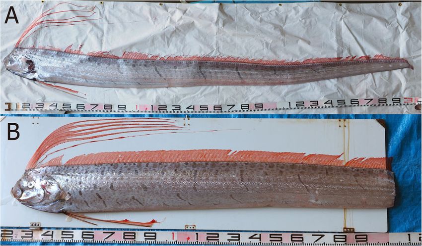
Oka et al. Zoological Letters        (2020) 6:4                                                                                     Page 2 of 6

Materials and methods                                                             reduce mortality risk. Water temperatures were kept at
Adult fishes                                                                      20.5–22.5 °C. Observations of fertilized eggs throughout de-
During early morning of January 29, 2019, two living Rega-                        velopment were performed under a microscope from 1 to 3
lecus russelii (Fig. 1) were captured in a set net (c.a. 360                      DAF and then daily after 7 DAF until hatching.
m × 60 m) for commercial fishes on the coast of Okinawa-
jima Island (26° 22′ 48″ N, 127° 42′ 45″ E; 40 m depth).
They were transferred to a car equipped with a seawater                           Larvae care and treatment
tank. By that point, one fish had already died; the other was                     The 19 newly hatched larvae were separated into three
alive, but had lost its tail during capture. Within an hour of                    tanks: nine and seven larvae were kept in two 30 L tanks,
being conveyed to the Okinawa Churaumi Aquarium, the                              while the remaining three were kept in a 100 L tank.
remaining specimen also died. These individuals were iden-                        The water temperature was 22 °C. Rotifers (Brachionus
tified as R. russelii from the number of gill rakers (male: 52,                   plicatilis) and brine shrimp (Artemia nauplii), enriched
female: 56) and pre-anal dorsal fin rays (male: 67, female:                       with highly unsaturated fatty acids, were provided as
70) following a previous study [1]. The two individuals                           food for the larvae. The larval behavior was filmed daily
measured 1220 mm in pre-anal length. They were dis-                               using a compact digital camera (OLYMPUS TG-5).
carded because there were no tanks of adequate size to pre-                       Total video length was 29 min 23 s; swimming, body
serve these two large specimens. For future genetic                               posture, and other characteristic behaviors were visible.
identification, the muscle tissue of both fish were deposited                     Analysis used a video (19 min and 35 s long) taken 0–2
in the collection of the Okinawa Churashima Foundation                            days after hatching (DAH). The videos from 3 and 4
(OCF) as OCF-P4061 and OCF-P4062.                                                 DAH were excluded because the larvae appeared unable
                                                                                  to swim and did not exhibit any characteristic behaviors.
Artificial insemination and egg incubation                                           Except for one larva that died on the day of hatching,
The female possessed one pair of ovaries; upon dissection, a                      those that appeared close to dying or had died were
large amount of transparent eggs spilled from the end of the                      taken as specimens for morphological observations. All
oviduct. The other individual was a male with a pair of testes                    living specimens were euthanized via anesthesia over-
containing white liquid, which was confirmed under micro-                         dose before fixing. Specimens were preserved in 5% for-
scopic observation to be sperm with high motility. Artificial                     malin and deposited in the OCF collection. Total length
insemination was performed using the dry method, mixing                           (TL), notochord length (NL), pre-anal length (PAL),
eggs and sperm in a 4 L container before adding seawater.                         head length (HL), and eye diameter (ED) were measured
After treatment, non-floating eggs were removed and the re-                       under a microscope (KEYENCE VHX-1000), from five
mainder incubated at 20 °C. The fertilized eggs (about 400)                       specimens that did not flex when fixed.
were maintained with weak aeration in a 30 L tank from 1                             The larval specimens (OCF-P04196) were cleared and
day after fertilization (DAF) to 7 DAF. Subsequently, ap-                         double-stained for bone and cartilage following pub-
proximately 100 eggs were transferred to a 100 L tank to                          lished protocol [10]. Photographs of clear and double-

 Fig. 1 Regalecus russelii pair used for artificial insemination. a: female, b: male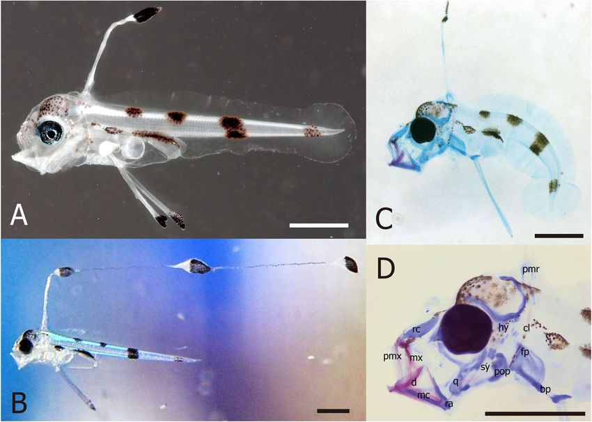
Oka et al. Zoological Letters (2020) 6:4 Page 3 of 6
stained specimens were obtained using the microscope’s proportion of first dorsal and pelvic fin rays in the noto-
filming function. chord length exceeded by 0.31 ± 0.05 and 0.23 ± 0.01
times (mean ± SD, n = 5), respectively. With the excep-
Results tion of pectoral fins, other fin rays were not developed.
Development of fertilized eggs The single dorsal fin ray had elongated into a filament
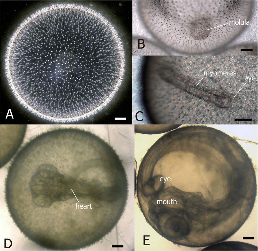
The eggs were spherical and 2.07–2.20 mm in diameter (averaging 1.8 times the notochord length, measured
(n = 38), with homogeneous yolk, no oil globules, and a from videos, n = 6) with three large bulges exhibiting
surface covered with numerous conical short spines granule-like pigmentation (Fig. 3). The proportions of
(Fig. 2a). About 400 floating eggs had developed into a PAL/NL, HL/NL, and ED/HL were 0.46 ± 0.02, 0.19 ±
morula at 1 DAF (Fig. 2b). At 7 DAF, 300 eggs 0.01, and 0.40 ± 0.03 (means ± SD), respectively (Table
remained, while the head, eyes, notochord, and myo- 1). None of the specimens had any head spination (Fig.
meres developed (Fig. 2c). The tip of the tail was re- 3). The myomere count was 27 in the pre-anal area and
leased from the yolk at 9 DAF, and the heart beats were 101–107 in the tail; however, the myomeres in the tail
confirmed with the body development at 11 DAF, when tip were unclear. The yolk sac was completely absorbed
270 eggs remained (Fig. 2d). This number declined to 40 by 3 DAH. Melanophore pigmentation was scattered on
by 15 DAF, when embryo mouth opened and the small head and snout; dorsally located over air bladder and
surface spines became inconspicuous. At 16 DAF, pig- hindgut; in two patches on dorsal trunk, one dark bar on
mentation appeared in the eyes and the dorsal fin ray mid-tail, one faded bar on tail tip; present on tip of pel-
elongated (Fig. 2e). The first individual hatched at 17 vic fin, and on swellings of the elongated dorsal fin ray.
DAF, while 18 larvae hatched at 18 DAF; the remaining
eggs failed to hatch. Osteological development
Observations of the transparent larval specimens showed
Larval morphology no bones or related vertebrae from the posterior part of
Larval measurements are shown in Table 1, and mor- the trunk (Fig. 3c, d). However, the bones around the
phological characters are shown in Fig. 3a and b. Newly head were well developed while connective tissue in jaws
hatched larvae (5.5–6.3 mm NL) had moderately elon- developed as cartilage, with ossification occurring only
gated and laterally compressed bodies (Table 1). The in premaxilla, maxilla, dentary, and part of the
Fig. 2 Fertilized eggs of Regalecus russelii. a: immediately after fertilization, b: morula at 1 day after fertilization (DAF), c: embryo at 7 DAF, showing
eye and myomere development, d: egg at 11 DAF, E: egg just before hatching (16 DAF). Scale bars: 0.2 mmOka et al. Zoological Letters (2020) 6:4 Page 4 of 6
Table 1 Measurements of larvae obtained from the first artificial insemination of Regalecus russelii
Specimen No. DAH TL (mm) NL (mm) PAL (mm) HL (mm) ED (mm)
OCF-P04185 0 6.40 6.10 2.90 1.12 0.46
OCF-P04186 1 6.50 6.30 2.90 1.18 0.45
OCF-P04188 2 5.70 5.50 2.55 1.16 0.43
OCF-P04191 3 6.10 5.95 2.70 1.08 0.48
OCF-P04192 4 5.80 5.53 2.35 1.03 0.43
symplectic bones. The pectoral fin, elongated dorsal fin, multiple times to escape entanglement if their elongated
and pelvic fin were also developed. The cleithrum was dorsal fin wrapped around their bodies. They also slowly
ossified, while other elements (pectoral fin plate, prox- rotated when changing direction. Such rotational behav-
imal middle radial, and basipterigium) were cartilage. ior occurred 19.8% of the time. Although we observed
larvae opening their mouth six times, it was unclear
whether this represented attempts to eat prey plankton,
Larval survival and behavior and we did not notice direct feeding behavior.
A total of 19 larvae hatched, but the number of living in-
dividuals decreased to 17 at 1 DAF, 12 at 2 DAH, 8 at 3
DAH, and 0 by 4 DAH. Discussion
Video analysis (Movie S1) showed that larvae swam Eggs hatched 18 DAF, in line with a previous study indicat-
mainly using the pectoral fin, although they sometimes ing that lampridiform fishes hatch after 10–20 days [11].
moved quickly using their tails when bait plankton Ours is the first study to track lampridiform eggs from
touched their bodies. Larvae often (56.3% of the time) fertilization to hatching. Our observations revealed that oar-
maintained a downward-facing body posture, while fish eggs develop much more slowly than eggs of other pela-
spending 4.7% of the time oriented slightly upward and gic teleostean fishes. The eggs we describe here shared
4.2% upside down. They were horizontally oriented and similar morphological characteristics to those of oarfish eggs
moved laterally 13.6% of the time. Larvae quickly rotated obtained from the ocean and identified through DNA
Fig. 3 Larvae of Regalecus russelii. a: larval specimen (5.5 mm notochord length (NL); 2 days after hatching (DAH); OCF-P04188, dorsal elongated
filament was lost), b: living larvae ca. 6 mm NL (1 DAH), c: transparent specimen (5.5 mm NL; 4 DAH; OCF-P04196), d: close-up of transparent
specimen head. Anatomical abbreviations: bp, basipterygium; cl, cleithrum; d, dentary; fp, fin plate cartilage; hy, hyomandibular; mc, Meckelian
cartilage; mx, maxilla; pmr, proximal middle radial; pmx, premaxilla; pop, preopercle; q, quadrate; ra, retroarticular; rc, rostal cartilage; sy, symplectic.
Scale bars: 1.0 mmOka et al. Zoological Letters (2020) 6:4 Page 5 of 6
barcoding [8]. Although we caught the two mature oarfish in consume. In addition, however, we also cannot exclude
winter, a previous study identified oarfish eggs around the the possibility that the captive environmental conditions
Marshall Islands in summer [8]. Thus, the spawning season caused this feeding dysfunction.
of this species may be relatively long (summer–winter), al-
though the exact duration remains unclear. Our capture of Conclusions
the matured pair in a set net, along with the discovery of This is the first report of successful artificial insemin-
stranded individuals oarfish with matured gonads [12], sup- ation in oarfish and provides the first reliable record of
port the hypothesis that this species rises to the surface for development from fertilized eggs to early larval stages in
spawning. Currently, over 40 records of stranded oarfish Lampridiformes. Eggs and sperm from a recently dead
were found between the temperate and tropical regions of pair of matured fishes were used for artificial insemin-
Japan [4]. Future studies should examine gonads from these ation. The eggs developed more slowly than other tele-
specimens to further clarify oarfish reproductive biology. ostean pelagic eggs and required 18 days for hatching.
Oarfish larvae had similar morphological features as Hatched larvae developed head composition and elon-
other lampridiform larvae (Lophotus lacepede, Trachip- gated dorsal and pelvic fin rays. These morphological
terus spp., and Zu cristatus) [13, 14]. However, previous features, including the pigmentation pattern, were simi-
reports of lampridiform larvae are unreliable, as they con- lar to characteristics reported for other lampridiform lar-
tain the multiple errors [1]. The present study provides vae in previous studies.
the first reliable description of lampridiform larvae, specif- Striking larval behaviors included maintenance of a
ically from R. russelii. The bodies of newly hatched oarfish downward-facing position and swimming using pectoral
larvae are more compressed than those of adults, but both fins. We were unable to determine the function of the
larvae and adults possess elongated dorsal and pelvic fins. remarkably elongated dorsal-fin filament and its orna-
The smallest juvenile ever captured in the ocean (13.7 mm ments. Larvae did not feed, and all larvae died within 4
NL) similarly had an adult-like elongated body and devel- DAH in spite of possessing a well-developed mouth.
oped dorsal fin rays on the anterior half of the body [9]. These findings contribute significantly to our under-
This similarity between hatched larvae and juveniles sug- standing of early oarfish life history, although the captive
gests that the larval body elongates soon after hatching. conditions of this study may differ greatly from natural
Larval and adult swimming behaviors were completely environmental conditions. In addition, the fact that the
different. Adults swim using their dorsal fins in a head- mature male and female oarfish were caught with a
up position [15], whereas larvae often maintained a coastal set net suggests that this species may migrate to
downward position and swam using their pectoral fins. the surface for spawning.
We do not know the reason for this larval posture, nor
do we know the function of their remarkably elongated Supplementary information
dorsal-fin filament and its ornaments; we did not ob- Supplementary information accompanies this paper at https://doi.org/10.
serve any situations in which larvae used the filament. 1186/s40851-020-00156-6.
Furthermore, the filament often tangled around the body
Additional file 1 Movie S1. Recording of oarfish larval behavior
and appeared to impede movement. We note, however,
that these behaviors occurred in captive conditions and
may differ from behaviors in the wild. Acknowledgments
The authors would like to thank Shohei Matsuzaki, Yuji Ashida, Naomi
Our osteological observations revealed a lack of ossifi- Tanaka, Rui Matsumoto, Kiyomi Murakumo, Taketeru Tomita, and other staff
cation in connective tissue at the posterior part of the of the Okinawa Churaumi Aquarium for assisting us with rearing
head, including vertebrae. However, upper and lower management and data collection. We also thank the fishermen in Yomitan
Fishery Cooperatives for providing us with the adult specimens.
jaws were well-ossified, and tissue related to opening the
mouth were developed as cartilage. We also observed os- Consent for publications
sification of the cleithrum, which supports the pectoral Not applicable.
fin. These results are consistent with the observation
Authors’ contributions
that larvae frequently opened their mouths and swam SO managed the present study, performed artificial insemination, reared
using their pectoral fins. The biggest mystery in our eggs and larvae, conducted data collection and analysis, and wrote the
study was that the larvae ate nothing, despite possessing manuscript. MN, RN, and KM assisted with artificial insemination. All authors
edited and confirmed the final manuscript.
mouths with apparently sufficient feeding function. This
observation suggests that, unlike other teleosts, oarfish Funding
do not feed on active plankton at the sea surface. Never- This study was funded by the Okinawa Churaumi Aquarium.
theless, we observed frequent “yawning” behavior that
Availability of data and materials
may be related to a very specific feeding ecology, al- The datasets used and analyzed during the current study are available from
though we do not know what type of food the larvae the corresponding author on reasonable request.Oka et al. Zoological Letters (2020) 6:4 Page 6 of 6
Ethics approval and consent to participate
Maintenance and handling of the fish, as well as all experiments, were in
strict accordance with the “Guide for Care and Use of Laboratory Animals of
Okinawa Churashima Foundation.”
Competing interests
The authors declare that they have no competing interests.
Received: 19 November 2019 Accepted: 20 March 2020
References
1. Roberts T. Systematics, biology, and distribution of the species of the
oceanic oarfish genus Regalecus (Teleostei, Lampridiformes, Regalecidae).
Paris: French National Museum of Natural History; 2012.
2. Schmitter-Soto JJ. The oarfish, Regalecus glesne (Teleostei: Regalecidae), in
the Western Caribbean. Caribb J Sci. 2008;44:125–8.
3. Feeney RF, Lea RN. California Records of the Oarfish, Regalecus russelii
(Cuvier, 1816) (Actinopterygii: Regalecidae). Bull South Calif Acad Sci. 2018;
117:169–79.
4. Hata H, Fujii T, Motomura H. Record of Regalecus russelii (Lampridiformes:
Regalecidae) from Amami-oshima island, Kagoshima prefecture, Japan. Nat
Kagoshima. 2018;45:123–7.
5. Senou H. Ryugunotsukai. In: Nakabo T, editor. The natural history of the
fishes of Japan. Tokyo: Shogakukan; 2018. p. 154–5.
6. Sanzo L. Uova e larve di Regalecus glesne Asc. Memoria R Comitato
Talassografico Italiano. 1925;118:1–7.
7. Sparta A. Ordine: Allotriognathi, in Lo s larva e stadi giovanili di Teleostei.
Fauna Flora Golfo Napoli Monografia. 1933;38:266–79.
8. Kawakami T, Aoyama J, Tsukamoto K. Morphology of pelagic fish eggs
identified using mitochondrial DNA and their distribution in waters west of
the Mariana Islands. Environ Biol Fish. 2010;87:221–35.
9. Okiyama M, Senou H. Regalecidae. In: Okiyama M, editor. An Atlas of Early
Stage Fishes in Japan. 2nd ed. Tokyo: Tokai University Press; 2014. p. 392–3.
10. Dingerkus G, Uhler LD. Enzyme clearing of Alcian blue stained whole small
vertebrates for demonstration of cartilage. Stain Technol. 1977;52:229–32.
11. Mito S. Pelagic fish eggs from Japanese waters 2. Lamprida, Zeida, Mugilina,
Scombrina, Carangina and Stromateina. Sci Bull Fac Agricul Kyushu Univ.
1961;18:451–66.
12. Forsgren KL, Jamal H, Barrios A, Paig-Tran EWM. Reproductive morphology
of oarfish ( Regalecus russellii ). Anat Rec. 2017;300:1695–704.
13. Charter S, Moser H. Lampridiformes. In: HG HGM, editor. The early stages of
fishes in the California current region. Kansan: Alle Press; 1996. p. 659–77.
14. Okiyama M. Lampridiformes. In: Okiyama M, editor. An atlas of early stage
fishes in Japan. 2nd ed. Tokyo: Tokai University Press; 2014. p. 385–92.
15. Benfield MC, Cook S, Sharuga S, Valentine MM. Five in situ observations of
live oarfish Regalecus glesne (Regalecidae) by remotely operated vehicles in
the oceanic waters of the northern Gulf of Mexico: in situ observations of
regalecus glesne. J Fish Biol. 2013;83:28–38.
Publisher’s Note
Springer Nature remains neutral with regard to jurisdictional claims in
published maps and institutional affiliations.You can also read