MRI Depicts Olfactory Bulbs and Cortical Involvement in COVID-19 Patients with Anosmia
←
→
Page content transcription
If your browser does not render page correctly, please read the page content below

MAGNETOM Flash (78) 1/2021                                                                         Neurologic Imaging · Clinical

MRI Depicts Olfactory Bulbs and
Cortical Involvement in COVID-19 Patients
with Anosmia
Letterio S. Politi, M.D.1,2; Marco Grimaldi, M.D.1; Luca Balzarini, M.D.3

1
  Department of Neuroradiology, IRCCS Humanitas Research Hospital, Rozzano, Milan, Italy
2
  Department of Biomedical Sciences, Humanitas University, Pieve Emanuele, Milan, Italy
3
  Department of Radiology, IRCCS Humanitas Research Hospital, Rozzano, Milan, Italy

    Abstract                                                    Key points
    Anosmia and ageusia are very common symptoms                • In COVID-19 patients presenting with anosmia, T2-
    in SARS-CoV-2 infections. Here we present magnetic            FLAIR hyperintensity can be depicted in the olfactory
    resonance imaging evidence of brain signal alterations        bulbs and anterior piriform cortex, suggesting possi-
    in the olfactory bulbs and the piriform cortex, presum-       ble viral invasion of the brain.
    ably caused by SARS-CoV-2.                                  • The signal alteration is reduced when patients
                                                                  recover from the symptoms.
                                                                • Anosmia can be the predominant COVID-19 mani-
                                                                  festation, and this should be taken into account
                                                                  when identifying and isolating infected patients in
                                                                  order to avoid disease spread.

The neurotropism of human coronaviruses has already             using a 1.5 Tesla scanner (MAGNETOM Aera, Siemens
been demonstrated in small animals [1]. In autoptic             Healthcare, Erlangen, Germany) equipped with a 20-
studies, the severe acute respiratory syndrome coronavirus      channel phase array head and neck coil. On 2D and 3D
(SARS-CoV), which was responsible for the SARS outbreak         fluid-attenuated inversion recovery (FLAIR) images,
in 2002–2003, was found in the brain of infected patients       hyperintensity was evident in the bilateral olfactory bulbs
[2]. It has been proposed that the neuroinvasive potential      (red arrows) and in the right rectus gyrus (yellow arrows).
of the novel SARS-CoV-2 virus, which causes coronavirus         This signal alteration in the cortex of the brain area that
disease 2019 (COVID-19), might be at least partially            is responsible for olfaction is highly suggestive of viral
responsible for respiratory failure in COVID-19 patients [3].   infection. Since many patients in the Italian outbreak
In this paper, we will share magnetic resonance imaging         complain of anosmia [4], a swab was performed and
(MRI) evidence of in vivo brain alteration presumably           RT-PCR analysis was positive for COVID-19. In a follow-up
caused by SARS-CoV-2, and we will stress that anosmia           MRI performed 28 days later, the signal alteration had
can be the predominant symptom of COVID-19.                     almost completely disappeared and the patient had recov-
     A 25-year-old female radiographer with no significant      ered from anosmia.
prior medical history who had been working in a COVID-19             Similar but less-obvious MRI findings were depicted
ward presented with a mild dry cough that lasted for one        in a 39-year-old female COVID-19 patient who presented
day. This was followed by persistent and severe anosmia         only with anosmia. In this case, the brain MRI was per-
and ageusia. She was always nonfebrile. Three days later,       formed eight days after symptom onset. Furthermore,
a nasal fibroscopy was unremarkable, and non-contrast           no brain abnormalities were seen in two other patients
chest and maxillofacial computed tomography were nega-          who presented with anosmia and underwent an MRI exam
tive. On the same day, a brain MRI was also performed,          at 12 and 25 days from symptom onset, respectively.

                                                                               siemens-healthineers.com/magnetom-world       43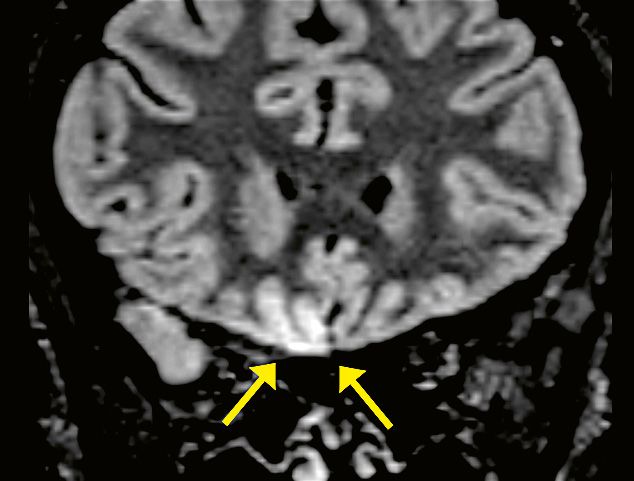
Clinical · Neurologic Imaging                                                                               MAGNETOM Flash (78) 1/2021

1A                                                                      Here we report on human brain involvement in patients
                                                                        who tested positive for COVID-19. We show signal alter-
                                                                        ations that are consistent with viral brain invasion in
                                                                        regions that are congruent with the patients’ symptoms.
                                                                        It should be noted that the posterior part of the gyrus
                                                                        rectus and medial orbital gyrus encompasses the so-called
                                                                        anterior piriform cortex, which receives input from the
                                                                        olfactory bulb through the lateral olfactory tract. The ante-
                                                                        rior piriform cortex and the posterior piriform cortex (the
                                                                        latter is located in the temporal lobe) are considered the
                                                                        most important olfactory cortical areas. Curiously, in hu-
                                                                        mans, the anterior piriform cortex seems to play a crucial
                                                                        role in encoding the difference between groups of odors.
                                                                        In anosmic animals like the dolphin, this entry zone is
                                                                        so undeveloped and flat that Broca called the area the
                                                                        “olfactory desert” (désert olfactif) [5].
                                                                             Although the presence of SARS-CoV-2 in the cortical
1B
                                                                        FLAIR hyperintense areas is not demonstrated by the MR
                                                                        images, we believe that the congruence between the
                                                                        clinical manifestations in our patient (i.e., olfactory
                                                                        dysfunction) and the cortical brain MRI abnormalities is
                                                                        highly suggestive of possible viral invasion; similar focal
                                                                        abnormalities are definitely unusual and alternative
                                                                        diagnoses are very difficult to find, especially in the clinical
                                                                        setting of anosmia (for instance, anti-NMDAR encephalitis
                                                                        can cause transient FLAIR hyperintensities, but the clinical
                                                                        manifestations rule out this entity; status epilepticus may
                                                                        cause transient gyral edema with T2-FLAIR hyperintensity,
                                                                        but olfactory aura continua / simple partial status epilepti-
                                                                        cus is exceptional, typically originates from the mesial
                                                                        temporal lobe, and causes olfactory hallucinations but not
                                                                        loss of smell). By contrast, viral infections are commonly
                                                                        considered potential causes of transient or permanent
1C
                                                                        sensorineural olfactory dysfunctions. In the presented
                                                                        cases, no analysis of cerebrospinal fluid (CSF) was per-
                                                                        formed; however, it should be noted that the clinical
                                                                        sensitivity of CSF analysis with molecular testing for
                                                                        intraparenchymal brain diseases remains undefined
                                                                        (with the exception of herpes simplex (HSV) encephalitis),
                                                                        negative results may not exclude infection, and in some
                                                                        cases cerebral biopsy may be necessary to confirm the
                                                                        diagnosis. Overall, we believe that the abovementioned
                                                                        MRI findings support the hypothesis that the SARS-CoV-2
                                                                        virus can invade the brain. Based on the MRI findings, our
                                                                        hypothesis is that, following initial replication in the nasal
                                                                        mucosa, SARS-CoV-2 may, as is the case with other corona-
                                                                        viruses, spread from the olfactory epithelium to the olfac-
                                                                        tory bulb, and subsequently to the posterior gyrus rectus
 1  Brain MRI alterations in a COVID-19 patient presenting with        through the lateral olfactory tract [1, 2].
     anosmia (three days after symptom onset).                               Overall, the presence of MRI abnormalities in the
     Coronal 2D FLAIR (1A, B) and axial reformatted 3D FLAIR images
                                                                        posterior part of the gyrus rectus could conceivably be
     showing hyperintensity in bilateral olfactory bulbs (red arrows)
     and in the right rectus gyrus (yellow arrows).
                                                                        related to the centripetal spreading of SARS-CoV-2 through
                                                                        the lateral olfactory tract, and the olfactory dysfunction
                                                                        experienced by our patient may have a sensorineural

44      siemens-healthineers.com/magnetom-world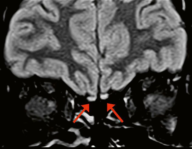
MAGNETOM Flash (78) 1/2021                                                                                                                 Neurologic Imaging · Clinical

origin. Our own and others’ observations of normal                                        References
brain imaging in subjects with COVID-19-related olfactory                                 1 Dubé M, Le Coupanec A, Wong AHM, Rini JM, Desforges M,
dysfunctions [6], and the disappearance of the MRI abnor-                                   Talbot PJ. Axonal Transport Enables Neuron-to-Neuron Propagation
malities in a follow-up study in one of our patients suggest                                of Human Coronavirus OC43. J Virol. 2018;92(17):e00404–18.
that these imaging changes might not always be present                                    2 Gu J, Gong E, Zhang B, Zheng J, Gao Z, Zhong Y, et al.
                                                                                            Multiple organ infection and the pathogenesis of SARS.
in COVID-19, or are limited to the very early phase of
                                                                                            J Exp Med. 2005;202(3):415–24.
the infection. Further, anosmia can be the predominant                                    3 Li YC, Bai WZ, Hashikawa T. The neuroinvasive potential of
COVID-19 manifestation, and this should be taken into                                       SARS-CoV2 may play a role in the respiratory failure of COVID-19
account when identifying and isolating infected patients                                    patients. J Med Virol. 2020;92(6):552–555.
in order to avoid disease spread.                                                         4 Giacomelli A, Pezzati L, Conti F, Bernacchia D, Siano M, Oreni L,
                                                                                            et al. Self-reported Olfactory and Taste Disorders in Patients With
                                                                                            Severe Acute Respiratory Coronavirus 2 Infection: A Cross-sectional
Acknowledgments                                                                             Study. Clin Infect Dis. 2020;71(15):889–890.
                                                                                          5 Schiller F. A memoir of olfaction.
We are grateful to Simona Superbi for providing us                                          J Hist Neurosci. 1997;6(2):133–46.
with technical support.                                                                   6 Eliezer M, Hautefort C, Hamel AL, Verillaud B, Herman P, Houdart E,
                                                                                            et al. Sudden and Complete Olfactory Loss of Function as a Possible
                                                                                            Symptom of COVID-19.
                                                                                            JAMA Otolaryngol Head Neck Surg. 2020;146(7):674–675.

                                                                                                                                  Contact
                                                                                                                                  Letterio S. Politi, M.D.
                                                                                                                                  Department of Neuroradiology
                                                                                                                                  IRCCS Humanitas Research Hospital
                                                                                                                                  Via Alessandro Manzoni 56,
                                                                                                                                  Rozzano (MI), 20089
                                                                                                                                  Italy
                                                                                                                                  letterio.politi@hunimed.eu

                                         Luca Balzarini, M.D.          Marco Grimaldi, M.D.         Letterio S. Politi, M.D.

                                                                                                                                                         Advertisement

View one of our most popular sessions
from the 11th MAGNETOM World Summit: Quantitative, Robust Neuro MRI
Multi-parametric MRI in Stroke Imaging
Shan Shan Lu (First Affiliated Hospital of Nanjing Medical University,
Nanjing, Jiangsu, China)

Clinical Benefits of MR Fingerprinting1
Meiyun Wang (Henan People’s Hospital, China)

MRI Techniques in Multiple Sclerosis:
New Diagnostic and Research Developments
Matilde Inglese (Università degli Studi di Genova, Italy)

Morphometry. Measuring Brain Maturation with Quantitative MRI
Baptiste Morel (Service de Radiologie Pédiatrique, Hopital Clocheville,
CHRU, Tours, France)

Wave CAIPI for Routine Clinical Brain MRI
Susie Huang (Massachusetts General Hospital, Boston, MA, USA)

Clinical Translation of 7T MRI: Epilepsy Imaging
Srinivasan Mukundan (Brigham and Women’s Hospital, Boston, MA, USA)

Siemens-Healthineers.com/MWS2020-recordings
                                                                                                                        Graphic recording: www.gabriele-heinzel.com

1
     R Fingerprinting is not commercially available in some countries. Due to regulatory reasons its future availablility cannot be ensured.
    M
    Please contact your local Siemens Healthineers organization for further details.

                                                                                                                siemens-healthineers.com/magnetom-world              45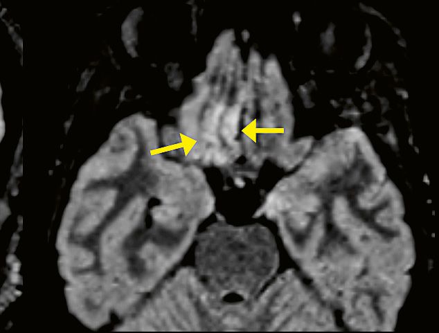


You can also read