MULTIDRUG-RESISTANT ENTEROBACTERALES IN OMAN: MOLECULAR EPIDEMIOLOGY AND THERAPEUTIC INSIGHTS - Hissa M. Al-Farsi
←
→
Page content transcription
If your browser does not render page correctly, please read the page content below
From the DEPARTMENT OF LABORATORY MEDICINE
              Karolinska Institutet, Stockholm, Sweden

MULTIDRUG-RESISTANT ENTEROBACTERALES IN
 OMAN: MOLECULAR EPIDEMIOLOGY AND
         THERAPEUTIC INSIGHTS

                      Hissa M. Al-Farsi

Stockholm 2021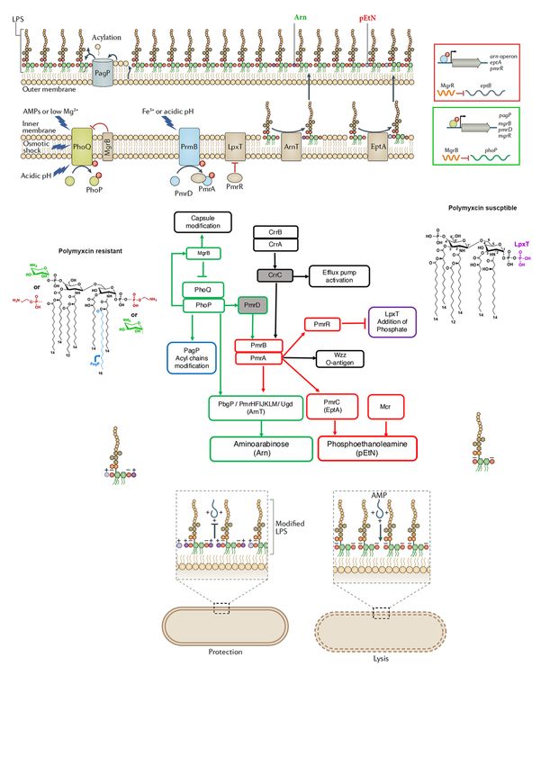
Cover image: A scanning electron microscopic (SEM) of extended-spectrum β-lactamase-
producing (ESBL) Escherichia coli. The image was created by Alissa Eckert (2013) and
obtained from the Public Health Image Library (PHIL) with no copyright restrictions.

All previously published papers were reproduced with permission from the publisher.
Published by Karolinska Institutet.
Printed by US-AB, Stockholm, Sweden.
© Hissa M. Al-Farsi, 2021
ISBN 978-91-8016-088-9
Multidrug-resistant Enterobacterales in Oman:
Molecular Epidemiology and Therapeutic Insights

THESIS FOR DOCTORAL DEGREE (Ph.D.)

By

Hissa M. Al-Farsi

Principal Supervisor:                 Opponent:
Prof. Christian Giske                 Dr. Teresa Coque
Karolinska Institutet                 Ramón y Cajal Institute for Bio Health Research
Department of Laboratory Medicine     (IRYCIS), Madrid, Spain.
Division of Clinical Microbiology     Department of Microbiology
                                      Division of Microbial Biology and Infections
Co-supervisor(s):
Assoc. Prof. Peter Bergman            Examination Board:
Karolinska Institutet                 Assoc. Prof. Åsa Sjöling
Department of Laboratory Medicine     Karolinska Institutet
Division of Clinical Microbiology     Department of Microbiology, Tumor and Cell
                                      Biology. Stockholm, Sweden.
Dr. Maarten Coorens
Karolinska University Hospital        Prof. Mikael Rhen
Department of clinical Microbiology   Karolinska Institutet
                                      Department of Microbiology, Tumor and Cell
Dr. Saleh Al-Azri                     Biology, Stockholm, Sweden.
Ministry of Health
Central Public Health Laboratories,   Prof. Ann-Beth Jonsson
Muscat, Oman.                         Stockholm University
                                      Department of Molecular Biosciences,        The
Dr. Amina Al-Jardani                  Wenner-Gren Institute, Stockholm, Sweden.
Ministry of Health
Central Public Health Laboratories,
Muscat, Oman.
To the loud peacemakers

               paving the way for

                          the quite peacekeepers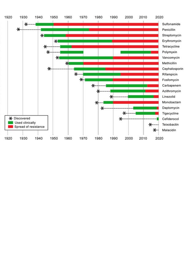
The Red Queen told Alice

“      Now, here, you see, it takes all the running you can do, to keep
                     in the same place”

- Lewis Carroll

From the book Through the Looking Glass (1871)
ABSTRACT The spread of antibiotic resistance is a concerning issue causing limited treatment options for bacterial infections, particularly with Gram-negative bacteria. Surveillance and epidemiological studies help to determine the magnitude of the problem as well as to establish early measures to slow down the spread of resistance and consequently increase antibiotic lifespan. Currently, there is a visible paucity of published data about resistance from the Arabian Peninsula. In this thesis, we studied a collection of carbapenem non- susceptible E. coli (n=35) and K. pneumoniae (n=237) isolated in 2015 from various hospitals in Oman. We aimed at identifying resistance mechanisms, mapping the bacterial population structure, investigating bacterial fitness, and studying potential treatment options available to tackle infections caused by such multidrug-resistant strains. These aims were addressed in five papers as discussed below. NDM and OXA-48 were the only carbapenemases we found in this collection, both among E. coli (Paper I) and K. pneumoniae (Paper II). The pattern of resistance among the isolates from Arabian Peninsula mimics the pattern reported from the Indian subcontinent, most likely due to the close socioeconomic interactions between them. Both regions lack KPC enzymes, which are commonly seen in China and the US from strains belong to ST11 and ST258, respectively. Despite ST11 being predominant in this collection, we did not detect KPC. Yet, we detected a high-risk clone of E. coli, ST131-H30Rx-CTX-M-15. Additionally, we identified newly emerging clones of K. pneumoniae and E. coli such as ST231 and ST1193-H64RxC, respectively. Nearly 10% of the K. pneumoniae isolates in our collection were colistin resistant which prompted us to study the mechanisms of colistin resistance (Paper III). MgrB-inactivation by insertion elements was seen in 8 isolates while other mutations were seen in other chromosomal genes known to be involved in colistin resistance e.g. pmrB, phoPQ and crrB. However, we did not detect mcr genes. Collectively, the genetic alterations are thought to reduce the net negative charge in bacterial cell wall, hence lowering the binding affinity of colistin. Our data underscores that there is no reduction in the surface charge in colistin- resistant K. pneumoniae, due to the MgrB-insertion (Paper IV). The genetic alteration might lead to other structural changes in the cell wall such as altering hydrophobicity, which required further investigation. Also, our data shows no difference in the survival rates of colistin resistant and susceptible strains in blood, serum and zebrafish model. Thus, gaining resistance against colistin does not infer a fitness cost in K. pneumoniae with MgrB-insertion (Paper IV). Additionally, colistin and LL-37 share similar binding mechanism which suggest there might be a cross-resistance between them. Our data supported this hypothesis, but only at high concentrations of LL-37 ( ≥ 50 mg/L) (Paper IV). Finally, we studied available options to treat infections caused by multidrug-resistant strains. Combining colistin and rifampicin showed good in vitro activity against multidrug-resistant strains of E. coli (Paper V) and K. pneumoniae (Paper III). To summarise, we conducted comprehensive genomic analysis of E. coli and K. pneumoniae isolates from Oman to reveal the resistance mechanism, their impact on bacterial cell structural and if there is a fitness cost inferred by the resistance mechanisms. Finally, we studied combination therapy as an available option at hand for tackling infections caused by multidrug-resistant strains.
LIST OF SCIENTIFIC PAPERS

  I.   Hissa M. Al-Farsi, Angela Camporeale, Karolina Ininbergs, Saleh Al-Azri, Zakariya Al-
       Muharmi, Amina Al-Jardani, Christian G. Giske. Clinical and molecular characteristics of
       carbapenem non-susceptible Escherichia coli: A nationwide survey from Oman. PLOS
       ONE 2020; 15: e0239924. DOI: 10.1371/journal.pone.0239924

 II.   Hissa M. Al-Farsi*, Maarten Coorens*, Isak Sylvin, Saleh Al-Azri, Zakariya Al-Muharmi,
       Amina Al-Jardani, Christian G. Gisk. Nationwide characterization of carbapenem non-
       susceptible Klebsiella pneumoniae in Oman. Manuscript. *shared first author

III.   Hissa M. Al-Farsi, Maarten Coorens, Karolina Ininbergs, Saleh Al-Azri, Zakariya Al-
       Muharmi, Amina Al-Jardani, Peter Bergman, Christian G. Gisk. Colistin resistance from
       clinical isolates of Klebsiella pneumoniae in Oman: Mechanism of resistance and
       synergism of colistin with azithromycin and rifampicin. Manuscript.

IV.    Hissa M. Al-Farsi, Salma Al-Adwani, Sultan Ahmed, Carmen Vogt, Anoop T. Ambikan,
       Anna Leber, Amina Al-Jardani, Saleh Al-Azri, Zakariya Al-Muharmi, Muhammet S.
       Toprak, Christian G. Gisk, Peter Bergman. Effects of the antimicrobial peptide LL-37 and
       innate effector mechanisms in colistin-resistant Klebsiella pneumoniae with mgrB
       insertions. Frontiers in Microbiology 2019; 10: 2632. DOI: 10.3389/fmicb.2019.02632.

V.     Anna Olsson, Marcus Hong M, Hissa M. Al-Farsi, Christian G. Giske, Pernilla Lagerbäck,
       Thomas Tängdén. Evaluation of the interactions of polymyxin B in combination with
       aztreonam, minocycline, meropenem and rifampicin against NDM- and OXA-48-like
       producing Escherichia coli. Manuscript.
CONTENTS
Preface__________________________________________________________________ 1
Introduction ______________________________________________________________ 3
1.1 Multidrug resistance as a public health issue ________________________________ 3
1.1.1 The pride comes before the fall ___________________________________________________ 3
1.1.2 The bright past just passed _______________________________________________________ 3
1.1.3 Back to the future ______________________________________________________________ 5
1.2 Antibiotics: Action and resistance _________________________________________ 7
1.2.1 The outer membrane when negativity is a merit _______________________________________ 9
1.2.2 Peptidoglycan when less is more: β-lactam target ____________________________________ 15
1.3 Bacterial fitness: The cost to exist _______________________________________ 16
1.4 Enterobacterales ______________________________________________________ 18
E. coli : An old yet turbulent friend ______________________________________________ 19
K. pneumoniae: An opportunist new foe __________________________________________ 19
Carbapenem resistant Enterobacterales __________________________________________ 19
1.4.3.1 K. pneumoniae carbapenemases (KPC) _________________________________________ 20
1.4.3.2 New-Delhi metallo-β-lactamases (NDM) ________________________________________ 21
1.4.3.3 OXA-48-like carbapenemase (OXA-48-like) ____________________________________ 21
Polymyxin Resistant Enterobacterales ____________________________________________ 22
1.5 Epidemiology of antimicrobial resistance__________________________________ 23
1.5.1 Carbapenem and colistin resistance globally ________________________________________ 23
1.5.2 Carbapenem and colistin resistance in Oman ________________________________________ 24
1.6 Typing bacterial strains ________________________________________________ 25
1.6.1 CRISPR-Cas system __________________________________________________________ 25
1.6.1.1 Components of CRISPR-Cas system ____________________________________________ 26
1.6.1.2 Classification _____________________________________________________________ 27
1.6.1.3 Function _________________________________________________________________ 28
1.7 The light at the end of the tunnel_________________________________________ 29
METHODOLOGICAL CONSIDERATION ________________________________________ 32
3.1 Ethical consideration __________________________________________________ 32
3.2 Dry lab work _________________________________________________________ 32
3.3 Wet lab work ____________________________________________________________ 34
3.3.1 Choice of medium _____________________________________________________________ 35
3.3.2 Bacterial inoculum _____________________________________________________________ 36
3.3.3 Time-kill assay ________________________________________________________________ 37
3.3.4 Zeta potential ________________________________________________________________ 37
3.3.5 Combination assays ___________________________________________________________ 38
RESULTS AND DISCUSSION _________________________________________________ 39
4.1 Population structure of resistant strains in Oman ______________________________ 39
4.1.1 E. coli : Paper I _______________________________________________________________ 39
4.1.2 K. pneumoniae : Paper II and Paper III _____________________________________________ 41
4.2 Interaction of innate effectors with colistin resistant strains: Paper IV _____________ 44
4.2.1 Interaction of colistin resistant strain with LL-37 _______________________________________ 44
4.2.1.1 Cross-resistance with LL-37 __________________________________________________ 44
4..2.1.2 Extracellular and intracellular impact of LL-37 on bacterial cell _____________________ 45
4.2.2 The mgrB-insertion did not impact surface charge ____________________________________ 46
4.2.3 Pathogenicity and virulence of colistin resistant strains _________________________________ 484.3 Impact of combination therapy onto resistant strains ___________________________ 48
4.3.1 Combination therapy in E. coli: Paper V ____________________________________________ 48
4.3.2 Combination therapy in K. pneumoniae : Paper III _____________________________________ 48
Conclusion ______________________________________________________________ 49
ACKNOWLEDGMENT _____________________________________________________ 50
REFERENCES ____________________________________________________________ 54LIST OF ABBREVIATIONS AmpC Ampicillinase type C AMPs Antimicrobial peptides OXA Oxacillinase ArnT Aminoarabinose glycosyotransferase BCA Background corrected absorption CaMHB Cation-adjusted media CaMHB Cas CRISPR-associated genes CFU Colony-forming unit CMS Colistinmethate sodium Col-R Colistin resistant Col-S Colistin susceptible CP-CRE Carbapenemase producing-CRE CRE Carbapenem-resistant Enterobacterales CRISPR Cluster regularly interspaced short palindromic repeats DIW Deionized water DR Direct repeats EptA phosphoethanolamine transferase ESBL Extended-spectrum β-lactamase GlcNAc N-acetylglucosamine HGT Horizontal gene transfer Hi-RC High-risk clone HMM High molecular mass IM Inner membrane IMP Imipenemase IS Insertion elements Kdo 3-deoxy-D-manno-oct-2-ulosonic acid KPC Klebsiella pneumoniae carbapenmase Arn Aminoarabinose (4-amino-4-deoxy-L-arabinose ) LB Lysogeny broth LPS Lipopolysaccharides MBL Metallo-β-lactamase MCR Mobile colistin resistant protein MHB Mueller Hinton Broth
MIC        Minimum inhibitory concentration
MLST       Multilocus sequence typing
MRSA       Methicillin resistant S. aureus
MurNAc     N-acetylmuramic acid
NDM        New Delhi metallo-β-lactamase
OM         Outer membrane
OMP        Outer membrane proteins
PBP        Penicillin binding proteins
PBS        Phosphate buffered saline
PG         Peptidoglycan
RPMI1640   Culture medium developed by Roswell Park Memorial Institute
SEM        Scanning electron microscopy
SNP        Single nucleotide polymorphism
ST         Sequence type
TEM        Transmission electron microscopy
VIM        Verona integron-brone metallo- β-lactamase
VRSA       Vancomycin resistant S. aureus
WGS        Whole genome sequencing
WHO        World health organisation
PREFACE
Long time ago, I thought it would be fun to attend a military training, so I did. I joined Nizwa
military academic school in Oman. No reasons were required there to yell at someone or treat
them like a slave. If the trainer did not like the way you walk, you might end having an extra
round (duplication) at the peak of the summer in July where temperature normally goes
beyond 40°C. When we were out for the physical training, the cells were inspected. If they
thought, the beds were not tidy, the cell will be turned upside down, the dustbin will be
emptied in the sleeping mattress. The number of participants dropped as some could not take
it anymore. I left the campus as well but only when the training period was over with ashy
skin and a pride being one of those who managed to stand the pressure.

The reason why me and others survived was not only attributed to us as individuals despite
that play a role as well since not all participants managed to finish the course. The main factor
was the induced pressure was not strong enough to break us but enough to stretch us and turn
us resilient. This is a sort of stress with good intention called hormesis. I tried to create such
positive stress throughout my PhD study by attending courses and conferences. I know it is
a motivation factor to go forward.

This is what we do with bacteria when we start taking an antibiotic yet we do not comply
with full treatment course. Bacteria become resilient as much as I did during the military
training. Hormetic effect explains what happens during the exposure to increment but low
dose of stress. Yet, what does happen if we withdraw the stress? After the military training I
reverted back to my old habits, no more waking up at 4:00 am to exercise yet still tidy my
bed religiously before I leave home! Why did I carry some of the habits but not others? It
depends on the cost I pay to maintain them and the rewards I gain from carrying them.

Generally, we as humans assume bacteria do not anticipate the future as we do but live in the
present moment as we fail to do. We assume as such bacteria will revert to be susceptible as
they lack the sense of foreseeing the future. A study showed that after 50% decrease in the
consumption of certain macrolide in Finland, there was 50% decrease in the prevalence of
erythromycin resistance in Streptococcus pyogenes between 1991 to 19961. While
withdrawal of cotrimoxazole and streptomycin in UK did not affect the prevalence of
sulfamethoxazole and streptomycin resistance in E. coli 2. This variation might be attributed
to the cost paid by bacteria to keep resistance attributes despite the absence of antibiotic
pressure. Much goes with the context of “The red Queen Theory” which was introduced by
Van Valen in 1973 that implies organisms must evolve and adapt to new challenges not to
extinct. Perhaps bacteria do think about the future, though not as intense as we humans do.

                                                                                                1
INTRODUCTION
1.1 Multidrug resistance as a public health issue
1.1.1 The pride comes before the fall
Not quite far, 100 years ago, we had no tools to fight back bacterial infections. We look at
pregnancy as a safe biological process to bring new lives now. Yet back then, it was a
biological process to trade mother’s life for infant’s life due to many reasons including failure
to treat post-partum infections. We as humans, did not have any tools to handle these
infections until someone thought outside the box.
Ingaz Philipp Semmelweis proposed the practice of washing hands with chlorinated lime
solution in 1847 to tackle the increased incidence of puerperal fever which was the main
cause of mortality in obstetrical clinics. He published a book “Etiology, Concept and
Prophylaxis of Childbed Fever” and showed results to support his recommendation but was
faced with rejection by the medical community then as they felt offended to be asked to wash
their hands. He suffered from mental breakdown from stress. In 1865, he passed away from
a gangrenous wound, due to infection after being beaten at an age of 473. These challenges
from the past continue until the present days. During the COVID-19 pandemic in 2020,
following the recommendation of wearing a mask and keeping physical distance, as simple
as they sound, were hard to implement. All in all, we see that relying on human behaviour as
a tool to fight infectious diseases is quite hard to sustain as such more reliable tools are
required such as antibiotics and vaccines.
At the beginning of 1910s, Paul Ehrlich introduced salvarsan as the first antimicrobial agent.
It was used to treat syphilis4. Followed by Fleming’s discovery of penicillin in 1928 and
many more antibiotics that gave us quite plenty of tools to fight bacterial infections and save
lives. In 1967, a remarkable announcement came from U.S. Surgeon General William
Stewart “The time has come to close the book on infectious diseases”5. That pride with plenty
of antibiotics at hand just faded after 1970s, since we entered a dry innovation season, while
bacteria fought back strongly.
1.1.2 The bright past just passed
The first serious battle we had was MRSA (Methicillin resistant S. aureus) in the beginning
of 1970s against Gram-positive bacteria. We opened our antibiotic closet looking for a
solution. Vancomycin, which was first sold in 1954 could be a desperate choice despite the
known side effects such as nephrotoxicity (Figure 1). Luckily, a purified form of vancomycin
was produced with less toxicity. In 1996, bacteria went ahead again in a second battle known
as VRSA (vancomycin resistant S. aureus). After this initial nightmare two new antibiotics
were introduced; linezolid and daptomycin in 2000 and 2003, respectively (Figure 1). As we
humans thought we had won the battle, Gram-negative bacteria pressed the replay button.
3Gram-negative bacteria are harder to treat since they have an extra protection layer in their cell wall which is a challenge for drugs to pass. Besides, the transmission of resistance genes between different species of Gram-negative bacteria is frequently high as compared to Gram- positive bacteria. Despite plasmid-encoded penicillinase was first been observed in Staphylococci, there were limited intra-species transmission compared to Gram-negative bacteria6. The first clinical impact of resistance in Gram-negative bacteria to β-lactam was in 1974 by penicillinases, when researchers reported ampicillin treatment failure in meningitis patients due to Haemophilus influenzae type b producing TEM β-lactamase7. The plasmid encoding blaTEM was transmitted to Neisseria gonorrhoeae8. This resistance mechanism spread widely between different continents due to naval activity between Asia, western Africa and England9. The drug development industry came up with a set of 24 novel modified β-lactams with extended-spectrum including β-lactamase inhibitors, third generation cephalosporins, monobactams, and carbapenems in the 1980s9. Figure 1. Timeline for key antibiotics. For each antibiotic three dates are represnted; the discovey date, the date when the antibiotic was used clinically and the date when resistance spread. The clinical use of polymyxin was discontinued for concern regarding side effects by 1970s. However, due to lack of treatment options, the drug was re-used again clinically in mid-1990s. Malacidin and teixobactin are not yet approved. Cefiderocol was recently approved in US (2019) and in Europe (2020). Adapted from Gautam and Sommer5. Copyright (2014) Sigma Xi, The Scientific Research Honor Society. 4
Soon bacteria responded by producing extended-spectrum β-lactamases (ESBLs). The first
ESBL in a clinical isolate was reported in 1983 from Klebsiella ozaenae in Germany10. By
the mid-1980s, outbreaks of Enterobacterales producing ESBL were reported in France due
to TEM derived CTX-1 and CAZ-1 enzymes. As their names imply CTX-1 conferred more
resistance to cefotaxime whereas CAZ-1 was more active against ceftazidime11. In the early
1990s, CTX-M-type ESBLs emerged and by the mid-1990s they caused large outbreaks of
Salmonella Typhimurium. More variants of CTX-types were detected including CTX-M-15
which proved to be a game changer regarding the spread of ESBL globally. By the 2000s it
became the predominant type of ESBL globally, replacing the TEM- and SHV-derived
ESBLs12. The access to the drug class carbapenems still kept us quite ahead of the bacteria.
However, the frequent use of carbapenems against ESBL-producers predicted carbapenem
resistance is the upcoming storm.

Type C ampicillinase (AmpC) enzyme was first discovered by Swedish investigators in 1940
in E. coli. As the name implies, it was initially thought to only affect ampicillin13. Another
report of this enzyme in Pseudomonas was published in 196514. Yet, it grabbed more
attention in 1976 when it was reported in the UK15. Now we know it is capable of not only
hydrolysing penicillins but also cephalosporins, cephamycins and monobactams, leaving
only carbapenems unaffected. However, AmpC could even hydrolyse carbapenems, if
combined with other mechanisms such as porin loss16. The threat was magnified when
AmpC-type enzymes were detected on plasmids in 1990, as this would enhance its propensity
to spread9.

The first carbapenemase was isolated from Pseudomonas in Japan in 1990 and was named
impenemase (IMP)17. Followed by the detection of Verona integron-borne metallo-β-
lactamase (VIM) in Italy from Pseudomonas as well in 199718. More notorious types of
carbapenemases were detected later such as the Klebsiella pneumoniae carbapenemase
(KPC), oxacillin-hydrolyzing carbapenemase (OXA-type) and New Delhi metallo-β-
lactamase (NDM)9. Here, we were literally trapped. We went back to our antibiotic closet
and took colistin out despite its limitation in both efficacy and toxicity19. As such colistin
resistance was foreseen. Multiple outbreaks were reported with colistin and carbapenem
resistance Enterobacterales (CCRE)20. Bacteria were again ahead of humans.

       1.1.3 Back to the future

We still lack a realistic estimation of lives lost yearly due to antibiotic resistance as the
number is overshadowed by main cause of death. For example, a cancer-patient if expired
due to a struggle with infection caused by antibiotic resistant bacteria, often the case will be
counted as a death caused by cancer rather than antibiotic resistance. A similar scenario is
seen in HIV and transplant patients. Thus, the numbers of death due to antibiotic resistance
is far underestimated. Also, many life-saving surgical procedures would cease if we have no
effective antibiotics to treat the inevitable infections at surgical sites which adds up in
underestimating the impact of resistance in public health. This will not only mask the burden

                                                                                              5
but will have a crucial impact on allocated resources for intervention. Consequently, the antibiotic resistance issue remains less studied with a big knowledge gap yet to be filled. A recent report in 2019 estimated the number of deaths attributed to antibiotic resistance only in the US, to be more than 35,000 while the number of infections was estimated to be more than 2.8 million each year21. This gives an estimation of less than 0.001% death rate. On the other hand, a study conducted in 2014 estimated the number of death due to antimicrobial drugs resistance, including antibiotics but not limited to them, would be 10 million yearly by 205022. Yet, they estimated the combined deaths caused by road traffic injuries, cancer and diabetes to be around 11 million annually in 205022,23. Bearing in mind diabetes, cancer and road injuries were listed within the top 10 causes of death in 2019 globally24. The paper received criticisms regarding uncertainty of assumptions to determine the rate of incidence, as it compiled two sets of data each of them used distinct assumptions and covered particular sources of infections e.g. community versus hospital acquired infections25. One advantage of the study was that it considered the impact of resistance on other medical procedures as a secondary effect for the problem. A better approach would be to lay down the estimation from each study separately rather than the risk of combining both, despite the methodological variations used in each. Also, it would be more informative to break the numbers down rather than counting HIV, malaria and TB. We know these diseases already cause high mortality. Yet, researchers in antibiotic resistance are looking for death cases caused by antibiotic resistance specifically. Additionally, listing death caused by primary or secondary attributes separately will give better insight to the magnitude of the problem. However, no doubt the report drove public attention and was a kick-off to think thoroughly about how to estimate the burden of antibiotic resistance and improve data collection as well as analysis in this regard. Additionally, political barriers and national health policy are other challenges. This has been evident after the discovery of NDM and the reaction from the Indian government to restrict sending biological samples abroad for research purposes. They saw NDM as a stigmatising factor to their country, especially after naming the enzyme by the capital city in India “New Delhi”. This impacted tourism which led to economic consequences26. Now, it is quite difficult to ship biological samples from India for research purposes and barriers to carry out epidemiological studies are becoming high to pass. Since it is a multifactorial issue with economic and political complications, the nomenclature of new genes could be revisited to minimise provoking logistic barriers for future studies. The awareness NDM brought to this matter was manifested by the neutral name given to the mobile colistin resistance (mcr) gene isolated from China in collaboration with some investigators who isolated blaNDM previously27,28. Antibiotics are social drugs with respect to their consumption by an individual will impact their effectiveness for others. Unlike the drugs that control high blood pressure or cholesterol level for example, where there is no such impact between individual consumption and long- term efficacy29. This creates another challenge related to social and cultural behaviours 6
towards consumption in different countries. Recent studies linked the increase of living
standard in low-income countries to the increase of antibiotic consumption30,31. This could
be due to the search for a fast fix that is affordable rather than the more time-consuming
solution such as building infrastructure for diagnostic facilities which enhance identifying
resistant strains prior to antibiotics consumption. Moreover, unregulated policy in private
health sectors could play a role, since they go immediately to the last treatment option to
satisfy the customer/patient and increase their reputation to cure infections fast andthereby
increasing their profits. In less economically fortunate nations the scenario differs, since the
majority of people cannot afford the expenses of health services such as visiting clinic and
performing diagnostic tests prior to buying the lifesaving antibiotic. Thus, implementing the
compulsory regulation for diagnosis and prescription prior to selling antibiotics might
prolong antibiotics lifespan but might indirectly cause loss of lives for those who cannot
afford paying for such services. Particularly, in the absence of social benefits to protect the
vulnerable groups. As we see, despite the global impact of the issue a “one size fits all”-
solution is not achievable and a tailored solution for each nation is needed bearing in mind
the local socioeconomic structure.
Additionally, the increase of income leads to higher demands for food consumption.
Consequently, the use of antibiotics in farming and agriculture are increasing to cover the
high demand for proteins30. The world health organisation (WHO) introduced the one health
concept to tackle this issue. Furthermore, the concept was nourished by other programs such
as Joint Programming Initiative on Antimicrobial Resistance (JPIAMR). It implies, the issue
of antibiotic resistance requires a collaborative attention from three sectors (human health,
animal and environment) as the consumption of antibiotics in any of them will impact the
utility and effectiveness of the drug in human health32. This expands the concept of antibiotic
from a social drug into a universal drug.
As researchers in the field of antibiotic resistance, we acknowledge having a problem that
requires intervention. Simultaneously, we acknowledge the multifactorial challenges in
estimating the real burden of resistance compared for example to other infectious diseases
such as HIV and malaria. I trust that epidemiological studies with ethical permits such as the
one presented herein will help mapping the burden of antibiotic resistance. Besides,
understanding the evolution of resistance may extend the life of the currently available
antibiotics and slow down resistance. Consequently, this would reduce the ultimate cost paid
by patients and society.
1.2 Antibiotics: Action and resistance
Antibiotics can be classified into different classes according to their chemical structures or
mechanisms of action. Based on their targets, antibiotics could be divided into two main
classes. A class of drugs that inhibits intracellular reactions such as proteins or nucleic acids
synthesis. Rifampicin and azithromycin belong to this class. The second class includes
antibiotics that target bacterial envelope such as β-lactams and polymyxins.
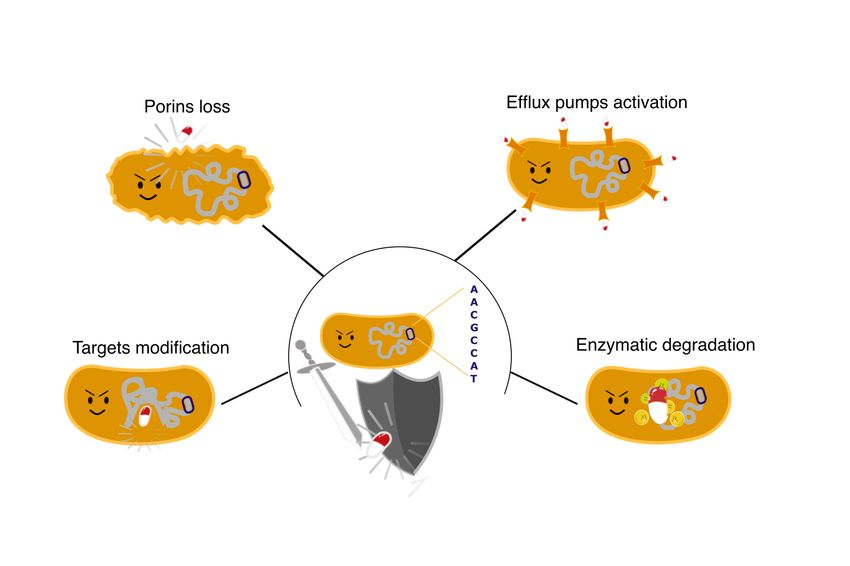
7As antibiotics have different targets, bacteria evolve to resist them differently. Some of the resistance mechanisms apply to a wide range of antibiotics while others are tailored for specific drugs based on their chemical structure or the structure of their targets. The physical mechanisms, also known as intrinsic mechanisms, aim to reduce antibiotics uptake through cell surface modification by restricting the uptake (influx) or enhancing the excretion (efflux). This is achieved by porins modification or efflux pumps activation, respectively. The specific resistance mechanisms involve modification of drug target or inactivation of the drug (Figure 2). An example of the former is resistance mechanism against colistin by altering lipid A structure whereas the production of β-lactamase is an example of drug inactivation5. The general intrinsic mechanisms are mainly attributed to chromosomal genes while the tailored mechanisms are generally carried in transferable elements such as plasmids. Especially this second type of resistance creates a challenge to control infections as it can be transmitted both vertically and horizontally. During vertical gene transfer, bacteria inherit their genetic contents from parental cells in the previous generation whereas during horizontal gene transfer (HGT) bacteria could gain genetic material from unrelated bacterial cells or their surroundings. There are three mechanisms of HGT; transformation, transduction and conjugation. They differ in the source of genetic material that bacteria uptake. In transformation bacteria obtain foreign DNA from the environment whereas for the remaining two mechanisms DNA will be obtained from a donor, either a phage or a plasmid by transduction or conjugation, respectively33. Conjugation plays a significant role in the spread of resistance genes within and between species. Yet, transduction is more involved in spreading virulence genes, and it is thus far unclear to which extent bacteriophages contribute to spreading resistance genes. Most of the genes involved in polymyxin (colistin) resistance are chromosomal, which limits their mobilities. In contrast, genes encoding β-lactamases are frequently found in plasmids, which allows for easy horizontal transmission and fast spreading. Both polymyxin and β- lactam antibiotics act by interfering with the biosynthesis of the outer membrane and the cell wall, respectively. Polymyxins compete with and replace divalent cations that usually bring nascent lipid A molecules together physically, while β-lactams chemically inactivate the enzyme responsible for cross-linking nascent peptidoglycans. The following section will describe different components within the bacterial envelope which are used as targets by these drugs as well as how bacteria alter these components to resist them. 8
Figure 2. Mechanisms of antibiotic resistnce. Bacteria aquire antibiotic resistance by modifying their DNA.
The genetic alteration could cause either a general mechanism of resistance such as porin loss or activation of
efflux pump. Alternatively, mutated bacteria could gain more specific mechanisms of resistance, for example
the ability to degrade antibiotics enzymatically or modify their targets so the binding would not occur.

The cellular envelope protects bacteria from hostile factors in their environment. In Gram-
negative bacteria there are three main layers; the outer membrane (OM), the peptidoglycan
(PG) and the cytoplasmic or inner membrane (IM). Among Gram-positive bacteria the
envelope has only two layers consisting of a PG and an IM. Generally, Gram-negative
bacteria are more resistant to antibiotics, due to the presence of the OM which restricts drug
uptake. The bacterial envelope plays a key role in resistance mechanism. Thus, understanding
the bacterial envelope is a key factor in slowing down resistance as well as developing new
therapeutic drugs to treat infections.

1.2.1 The outer membrane when negativity is a merit

The outer membrane in Enterobacterales is composed of phospholipids arranged in a bilayer
of inner symmetric leaflets and outer asymmetric leaflets. The outer leaflets are intercalated
by lipopolysaccharides (LPS) through Van der Waals forces. In addition, the OM bilayer is
embedded with outer membrane enzymes such as PagP and PagL in E. coli and Salmonella
enterica, respectively. These enzymes usually regulate and modify LPS by acylation
process34. Additionally, OM has outer membrane proteins (OMPs) such as porins which
modify cellular permeability (Figure 3).

                1.2.1.1 Outer membrane protein: gatekeeper

The outer membrane has β-sheet proteins which are wrapped into cylinders known as outer
membrane proteins (OMP). The main function of OMPs is to exchange small molecules
across outer membrane. Consequently, they enhance bacterial adaptability to various
environments through regulating permeability. Porins are the most abundant OMPs that

                                                                                                             9
A C
kdo
A C
kdo
MurNAC
GlcNAc
D
G lcN Ac
M ur NA c G lcN Ac M ur NA c G lcN Ac
M ur NA c G lcN Ac G lcN Ac
M ur NA c M ur NA c
B
G lcN Ac M ur NA c
G lcN Ac M ur NA c M ur NA c
G lcN Ac
M ur NA c G lcN Ac
G lcN Ac
M ur NA c G lcN Ac
M ur NA c
G lcN Ac
M ur NA c D
G lcN Ac
M ur NA c
G lcN Ac M ur NA c G lcN Ac
M ur NA c G lcN Ac G lcN Ac
M ur NA c M ur NA c
B
G lcN Ac
M ur NA c
G lcN Ac
M ur NA c M ur NA c
G lcN Ac
M ur NA c
G lcN Ac
G lcN Ac
M ur NA c G lcN Ac
G lcN Ac
M ur NA c M ur NA c
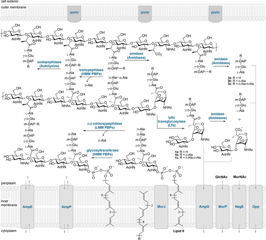
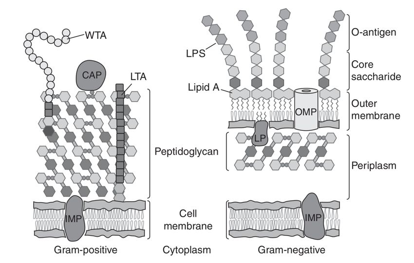
Figure 3. Depiction of cell-envelope components. A| Cell-envelope of Gram-positive and Gram-negative
bacteria. Gram-positve bacterium has an exposed yet thick peptidoglycan (PG) layer. Whlist Gram-negative
bacterium protects the PG layer by an outer membrane (OM). CAP; covalently attached protein; IMP, integral
membrane protein; LP, lipoprotein; LTA, lipoteichoic acid; WTA, wall teichoic acid. Adapted with permission
from Silhavy et al.35 Copyright (2010) Cold Spring Harbor Laboratory Press. B| Porin and efflux pump are
involved in antibiotic resistance by altering cellular influx and efflux, respectively. Efflux pump consists of
three main components; an outer membrane protein (OMP), a membrane fusion proteins (MFP) and a pump as
an inner membrane (IM) transporter. LPS; Lipopolysaccharides. Adapted with permission from Li et al.36
Copyright (2015) American Society for Microbiology. C| Chemical structure of LPS components; lipid A, core
oligosaccharide and O-antigen. The core oligosaccharide is located in the middle between lipid A and O-
antigen. It is attached to lipid A via Kdo (3-deoxy-D-manno-oct-2-ulosonic acid) molecule (framed in red).
Adapted with permission from Barkleit et al.37 Copyright (2008) The Royal Society of Chemistry. D| Final steps
involved in PG synthesis require two types of high-molecular-mass penicillin-binding proteins (HMM PBP).
Glycosyltransferase (HMM PBP) binds to the pre-formed disaccharide units; N-acetylglucosamine (GlcNAc)
and N-acetylmuramic acid (MurNAc) to form a linear glycan layer. While transpeptidase (HMM PBP) cross-
links glycan linear layers by utilising acyl-D-alanine-D-alanine (D-Ala-D-Ala) in the side chain of a MurNAc
unit as a substrate (Figure 5). Thus linking 2 MurNAc units to form a solid mesh-like structure. Adapted with
permission from Dik et al.38 Copyright (2018) American Chemical Society.
affect membrane permeability. Thus, genetic alterations or total loss of porins will enhance
bacterial resistance to antibiotics. For example, it is well established that alteration or total
loss of specific porins in E. coli (OmpF and OmpC) or K. pneumoniae (OmpK35 and
OmpK36) causes resistance to a wide range of antibiotics including β-lactams39. Porins
alteration could be achieved by gaining mutations directly into genes encoding porins or the
regulatory genes such as envZ and ompR in E. coli40.
Additionally, outer membrane vesicles (OMV) are produced by most Gram-negative
bacteria. These vesicles are released from the cell envelope. They are simply sacs with high
profile of proteins and lipids and have been linked to antimicrobial resistance due to their
neutralising capacity. Studies showed mutant E. coli that over express OMV can resist
polymyxin39.
10Besides the role of the outer membrane in antibiotic resistance, it can enhance bacterial
pathogenicity. For example, OmpX in E. coli and OmpK17 in K. pneumoniae are known to
help in adhesion and invasion as well as promoting bacterial resistance to serum by
inactivating the complement system41. Also, OmpA in K. pneumoniae can clear infection and
proposed as a potential target for vaccine development42. The loss of OmpA in K.
pneumoniae has been found to reduce capsule expression thereby increase susceptibility to
polymyxin43. In the other hand, OMPs play an indirect role in active excretion of harmful
molecules such as antibiotics as being part of the efflux-pump system.
1.2.1.2 Efflux pump: dealing with gatecrashers
The efflux pump consists of three units; an inner membrane transporter (pump), a periplasmic
fusion protein and an outer membrane (OM) channel protein (Figure 3B). The main function
of the efflux pump is to expel toxic substances such as antibiotic from passing to bacterial
cell. Also, it plays a role in virulence mainly by enhancing colonization through protecting
bacteria from antimicrobial compounds present naturally in mucosal surfaces. For example,
over expression of the AcrAB-TolC pump is known to cause resistance against β-lactams
and polymyxins in E. coli and K. pneumoniae40,44,45.
1.2.1.3 Lipopolysaccharides (LPS): polymyxin target
Structurally, LPS is composed of three units; the hydrophobic lipid A, two hydrophilic chains
of oligosaccharides and O-antigen. The conserved lipid A anchors LPS in the outer leaflets
of outer membrane enhancing its asymmetric structure compared to the symmetric structure
of the inner leaflets. The oligosaccharide is located in the middle between lipid A and O-
antigen and arranged into two cores; the inner being attached to lipid A and made of 3-deoxy-
D-manno-oct-2-ulosonic acid (Kdo), while the outer core being attached to the O-antigen and
is made up of variable hydrophilic repeated saccharide units34,46,47(Figure 3C).
LPS enhances the anionic charge of the outer membrane due to the presence of phosphoester
moieties in lipid A as well as carboxylate and phosphate groups within the core
oligosaccharide and the O-antigen units46. Thus, LPS restricts the entry of big negatively
charged molecules. Besides, LPS affects the bacterial ability to respond to the immune
system and plays a role in adhesions47.
Since LPS is an important component with multiple functions, bacteria possess many
enzymes to modify its features as an adaptation process to different surrounding34. LPS
synthesis starts with the Raetz pathway in the inner membrane facing the cytoplasm. It
involves a series of reactions with highly conserved enzymes. Eventually, LPS is transported
across the inner membrane to the outer membrane. However, adjustment of LPS contents
continues even when it reaches its final destination. This is achieved through modification of
the variable O-antigens or mainly the conservative lipid A34,47,48.
11Structurally, lipid A is an anionic hydrophobic disaccharide of glucosamine (GlcN) with four
to seven acyl chains (fatty acids) and phosphate groups. It is attached to the oligosaccharide
by two molecules of Kdo sugars49 (Figure 3C). Divalent cations (magnesium and calcium)
attach to phosphate groups in lipid A and bridge adjacent LPS molecules to add stability50.
Lipid A exhibits different degrees of acetylation and phosphorylation in different bacteria.
The extent of phosphorylation determines the net charge of the membrane. This is affected
by different mechanisms like masking phosphate groups from lipid A with positively charged
molecules such as phosphoethanolamine (pEtN by EptA) and 4-amino-4-deoxy-L-arabinose
(Arn by ArnT) or adding more phosphate groups to increase negativity via LpxT34. The net
charge is targeted as a binding site by polymyxins and antimicrobial peptides such as LL-37.
1.2.1.4 Polymyxin mechanism of action
Polymyxins are cationic, hydrophobic and lipophilic molecules. The last two properties give
polymyxins amphipathic chemophysical properties which is the core of their antibacterial
activity46. Polymyxin, as positively charged molecule binds to the negatively charged
phosphate group in lipid A electrostatically. The binding will consequently, displace the
divalent cations (magnesium and calcium) from phosphate groups in lipid A which will
disrupt outer membrane stability as these cations bridge LPS. Additionally, polymyxin
hydrophobic regions bind to LPS. The detailed mechanism of polymyxin antibacterial
activity is still unknown, yet we know electrostatic and hydrophobic interactions are essential
steps in polymyxins initial encounter with the bacterial cell20,46. As such it was hypothesized
that bacteria resist polymyxin by reducing the net negative charge of lipid A20.
One might wonder, if bacteria aim to reduce the net negative charge, the affinity of divalent
cations to bind and bridge lipid A will be lost as well. To what extent these cations are
important for the stability of bacterial cell requires more research. Clearly, there is still a gap
in our understanding of polymyxin action and resistance mechanisms. Below the known
mechanisms of resistance are described.
1.2.1.5 Polymyxin mechanism of resistance
Bacteria develop resistance against polymyxin by decreasing the net negative charge of lipid
A. This is achieved by adding positively charged aminoarabinose (Arn) molecules (by ArnT)
and phosphoethanolamine (pEtN) molecules (by EptA). Hence lowering the affinity of
colistin to lipid A. The modification of LPS is upregulated by the two-component regulatory
systems PhoPQ and PmrAB. Upregulation of these systems will increase the expression of
genes encoding proteins for the biosynthesis of aminoarabinose and phosphoethanolamine.
The two-component systems usually become activate in response to stimuli such as metal
concentration or low acidity34 (Figure 4).
The PmrAB two-component system consists of the pmrABC operon that encodes the
membrane bound sensor kinase (PmrB), regulatory protein (PmrA) and transferase (PmrC
also known as EptA). Low pH or high iron concentration will activate PmrB. PmrB will
12Figure 4. Modification of lipopolysaccharides (LPS). Modification of LPS involves two-component systems;
PhoPQ and PmrAB. They work independently or together mediated by PmrD. The activation of PhoPQ or
PmrAB eventually reduces the net negative charge in LPS through addition of aminoarabinose (Arn) by ArnT
(aminoarabinose transferase) enzyme (pathway indicated by green lines) or phosphoethanolamine (pEtN) by
EptA (phosphoethanolamine transferase) enzyme (pathway indicated by red lines), respectively. Also,
phosphated PhoP activates PagP, MgrB, PmrD and MgrR. PagP (blue lines) modifies the acyl chain in lipid A
by the addition of palmitate chain. MgrB regulates PhoQ negatively. Mutated mgrB gene, will allow continuous
expression of PhoPQ. Also, mutated mgrB could modify capusle structure in K. pneumoniae. Whlist PmrAB
two-component system activates EptA (PmrC) and PmrR. The latter inactivates LpxT (purple line) to cease the
addition of phospahte group, thus lowering the net negative charge. Binding of cationic antimicrobial peptides
(AMPs) such as LL-37 to bacterial cell will be reduced as a consequence of LPS modification with reduced net
negative charge. In the schematic diagram, mediatores are shaded with gray boxes. The inverted and horizontal
“T” shape indicates inhibition. Other mechansims apart from the addition of pEtN (red), Arn (green) or
palmitate chain via Pagb (blue) are depicted in black lines. Adapted with permission from Simposon and Trent34.
Copyright (2019) Springer Nature and Nowicki et al.51Copyright (2014) John Wiley and Sons.
13phosphorylate and activate PmrA. Activated PmrA could upregulate PbgP (also known as PmrHFIJKLM or ArnBCADTEF) directly which will eventually add aminoarabinose by ArnT. Also, PmrA could activate PmrC (EptA) which will eventually produce phosphoethanolamine (pEtN). Mutated pmrB turns on these series of reactions continuously, independent of the external stimuli40. Likewise, the PhoPQ system is made up of a membrane bound sensor kinase (PhoQ) and a regulatory protein (PhoP). A stimulus such as low concentration of magnesium or the presence of antimicrobial peptides (AMPs) will activate PhoQ which will phosphorylate PhoP. Activated PhoP will upregulate pbgP operon either directly or indirectly by binding to PmrD which will eventually upregulate pbgP operon. In either way, activated pbgP will activate ArnT. Since PmrD is a mediator between the two-component systems PhoPQ and PmrAB, its activation will activate PmrAB. The later will increase the expression of pmrC gene which yields more pEtN. Hence, PhoPQ activation upregulates both ArnT and EptA enzymes. It is worth noting that PhoPQ is downregulated by the mgrB gene. Hence mutated mgrB will turn the PhoPQ system on and decrease the net negative charge in lipid A which is a common mechanism to resist colistin in K. pneuomoniae52. Additionally, K. pneumoniae has another two-component system known as CrrAB. It encodes a kinase (CrrB), a regulatory protein (CrrA) and a modulator (CrrC). It involves indirectly in modifying LPS by activating PmrAB via CrrC as a mediator between CrrAB and PmrAB. PmrA will activate PmrC and PbgP (Figure 4). Additionally, CrrC could activate a putative efflux pump that cause polymyxin resistance53. Still much needs to be revealed about what activates this system. Yet, we know once CrrAB is activated, the production of both aminoarbinose and phosphoethanolamine will increase40. Besides the activation of the two-component systems, bacteria have other ways to reduce the negative charge of lipid A. The expression of enzymes in the outer membrane is one of them. Activated PhoP regulates PagP production in E. coli which plays a role in modifying acyl chains by adding palmitate chain to maintain asymmetric outer membrane layer. Asymmetric layer protects bacteria from antibiotics entry yet without compensating much by lowering their nutrients uptake34,54 (Figure 4). Also, LpxT is an inner membrane enzyme responsible for adding phosphate group into lipid A. PmrR inhibits LpxT thereby reducing the net negative charge (Figure 4). Another pathway to add phosphoethanolamine is seen in bacteria that carry a mobile colistin resistance (mcr) gene, with a similar effect as the chromosomal mutations mentioned above27 (Figure 4). Also, mutations in mgrB gene seem to modify capsule structure in K. pneumoniae and increase resistance to polymyxin as well as affect the expression55. Additionally, alterations of O-antigen such the length of the chain can induce polymyxin resistance. In Salmonella enterica, PmrA activates wzz gene that promotes O- antigen production thereby increases resistance to polymyxin56(Figure 4). 14
1.2.1.6 Polymyxin cross-resistance with LL-37

The binding mechanism of colistin through electrostatic interaction with the negatively
charged LPS resembles the mechanism used by antimicrobial peptide such as LL-37. LL-37
is part of the innate immune system and has antimicrobial properties against a wide range of
microbes including bacteria57. It resides in different cell types, most notably in neutrophils58.
A concern was raised regarding the cross resistance between colistin and LL-37, since they
have similar binding mechanism59,60. If a bacterial pathogen can resist colistin, it might
eventually resist the killing by LL-37 which would lead them to escape host immunity (Figure
4).

       1.2.2 Peptidoglycan when less is more: β-lactam target
The bacterial cell wall is made of peptidoglycan (PG) in both Gram-negative and Gram-
positive bacteria. The PG layer in Gram-negative is thin (35-40 nm) and hidden beneath an
outer membrane compared to the exposed thicker (40-60 nm) PG in Gram-positive bacteria
(Fig 3A). Disrupting the construction of PG is the mechanism of action of β-lactam drugs.
Hence, penicillin is more effective against Gram-positive bacteria61.

The building unit of PG is a glycan strand which consists of an alternating saccharide of N-
acetylglucosamine (GlcNAc also known as NAG) and N-acetylmuramic acid (MurNAc also
known as NAM). The construction of PG is a complex process involving different types of
penicillin-binding proteins (PBPs). Two of them are of interest in this context,
glycosyltransferase high molecular mass (HMM PBPs) and transpeptidase (HMM PBPs).
Glycosyltransferase high molecular mass (HMM PBP) builds linear glycan strand by
covalently linking the two saccharides units (GlcNAc and MurNAc) via β-1,4-glycosidic
bond. This will result in a linear glycan strand made up of disaccharides. One of them
(MurNAc) has a stem made of pentapeptide with acyl-D-Ala-D-Ala end. Acyl-D-Ala-D-Ala
is utilised as a substrate by transpeptidase to produce acyl enzyme with transient nature to
cross-link two MurNAc units in different linear glycan strands forming eventually a stable
mesh structure (Fig 3C-D). In short, glycosyltransferase creates linear glycan strand between
GlcNAc and MurNAc, while transpeptidase cross-links two MurNAc units in different
glycan strands to form rigid cell wall. β-lactam interferes with the cross-linking process as
elaborated below.

              1.2.2.1 β-lactamase: when having a ring does matter

β-lactam chemical structure mimics acyl-Ala-D-Ala, as both have a central amide bond. β-
lactam will bind and lock transpeptidase (HMM PBPs) and as such interferes with the mesh
formation. This will prevent the final crosslinking of the nascent peptidoglycan layer (Figure
5). Thus, structural integrity of the cell wall decreases, which results in osmotic lysis of the
bacterial cell38.

                                                                                              15
Figure 5. Mechanisms of action and resistance of β-lactam. A| Transpeptidase is a penicillin-binding protein (PBP). It utilises the native D-Ala-D-Ala terminal in MurNAc (N-acetylmuramic acid) saccharide as a substrate to produce acyl enzyme with transient nature. This eventually crosslinks with another MurNAc saccharide in a nascent units of peptidoglycan to form mesh-like structure. B| β-lactam, here represented by penicillin, mimics D-Ala-D-Ala of MurNAc molecule as both have amide bond, yet penicillin has the amide in β-lactam ring. PBP will bind to penicillin instead of D-Ala-D-Ala substrate, thus an irreversible complex acyl enzyme will be formed. This will cease the following step of cross-linking and eventually rupture the cell wall. However, in the presence of β-lactamase (βL), the enzyme will hydrolyse β-lactam ring rendering the drug inactive. Adapted with permission from Astrid Zervosen et al.62Copyright (2009) American Chemical Society. 1.3 Bacterial fitness: The cost to exist The most prevalent strain in nature is known as wild-type strain. Such strain fits well in nature as their genetic content is stable. However, under selective pressure they lack the tools to adapt and survive. Under such unfavourable conditions and to avoid extinction, wild-type strain is forced to adapt. In the pre-antibiotic era, the main challenge faced by bacteria is to survive in a host. Thus, the wild-type strains deviated by gaining a set of tools “virulence factors” that enabled them to establish an infection as well as to escape host immunity. 16
Furthermore, antibiotics added a new challenge that required a new set of tools, “resistance
factors”. Acquiring these tools will cost wild-type a reduction in their fitness.

Resistant strains can be weakened by altering their structures in a way to bypass antibiotic
activity. This can lead to reduced survival strength when competing with wild-type strains in
the absence of antibiotic33. Therefore, resistant strains usually invade immunocompromised
patients with impaired immune systems. They are widely recognized in hospitals causing
healthcare-associated infections compared to virulent strains that are usually associated with
community acquired infection.

A general model for antibiotic evolution illustrates that wild-type lineages will gradually
evolve in three distinct stages under continuous antibiotic pressure (Figure 6A). Initially
wild-type lineages will trade their fitness by developing new mutations for survival. At this
initial stage, the cost of fitness has raised the hope as if the consumption of antibiotic reduced,
the bacteria will revert back to the susceptible phenotype33. Additionally, it opens the door to
fight bacteria not only by finding new antibiotics but also looking to enhance the immune
system. Yet if antibiotic pressure continues, lineages will enter a second stage. There, bacteria
would aim to stabilise the newly gained mutant-type by acquiring compensatory mutations
to restore fitness and become as competent as the wild-type strains. Another approach to
restore fitness is by discarding genes that do not serve the updated genetic structure. When
such generation of resistant bacteria emerges, it is unlikely that withdrawal of antibiotic will
revert to a susceptible phenotype33. What is more concerning is the possible marriage of both
virulent and resistant strains. At this stage, bacteria could gain mutations that increase their
fitness while maintaining their resistance pattern. For instance, they upgrade advantageous

   A
                                                          B
                                                                              1.0
                                                                                       WT
                                                           Relative fitness

                                                                              0.9

                                                                                                                      mgrR
                                                                                                                      gyrA
                                                                                                                      parC
                                                                              0.8      mgrR

                                                                                                       mgrR
                                                                                                       gyrA

                                                                              0.7
                                                                                0.01          0.1         1      10
                                                                                                    MIC (mg/L)

Figure 6. Bacterial fitness. A| General evolutionary model for lineages under antibiotic selection. Adapted
from Marcusson et al.(2009)63. Copyright via CC BY licence, PloS. B| Resistance to fluoroquinolones in E.
coli. High resistance is achieved when three mutations were acquired and this causes a significant increase in
MIC-value from 1 mg/L in double mutations (mgrR, gyrA) to >32 mg/L in the lineage with triple mutations
(mgrR, gyrA, parC). MIC; minimum inhibatory concentration. Adapted with permission from Anderson and
Hughes33. Copyright (2010) Springer Nature.

                                                                                                                             17
You can also read