New and promising imaging technologies in gynecology - Paul van Kesteren OLVG Oost Amsterdam
←
→
Page content transcription
If your browser does not render page correctly, please read the page content below
New and promising imaging
technologies in gynecology
        Paul van Kesteren
      OLVG Oost Amsterdam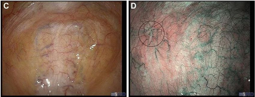
• New technology in ultrasound
  imaging

• New MRI and CT technology

• Intra operative imaging
Compression Elastography
• Measures stiffness/elasticity
  of tissue

• Diagnosis of fibroids and
  adenomyosis

• Hard tissue: dark colours
  Soft tissue: bright colours

Difference in tissue
stiffness between fibroids
and adenomyosis can be
shown by elastography
Compression Elastography vs gray scale in the
      diagnosis of fibroids and adenomyosis
Prospective diagnostic evaluation study:
- MRI vs Compression Elastography vs Gray Scale (cine loops 1,5 min)
- Residents vs gynecologists

  - 10 women with fibroids
  - 10 women with adenomyosis

      Stoelinga et al, Ultrasound in Med. & Biol., Vol. 44, No. 8, pp. 1654–1663, 2018
Compression Elastography vs Gray scale US in the
    diagnosis of fibroids and adenomyosis
• Better than gray-scale with color/power doppler ?

• Difference in vascularization pattern in fibroids and
  adenomyosis

 MUSA: Van den Bosch et al, Ultrasound Obstet Gynecol. 2015 Sep;46(3):284-98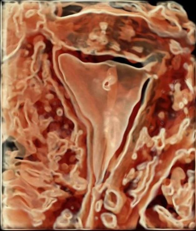
Contrast enhanced ultrasound for tubal patency

• Hysterosalpingo Contrast
  Sonography (HyCoSy)

• Hysterosalphingo Foam
  Sonography (HyFoSy)

• HyFoSy more accurate than
  HyCoSy
   – Sensitivity: 87,5% vs 57,8%
   – Specificity: 100% vs 66,6%
  Piccioni et al, J Clin Ultrasound. 2017 Feb;45(2):67-71
HyFoSy

• Accurate alternative for HSG
   – Up to 97,4 % concordance
   Rajesh et al, Int J Womens Health. 2016 Dec 28;9:23-32

• Less painfull than HSG
   – HyFoSy median VAS 1.7 (IQR: 2.1)
     vs HSG median VAS 3.7 (IQR: 4.2)
      Dreyer et al, Fertil Steril. 2014 Sep;102(3):821-5

• HyFoSy cost effective alternative
  for HSG?
   Study protocol: BMC Womens Health. 2018 May 9;18(1):64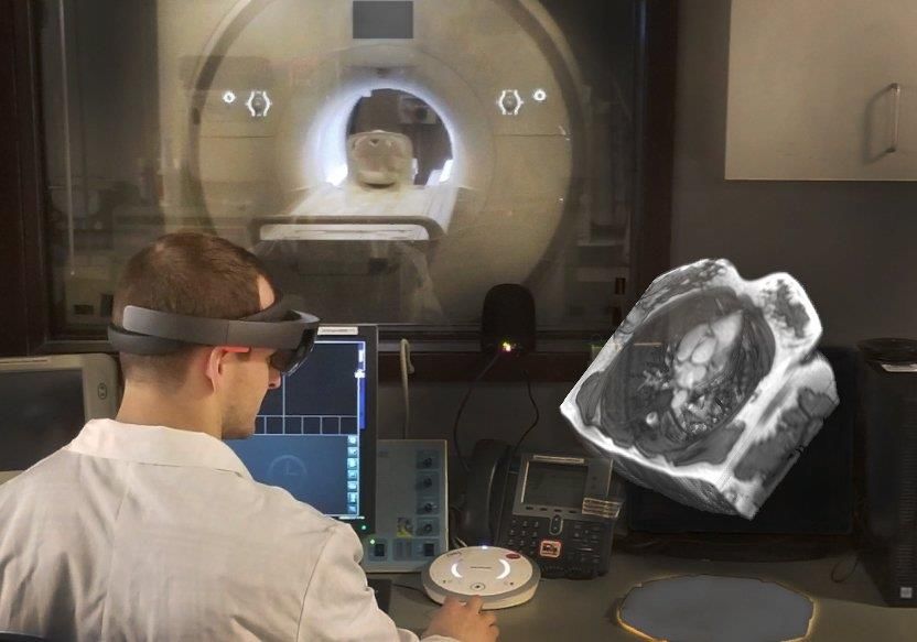
3D in HyCoSy and HyFoSy

• 3D HyCoSy and 3D HyFoSy is more
  accurate than 2D
   Alcazar et al, Gynecol Obstet Invest. 2016;81(4):289-95
   Ludwin et al, Ultraschall Med. 2019 Feb;40(1):47-54

                                                        3D HyFoSy

3D HyFoSy with colour doppler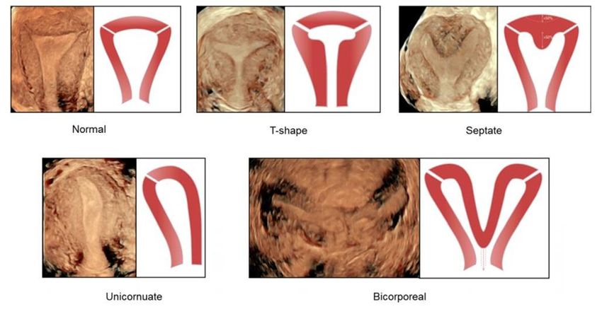
Clinical value of 3D ultrasound
• Off-line rendering and calculations of the 3D ultrasound
  dataset
3D Saline Infusion Sonography for intra cavitary
pathology
• Despite beautiful pictures 3D is not better than 2D because
2D SIS is already very accurate
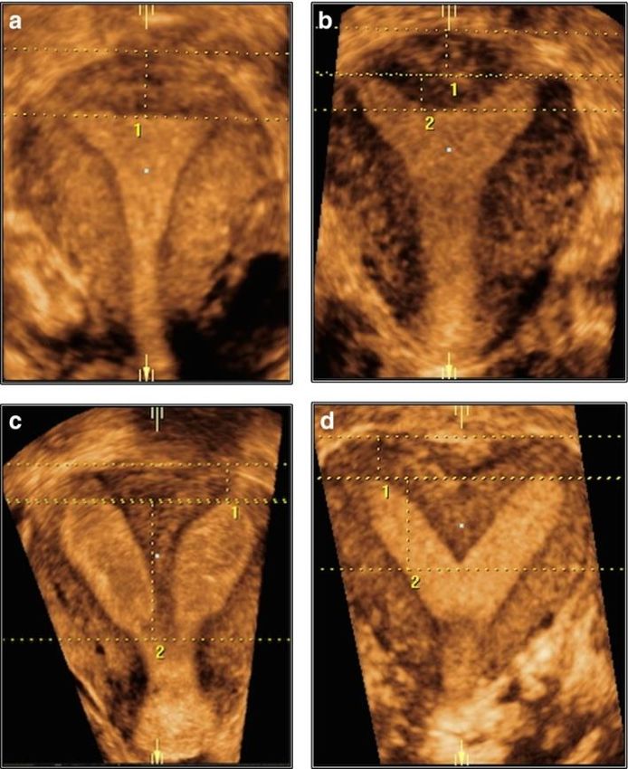
Nieuwenhuis et al, Cochrane Database Syst Rev. 2017 May 5;53D ultrasound in diagnosis of female congenital
anomaliesGraupera et al, Ultrasound Obstet Gynecol. 2015 Nov;46(5):616-22
The Thessaloniki ESHRE/ESGE consensus
• Three-dimensional (3D) US is recommended for
  the diagnosis of female genital anomalies in
  'symptomatic' patients belonging to high risk
  groups for the presence of a female genital
  anomaly and in any asymptomatic woman
  suspected to have an anomaly from routine
  evaluation

• Adolescents with symptoms suggestive for the
  presence of a female genital anomaly should be
  thoroughly evaluated with 2D US, 3D US, MRI
  and endoscopically

 Grimbizis et al, Hum Reprod. 2016 Jan;31(1):2-7
3D power doppler ultrasound in fibroid growth

• Relevant for clinical practise

• Well vascularized fibroids may
  grow faster

• Vascular Index, measure of
  fibroid vascularization
    – Calculated off-line with VOCAL
      software

   Nieuwenhuis et al, BJOG. 2018 Apr;125(5):577-584
3D power doppler ultrasound in fibroid growth
• Vascular Index predicts fibroid
growth
– N=66, Maximum fibroid number: 2,
maximum diameter: 8 cm
• Vascular index less accurate in
large and multiple fibroids
Nieuwenhuis et al, BJOG. 2018 Apr;125(5):577-584Automated volume calculation with SonoAVC
• SonoAVC (GE Medical Systems, Zipf,
Austria)
– Software that identifies and quantifies
hypoechogenic regions within a 3D
dataset
– Automatic estimation of their volume
– each different volume is color coded
separately
– Counting ovarian follikels
Raine‐Fenning et al, Ultrasound Obstet Gynecol 2007; 30: 1015– 1018
Froyman et al, Ultrasound Obstet Gynecol. 2018 Jan;51(1):147-149Follicle counting using Sonography-based Automated Volume Count software in three-dimensional volume of left ovary, depicting 67 antral follicles. Froyman et al, Ultrasound Obstet Gynecol. 2018 Jan;51(1):147-149
SonoAVC in ART

• Follicular monitoring in IVF:
  significant reduction in time and a
  good correlation with manual
  counts
   Singh et al, Int J Gynaecol Obstet. 2015 Nov;131(2):166-9

• Does not achieve better fertility
  outcomes than standard 2D
  sonography
   – number of retrieved oocytes, number and rate of
     mature oocytes, fertilization rate, and clinical
     pregnancy rate
   Wertheimer et al, J Ultrasound Med. 2018 Apr;37(4):859-866
SonoAVC in uterine anomalies Ludwin et al, Ultrasound Obstet Gynecol 2017; 50: 138–140 Ludwin et al, Ultrasound Obstet Gynecol 2019; 53: 139–143
Virtual hysteroscopy
• For diagnostic purposes only
– Added value next to ultrasound?
– Can it replace diagnostic hysteroscopy?
• Off-line technique, can not be used to visualize ‘live’ intra
uterine instrumentation
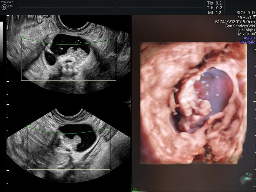
Cossi et al, Med Ultrason. 2017 Apr 22;19(2):216-217New rendering algorithms for 3D ultrasound • HDlive flow GE healthcare / Realistic Vue Samsung • Advanced skin rendering techniques, shadows, and a virtual light source • Increases in depth 3 dimensional perspective
New rendering algorithms for 3D ultrasound

• HDlive Silhouette GE healthcare /
  Chrystal Vue Samsung

• Clear outline of structures

• Semi transparent display of the
  inner core

• Promotes depth perception
HDLive flow: Saline Infusion Sonography of a
         normal and septate uterus
HDlive Silhouette normal and septate uterus
HDLive flow
Adnexal cysts
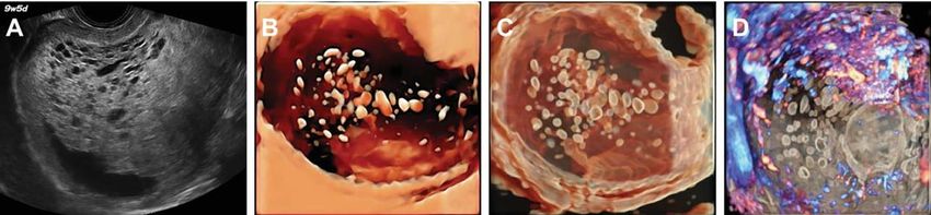
HDLive SilhoutteMolar pregnancy Hata et al, Ultrasound Obstet Gynecol 2018; 52: 552-554
Hdlive / Silhouette, Realistic / Chrystal Vue

• Amazing pictures with lots of detail and depth perception

• So far no comparative studies in gynaecology

• Promising technique
   – Uterine anomalies and pathology (niche, vascular anomalies,
     congenital malformations, polyps, fibroids, Asherman)
   – Early pregnancy (caesarean scar pregnancy, interstitial pregnancy)
   – Adnexal pathology
   – Difficult anatomy
New MRI and CT technologies • Diffusion Weighted Imaging • 3D MRI • CT rendering
MRI: Diffusion Weighted Imaging

• Diffusion (random motion) of water
  molecules generates contrast
• Reveals details about tissue
  architecture

• Indicate early pathologic change
• Early detector of brain ischemia
  (stroke)
• Leiomyosarcoma vs benign
  leiomyoma
Apparent diffusion coefficient (ADC)

• measure of the magnitude of
  diffusion (of water molecules) within
  tissue

• less than 1.0 to 1.1 x 10⁻³ mm2/s
  (low ADC) indicates diffusion           T2 benign fibroid
  restriction

• Diffusion restriction = pathology

                                          ADC benign fibroid, normal diffusion
T1 leiomyosarcoma ADC leiomyosarcoma, diffusion restriction shown as hypointens area
DWI for screening for leiomyosarcoma
                       (In combination with other markers)

•   Retrospective cohort 1960 women prior to fibroid resection
•   10 LMS
•   8 other sarcomas
•   identified LMS patients with 100% sensitivity and 97%
    specificity

Tong et al, J Magn Reson Imaging. 2019 Jan 13. [Epub ahead of print]
3D MRI
- Creates 3D models of MRI images
- Reconstruct fibroid uterus
- Surgical planning
Lee et al, J Korean Med Sci. 2018 Jan 8;33(2)CT rendering
Intra operative Imaging techniques

• Fluorescence techniques
   – Indocyanine Green

• Narrow band Imaging

• Enhanced reality
Near infra red fluorescence with Indocyanine
                 green (NIR ICG)
• Indocyanine green (ICG)
   – Cyanine dye used in medical diagnostics
   – IV or local injection

• ICG becomes fluorescent once excited with a specific
  wavelength light in the near-infrared (NIR) spectrum
NIR ICG • Visualizing perfusion (defects) of anatomical structures • Lymphatic system • Penetration depth of NIR light up to 10 mm
NIR ICG in gynaecology

• (Sentinel) lymph node dissection
   – Reduces surgical time
   – Improved SLN detection rate
   Ferreira et al, Surg Technol Int. 2019 Apr 29;34

• Visualize the ureter
NIR ICG in endometriosis surgery

• Identify occult hypervascular areas of the peritoneum

• Visualization of the ureter in endometriosis surgery; feasibility
  study in 10 patients
   –   Park, J Minim Invasive Gynecol. 2015 Nov-Dec;22(6S):S69.

• In bowel endometriosis
   – Select the transection line
   – Evaluate bowel perfusion of the neo-anastomosis

   – May prevent anastomosis-related complications
   – No firm evidence (colorectal surgery)
NIR ICG endometriosis surgery

Seracchioli et al, Fertil Steril. 2018 Jun;109(6):1135
Narrow Band Imaging

• Narrowband imaging (NBI)
   – mix of 415-nm and 540-nm light (peak light absorption of hemoglobin)
   – Enhaces tissue details
Narrow Band Imaging

• Blood vessels appear much darker than the nonvascular
  structures

• NBI has limited penetration (0.15-0.3 mm), which
  favorably limits visibility to the superficial peritoneum
  only
Narrow Band Imaging in Endometriosis
• Enhanced visualisation of endometriosis may:
    – Increase eradication during endometriosis surgery
    – Improve surgical outcome (less pain, better quality of life)
          • Or increase complications due to more extensive surgery?
    – Decrease recurrence

  Ma et al, J Minim Invasive Gynecol. 2019 Mar - Apr;26(3):427-433
NBI in endometriosis surgery Barrueto et al, J Minim Invasive Gynecol. 2015 Jul-Aug;22(5):846-52
•    NBI increased the detection of pathologically confirmed
                         endometriosis

Barrueto et al, J Minim Invasive Gynecol. 2015 Jul-Aug;22(5):846-52
The differences in pain reduction and quality of life improvement, at 3 and 6 months post surgery, were similar for WL/NBI compared to traditional WL conditions Gallicchio et al, J Minim Invasive Gynecol. 2015 Nov-Dec;22(7):1208-14
LITE study
Laparoscopic Imaging Techniques in Endometriosis Surgery

At random evaluation of 4
laparoscopic visualisation
techniques:
                                        N=20, ASRM grade III-IV endometriosis
                                        80% power to increase sensitivity from 72%
• 2D white light (reference)
                                        tot 85% : at least 81 endometriosis lesions
• 3D white light                        All techniques in randomised order per
• NBI                                   patient.
• NIR fluorescence with ICG
                                        Four quadrant evaluation of the pelvis

                                        Biopsies of for endometriosis suspected areas
                                        Control biopsies of healthy peritoneum

                                         Lier et al, Surg Endosc. 2019 Apr 26. [Epub ahead of print]
LITE studie
Laparoscopic Imaging Techniques in Endometriosis Surgery
Techniques Outcomes P value
2D-white light imaging
N = 176
Sensitivity 75.8%
Specificity 79.7% Statistics:
Narrow-band imaging
Sensitivity 81.3% p = 0.29 Sensitivity:
Specificity 70.6% p = 0.83 -McNemar’s
Near-infrared imaging/ICG
Sensitivity 36.1% p < 0.001 Specificity:
Specificity 89.0% p < 0.001 -Tango’s method
3D-white light imaging
Sensitivity 83.5% p = 0.005
Specificity 82.4% p < 0.001
Lier et al, Surg Endosc. 2019 Apr 26. [Epub ahead of print]LITE studie Laparoscopic Imaging Techniques in Endometriosis Surgery Posthoc analysis: Detected with 3D AND/OR NBI Yes 83 25 Sensitivity 91.2%
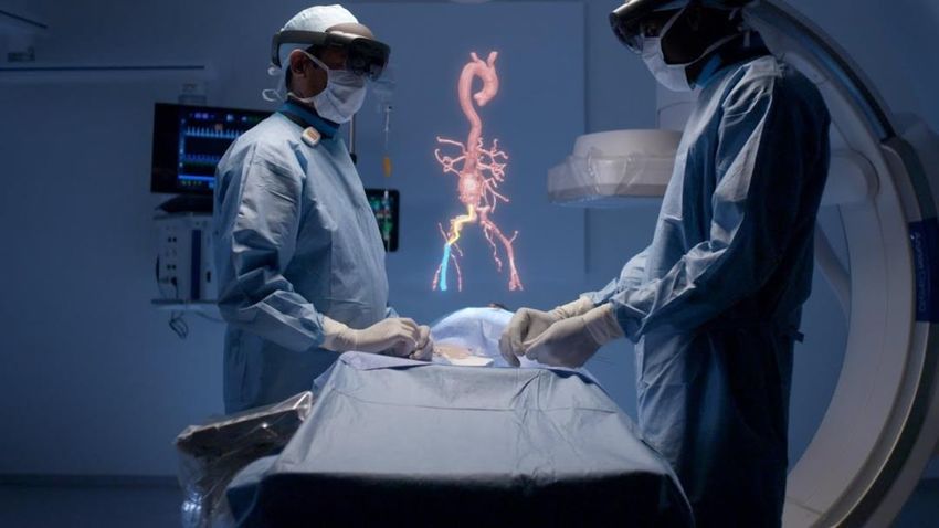
Enhanced reality in surgery: the next step

• Augmented reality: digital
  data projected in real
  environment

• Virtual reality: digital data
  as virtual environment

• Mixed reality: digital data
  in interaction with real
  environment
Augmented reality in myomectomy and
                    resection of adenomyosis

Bourdel et al, Fertil Steril. 2017 Mar;107(3):737-739 and Bourdel et al, J Minim Invasive Gynecol. 2019 Apr 6. [Epub ahead of print]
Microsoft Hololens 2
Hololens 2
Conclusions
• New technology in diagnostic imaging shows more detail and gives better
  perception of the anatomy
    –   New equipment with better resolution, rendering 3D dataset (ultrasound, MRI, CT)

• 3D ultrasound has clinical value in specific cases
    –   Coronal plane
    –   Post processing rendering and measurements
    –   Automated calculations (volume, vascularity index)

• Contrast enhanced ultrasound for diagnosis of tubal patency

• MRI Diffusion Weighted Imaging seems useful in the diagnosis of
  leiomyosarcoma

• Image guided surgery and enhanced reality improve visualization of
  anatomy and diseased structures, and may result in better surgical
  outcome and less complications
www.iceberg2021.org International Conference on Evidence Based Endoscopy and Research in Gynecology
You can also read