Acutus Medical
Clinical Compendium
Ventricular Tachycardia Abstracts
←
→
Page content transcription
If your browser does not render page correctly, please read the page content below
Acutus Medical Clinical Compendium Ventricular Tachycardia Abstracts
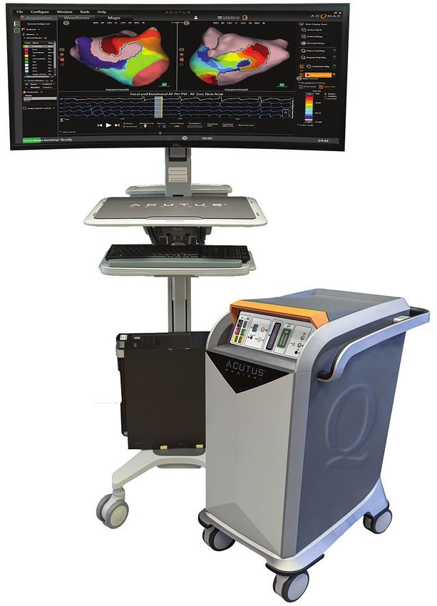
ACQMAP CLINICAL COMPENDIUM

                                 3D IMAGING, MAPPING & NAVIGATION
                                 SYSTEM
                                 ■   3D ultra-high resolution ultrasound
                                     imaging
                                 ■   Full chamber single beat mapping
                                     capability
                                 ■   Enable mapping of stable and unstable
                                     rhythms
                                 STANDALONE OPEN PLATFORM SYSTEM
                                 ■   Compatible with a wide range of
                                     diagnostic and therapeutic tools
                                 ■   Maintain customer preference for
                                     ablation and mapping catheters
                                 MULTIPLE MAPPING MODES — BUILT TO
                                 MAP EVERY BEAT
                                 ■   Contact mapping for simple anatomical
                                     ablations
                                 ■   Hover mapping with SuperMap for
                                     multi-morphic, repetitive rhythms
                                 ■   Single position non-contact mapping
                                     of unstable arrhythmias

                             i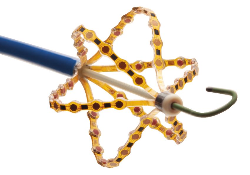
ACQMAP CLINICAL COMPENDIUM

       Next Generation AcQMap 3D Imaging and Mapping Catheter
                              Improved Handling and Deliverability

                                                                    High quality 3D ultrasound anatomy
                                                                    reconstruction with 48 dedicated
                                                                    piezoelectric sensors
                                                                            Non-contact mapping with 48
                                                                            dedicated engineered sensors

                                                                                           .035” (0.89 mm) J-tip
                                                                                           guidewire compatible

     • The world’s only integrated high-resolution ultrasound imaging and non-
       contact mapping catheter
     • Optimal maneuverability with the AcQGuide MAX steerable sheath
     • Improved handling1 with enhanced torquability
     • Enhanced deliverability with a .035” guidewire

                        The AcQMap catheter is not indicated for human use in the ventricle; preclinical work only.

                                                              ii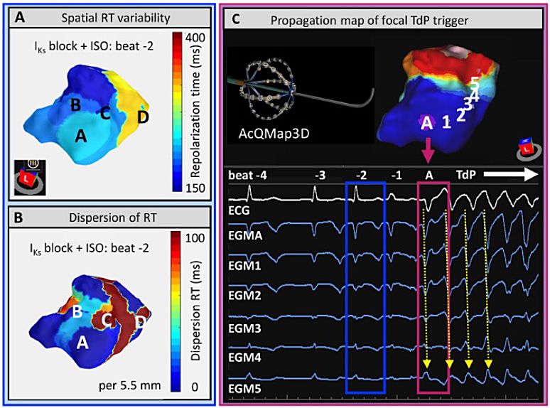
ACQMAP CLINICAL COMPENDIUM

   CONTENTS
   VENTRICULAR TACHYCARDIA ABSTRACTS
   1   High-resolution Real-time Left-ventricular Endocardial Activation-repolarization Mapping in an
       In-vivo Canine Model of Drug-induced Long-QT 1 Syndrome and Torsades De Pointes.

   2   Accuracy of Non-Contact Ultrasound-Based Left Ventricular Anatomy and Whole-Chamber
       Charge Density Mapping for Identifying Ischemic Scar in a Sheep Model

   3   Left Ventricular Substrate Characterization by Conduction Velocity in Sinus Rhythm, Ventricular
       Pacing, and Ventricular Fibrillation

   4   Comparison of Intracardiac Non-Contact Calculated Ventricular Electrograms to Measured
       Contact Electrograms During Mapping: Validating SuperMap Technology

                                                      iii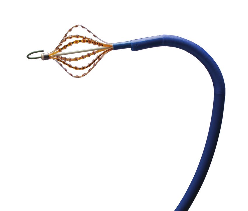
VENTRICULAR TACHYCARDIA ABSTRACTS

         ACQMAP CLINICAL COMPENDIUM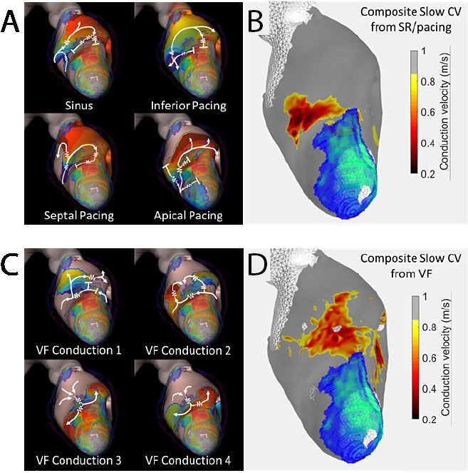
V E N T R I C U L A R TA C H Y C A R D I A A B S T R A C T S

              High-resolution Real-time Left-ventricular Endocardial
              Activation-repolarization Mapping in an In-vivo Canine
              Model of Drug-induced Long-QT 1 Syndrome and Torsades
              De Pointes.
              HRS Abstract 2021 D-XXXX-XXX. Heart Rhythm Vol. 18, No. 7, July Supplement 2021, SXXX.

              Rachel M.A. ter Bekke, MD, PhD, Nathan Angel, Annelies Vanderper, PhD, Tim Corvi, David J. Gallacher, Henk J. van der Linde, PhD and Paul G. A. Volders, MD,
              PhD. Maastricht University Medical Centre - Cardiology, , Acutus Medical, Carlsbad, CA, Janssen Pharmaceutica, Beerse, Belgium, CARIM/Cardiology, Maastricht,
              Netherlands.

             Background: Congenital loss of function or drug-induced inhibition of IKs            Results: IKs block prolonged QTc by 40% (386±22 ms) and mean RT by
             causes repolarization lability, predisposing to QT prolongation (LQT1) and           44% (309±20 ms), as recorded with the AcQMap3D catheter (AT/RT errors
             sympathetically-evoked torsades de pointes (TdP). High-resolution in-vivo            2.9±4.0/5.4±6.7 ms; cross-correlation 1.0±0.1; time difference 0.8±1.8 ms).
             mapping of endocardial activation-repolarization times (AT, RT), gradients           ΔRT increased by 23% (85±7 ms) and max local RT dispersion by 19%
             and focal excitation conspiring to TdP in LQT1 is lacking.                           (80±9 ms). Isoproterenol induced regional RT shortening (base>apex)
                                                                                                  and beat-to-beat instability, increasing ΔRT significantly by 122% (189±62
             Objective: To study arrhythmogenic mechanisms of TdP in a canine model
                                                                                                  ms) and local RT dispersion by 91% (153±51 ms; Figure). Focal triggers of
             of drug-induced LQT1 using realtime and high-resolution electroanatomical
                                                                                                  TdP emerged from regions bordering high RT dispersion, driving reentrant
             mapping.
                                                                                                  excitation.
             Methods: IKs inhibition (HMR1556 0.025-0.05 mg/kg/min) was applied
                                                                                                  Conclusion: High-resolution mapping in an in-vivo LQT1/TdP model uniquely
             in three fentanyl/etomidateanesthetized dogs; anesthesia to maintain
                                                                                                  identifies spatiotemporal RT instabilities, focal excitation and TdP.
             near-normal autonomic responsiveness. The AcQMap3D noncontact
             imaging and mapping catheter (Acutus Medical, US) was positioned in the
             LV. Isoproterenol (1.25-2.5 mcg/kg) induced TdP. AT, RT (Wyatt), RTmax-min
             (ΔRT), local RT dispersion
             (per 5.5 mm) and TdP
             initiation/propagation
             were studied.

                                                                                          1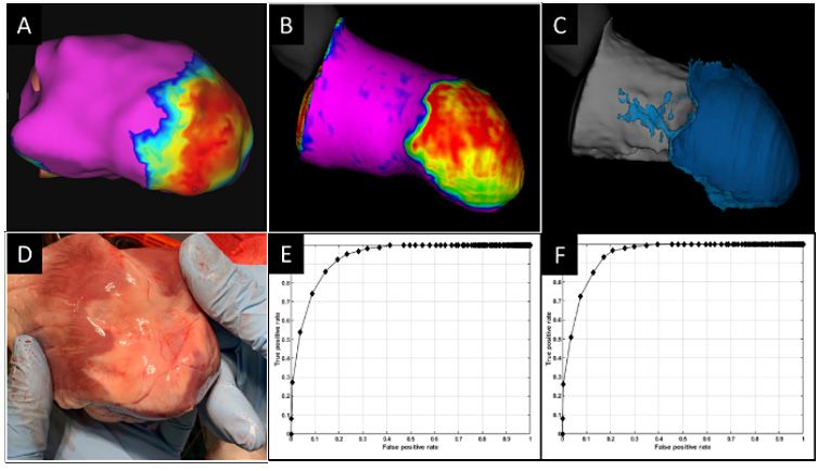
V E N T R I C U L A R TA C H Y C A R D I A A B S T R A C T S
Accuracy of Non-Contact Ultrasound-Based Left
Ventricular Anatomy and Whole-Chamber Charge Density
Mapping for Identifying Ischemic Scar in a Sheep Model
F. Daniel Ramirez, MD, MS, Jeffrey R. Winterfield, MD, FHRS, Pratik Shah, Annelies Vanderper, PhD, Tim Sorrell, Dave Robinson, Xinwei Shi, Derrick Chou, PhD, Eli
Ghafoori, Nathan Angel, Leo Mariappan, YOSUKE NAKATANI, MD, PhD, Bruno Sore, Virginie Loyer, Jean-Marc Peyrat, MBA, MSci, PhD, Hubert Cochet, MD, PhD and
Pierre Jais, MD. Hopital Cardiologique du Haut-Leveque, Bordeaux, France, MUSC, Mount Pleasant, SC, Acutus Medical, CA, Acutus Medical, Carlsbad, CA, Hôpital
Cardiologique du Haut-Lévêque-CHU de Bordeaux, Université de Bordeaux, Pessac, France, InHeart, Bordeaux, France, Liryc, France, inHEART, Pessac, France,
IHU LIRYC (CHU/Univ Bordeaux), Pessac, France, universite de Bordeaux, Bordeaux, France
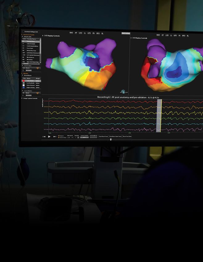
Background: Conventional electroanatomical mapping systems require Results: Total SuperMap acquisition time was 2:27 minutes, resulting in
serial collection of data points, which can be time-consuming and render 3,664 anatomical vertices. Scar areas were 17.62 and 18.06 cm2 on MRI and
characterizing certain arrhythmias challenging. Non-contact ultrasound- SuperMap, respectively. ROC curves demonstrated AUC values of 0.92 and
based anatomical reconstruction and whole-chamber charge density (CD) 0.93 for 25% transmural scar and total scar, respectively (accuracy 0.87 for
mapping has been used to guide ablation in patients with persistent atrial both) (Figure).
fibrillation. Its value in ventricular chambers has not been explored.
Conclusion: Non-contact ultrasound-based LV anatomy and CD mapping
Objective: To test the ability of the AcQMap High-Resolution Imaging and was feasible, rapid, and demonstrated a high level of correlation with MRI
SuperMap System to rapidly and accurately reconstruct left ventricular (LV) parameters for identifying ischemic scar in this preclinical model. This
anatomy and to delineate ischemic scar. technology could be valuable for the treatment of ventricular arrhythmias.
Methods: An anterior wall infarct was induced in a sheep. After 8 weeks,
cardiac MRI was performed and 3D reconstructions of the LV created. An
ultrasound and CD mapping catheter
was advanced into the LV via transseptal
access to generate SuperMap geometries.
Scar was defined by late gadolinium
enhancement and CD Laplacian amplitude
thresholds on MRI and SuperMap,
respectively. Corresponding points from
both geometries were compared via
pairwise analysis.
Figure. A LV SuperMap amplitude map (right lateral view). B, C. MRI-based tissue density (25% transmurality
and total scar, respectively). D. Infarct visible on gross anatomy. E, F. ROC curves of SuperMap amplitude and
MRI-based tissue density (25% transmurality and total scar, respectively).
2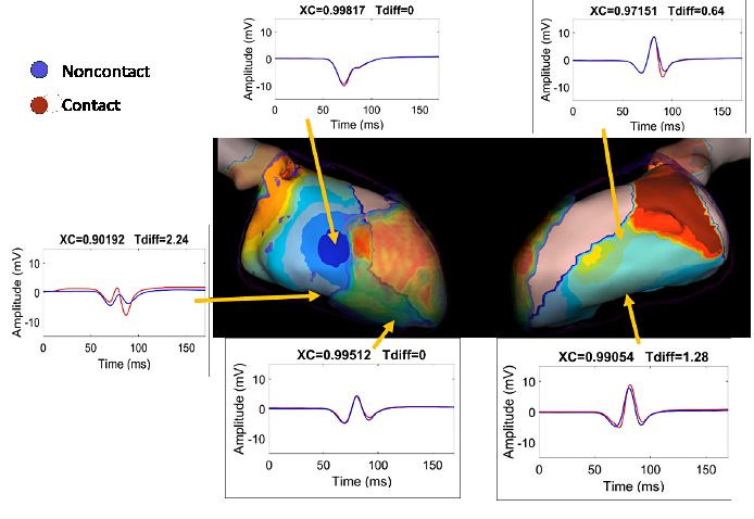
V E N T R I C U L A R TA C H Y C A R D I A A B S T R A C T S

             Left Ventricular Substrate Characterization by Conduction
             Velocity in Sinus Rhythm, Ventricular Pacing, and
             Ventricular Fibrillation

             F. Daniel Ramirez, MD, MSc, Jeffrey R. Winterfield, MD, FHRS, Hubert Cochet, MD, PhD, YOSUKE NAKATANI, MD, PhD, Jean-Marc Peyrat, MBA, MSci, PhD, Bruno Sore,
             Virginie Loyer, Nathan Angel, Annelies Vanderper, PhD, Tim Sorrell, Dave Robinson, Xinwei Shi, Eli Ghafoori, Pratik Shah, Derrick Chou, PhD, Leo Mariappan and
             Pierre Jais, MD. Hopital Cardiologique du Haut-Leveque, Bordeaux, France, MUSC, Mount Pleasant, SC, IHU LIRYC (CHU/Univ Bordeaux), Pessac, France, Hôpital
             Cardiologique du Haut-Lévêque-CHU de Bordeaux, Université de Bordeaux, Pessac, France, inHEART, Pessac, France, InHeart, Bordeaux, France, Liryc, France,
             Acutus Medical, CA, Acutus Medical, Carlsbad, CA, universite de Bordeaux, Bordeaux, France

             Background: Ventricular tachyarrhythmias after
             myocardial infarction remain a common cause of sudden
             death. Scar or replacement fibrosis is known to be an
             underlying substrate for these fatal rhythms. Substrate
             mapping during sinus rhythm is a low-risk approach to
             identifying ablation targets for these arrythmias.

             Objective: To explore whether shared areas of consistent
             conduction slowing or block could be identified
             irrespective of wavefront direction and rate (during
             sinus rhythm, ventricular pacing from multiple sites, and
             ventricular fibrillation [VF]).

             Methods: Anterior infarcts were induced in 3 sheep via
             intracoronary ethanol infusions. Conduction velocities
             (CV) were computed from charge density using a
             polynomial fitting method during sinus rhythm and
             during pacing from 3-5 sites as well as during 4-5
             seconds of VF (AcQMap, Acutus Medical). Regions of
             consistently slow CV were compared to areas of scar as
             identified on enhanced cardiac MRI.

             Results: Localized areas of consistently slow CV were
             observed in all animals during all rhythms, including
             VF (mean 24 wavefronts). These areas most often
             corresponded to border regions of ventricular scar
             identified on MRI (Figure).                                         Figure. Example of ventricular substrate mapping using multi-directions wavefronts,
                                                                                 revealing sites of abnormal conduction preset in SR, pacing, and VF. A, C. Propagation
                                                                                 sequence for 4 unique substrate-interrogating wavefronts: SR and pacing (panel A),
             Conclusion: Our study suggests that localized regions of            VF (panel C). Anterior scar density shown in color at apex (red: more dense). Snapshot
             the ventricle with consistently slow CV exist, including            of propagation shown in isochronal color (red: leading wavefront; other colors:
             during VF, and that they are often found near scar borders.         preceding wavefront locations, or ‘history’). Slow zones delineated by zig-zap lines. B,
                                                                                 D. Composite maps formed from multiple wavefronts of consistently slow CV from SR
             These regions could be identified through interrogative             and pacing (panel B) and during VF (panel D) (red: slowest). 90% scar transmurality
             substrate mapping during ventricular pacing from varied             from MRI overlaid (green: more dense).
             sites.

                                                                                       3
V E N T R I C U L A R TA C H Y C A R D I A A B S T R A C T S
Comparison of Intracardiac Non-Contact Calculated
Ventricular Electrograms to Measured Contact
Electrograms During Mapping: Validating SuperMap
Technology
F. Daniel Ramirez, MD, MS, Eli Ghafoori, Nathan Angel, Annelies Vanderper, PhD, Tim Sorrell, Dave Robinson, Xinwei Shi, Derrick Chou, PhD, Pratik Shah, Leo
Mariappan, Jeffrey R. Winterfield, MD, FHRS, YOSUKE NAKATANI, MD, PhD, Bruno Sore, Virginie Loyer, Jean-Marc Peyrat, MBA, MSci, PhD, Hubert Cochet, MD, PhD
and Pierre Jais, MD. Hopital Cardiologique du Haut-Leveque, Bordeaux, France, Acutus Medical, CA, Acutus Medical, Carlsbad, CA, MUSC, Mount Pleasant, SC,
Hôpital Cardiologique du Haut-Lévêque-CHU de Bordeaux, Université de Bordeaux, Pessac, France, InHeart, Bordeaux, France, Liryc, Bordeaux, France, inHEART,
Pessac, France, IHU LIRYC (CHU/Univ Bordeaux), Pessac, France, universite de Bordeaux, Bordeaux, France
Background: Complex ventricular tachycardias can be challenging to map Results: After creating ultrasound-based LV geometries, CD acquisition
due to time required to generate complete maps and the presence of low times were 2.9 ± 0.8 minutes per map. In total, 2285 unipolar voltage EGMs
amplitude electrograms (EGMs). SuperMap (Acutus Medical) is a novel were registered during electrode contact with LV tissue. EGM morphology
mapping method that temporally aligns multiple non-contact unipolar correlation between non-contact and contact EGMs was 0.93 ± 0.11 with a
EGMs detected from a roving multielectrode catheter (AcQMap, 0.70 mm2 mean temporal delay of 3.6 ± 6.3 msec in EGM onset (Figure). The degree of
electrode areas) then applies a charge density (CD) inverse solution to correlation did not differ based on direction of activation wavefront.
calculate and display global activation and amplitude maps.
Conclusion: Non-contact SuperMap-derived ventricular EGMs strongly
Objective: To compare EGMs derived from roving electrodes (not in contact correlate with conventional contact-based EGMs with respect to
with LV tissue) to those recorded during electrode contact with the LV wall. morphology and timing.
Methods: Anterior wall infarcts were induced via intracoronary ethanol
infusions in two sheep. After 8 weeks, ultrasound-based anatomical and
SuperMap CD maps of the LV were created using AcQMap multielectrode
catheters during sinus rhythm and pacing
from septal, lateral, and apical locations.
At locations where contact EGMs were
recorded, the closest corresponding
SuperMap-calculated EGM was used for
comparison.
Figure. Example of unipolar
contact and non-contact EGMs
comparisons with corresponding
locations indicated on an imported
MRI anatomy. MR-imaged infarct
area is overlaid in translucent color
at apex (red: denser). Snapshot of
propagation shown in isochronal
color (red: leading wavefront;
other colors: preceding wavefront
locations or ‘history’).
4For more information please contact your
Prior to using these devices, please review the Instructions for Use
Acutus Medical representative: for a complete listing of indications, contraindications, warnings,
ACUTUS MEDICAL, INC. ACUTUS MEDICAL NV precautions, potential adverse events, and directions for use. The
2210 Faraday Ave Ikaroslaan 25 AcQMap and AcQGuide devices are CE Marked and FDA cleared.
Suite 100 1930 Zaventem acutus.com/patents
Carlsbad, CA 92008 Belgium Acutus Medical®, Acutus®, AcQMap® and AcQGuide® are
United States Phone: +32 2 669 75 00 registered trademarks of Acutus Medical, Inc. This material
is the property of Acutus Medical and its subsidiaries.
Phone: 888-202-8151 FAX: +32 2 669 75 01 Please do not copy, forward, or distribute this document.
Copyright © 2020 Acutus Medical, Inc. All rights
acutus.com acutus.com reserved.
2021-06 MM-144 Rev. AYou can also read