NON-SMALL CELL LUNG CANCER - UNDERSTANDING UNDERSTANDING SERIES - A GUIDE FOR THE PATIENT
←
→
Page content transcription
If your browser does not render page correctly, please read the page content below
Understanding series

Understanding

Non-
Small
Cell
Lung
cancer

1-800-298-2436 | go2foundation.org

                I
a guide for the pati en t
ANATOMY
           OF THE LUNGS
 The following image shows different parts that make
 up the lungs. Please use this picture to help guide you
 through the topics discussed in this brochure.

                                                                   Lymph
                                                                    nodes                Alveoli
TRACHEA
(WINDPIPE)

UPPER
LOBE

MIDDLE                                                                               Bronchiole
LOBE
                                                                                             UPPER
                                                                                              LOBE

                                                                                            LOWER
                                                                                              LOBE
LOWER
LOBE                                      MAIN
                                         BRONCHI
                     Right                                              Left
                     Lung                                               Lung

The content of this publication is for informational purposes only and is not intended to be a substitute
for professional medical advice, diagnosis or treatment. Only your doctor can provide you with advice on
what is safe and effective for you. Models used in the brochure are for illustrative purposes only.
TABLE OF CONTENTS

Non-Small Cell Lung Cancer (NSCLC)....................................................................................... 2

Diagnosing NSCLC

Imaging............................................................................................................................................................................. 5
Biopsies............................................................................................................................................................................ 6
Histology and Subtype................................................................................................................................. 7
Staging.............................................................................................................................................................................. 8
The Lymphatic System............................................................................................................................ 10
Biomarker Testing.............................................................................................................................................. 11

Treatment Options

Surgery............................................................................................................................................................................ 13
Radiation Therapy............................................................................................................................................ 14
Chemotherapy...................................................................................................................................................... 15
Targeted Therapy.............................................................................................................................................. 15
Immunotherapy...................................................................................................................................................15
Clinical Trials............................................................................................................................................................ 17
Side Effects................................................................................................................................................................ 18
Your Treatment Team.................................................................................................................................. 21
Glossary......................................................................................................................................................................... 22
Resources................................................................................................................................................................... 24
non-small cell
 lung cancer

Cancer is a group of diseases in which normal
cells change, grow and divide out of control.
Cancer that begins in the lungs – lung cancer – is
one of the most commonly diagnosed cancers
in the United States. There are two main types:
non-small cell lung cancer (NSCLC) and small cell
lung cancer (SCLC). NSCLC makes up about 85%
of lung cancer diagnoses.

                        2
risk factors

a history of smoking is the main risk
factor for developing lung cancer. Cigarettes
contain many carcinogens, which are
substances that cause lung cancer.

Exposure to radon (an invisible, odorless,
tasteless radioactive gas that occurs naturally in
soil and rocks).

family history of lung cancer.

Radiation therapy to the chest area.

Other lung illnesses (such as
emphysema, chronic obstructive pulmonary
disease [COPD] or tuberculosis).

Exposure to industrial chemicals
including arsenic, asbestos, beryllium and
uranium.

Exposure to secondhand smoke
(or passive smoking).

                   3
Diagnosing
              NSCLC

                       imaging
A number of tests provide information on areas of the lungs
that do not appear normal. Doctors sometimes refer to these
areas as tumors, spots, lesions, nodules or masses. These
imaging techniques can provide information beyond what
can be seen with a chest X-ray. Imaging can help doctors learn
if a suspicious area is cancerous (malignant) or not cancerous
(benign). Some imaging tools include the following:

CT (computed tomography) or “CAT” scanning can show
tumors that may not be visible on a normal chest X-ray.

LDCT (low-dose computed tomography) scanning is used
to screen those who are at high risk every year to see if they
have lung cancer.

PET (positron emission tomography) scanning shows how a
tumor is using glucose (also known as sugar). Since tumors
typically use more glucose than surrounding tissue, they
appear as “hot spots” (bright areas) in these images.

MRI (magnetic resonance imaging) creates detailed images
of the body and can help determine whether a tumor has
spread beyond its original location. Used in NSCLC to check
for spread of cancer to the brain.

                               5
Biopsies
A biopsy is a procedure in which tissue is removed from the
body for testing. The tissue can help doctors diagnose cancer
and provide specific information about the suspicious area.
There are several types of biopsy procedures:
Fine
NEEDLE    needle
              BIOPSYaspiration
                          OR NEEDLE (FNA):            Tissue isBIOPSY:
                                                   SURGICAL        removed       using
                                                                             Tissue   is
ASPIRATION:
a   thin hollowAneedle. hollow needle
                                  Depending removed            during surgery.
                                                   on the location        of the
is inserted through the skin to
tumor, FNA is done during a bronchoscopy           BRONCHOSCOPY:   procedure      (inlighted
                                                                            A thin,
draw out tissue for testing. The
which       a camera-equipped                      tube   (bronchoscope)       is passed
procedure        is usually done withtube is used to view the windpipe
                                                   down the throat through the
and
the aid  other    airways)
            of imaging         or through
                            tests  such as skin. This procedure may be
                                                   mouth or nose and into the center
CT scans,by
guided         fluoroscopy,
                   an ultrasoundultrasound
                                        or a CT scan.
                                                   area of the lungs. A needle is
or MRI to determine where to
                                                   then inserted down the tube and
insert
Corethe         needle.biopsy:
            needle         There areTissue
                                        two     is samples
                                                   removed       using    a wider
                                                              are   removed     for testing.
types of this kind of biopsy:
needle. More tissue can be removed                  xE  with   this   procedure
                                                      ndobronchial Ultrasound
  x Finewith
than       Needle fineAspiration      (FNA):
                        needle aspiration.           (EBUS): Uses a bronchoscope
     Tissue is removed using a thin
                                                     and ultrasound (high
     hollow needle.
Surgical                 Depending
                    biopsy:     Tissueon   is removed      duringsounda surgical
                                                     frequency               waves) and
     the location of the tumor, FNA
procedure.          Smaller    tissue    samples     allowsbe
                                                     may      forremoved
                                                                   better examination
     is done during a bronchoscopy
surgically       during     a bronchoscopy           of the lymph
                                                    procedure;         nodessamples
                                                                     larger     and other
     procedure      (in which    a camera-
                                                     structures in the center of the
may  equipped
         require   tube   is used tosurgery.
                      traditional                    chest to see if cancer has spread.
     view the windpipe and other
     airways) or throughFluid
Thoracentesis:                  skin. is
                                       This
                                          removed   xE lectromagnetic
                                                      from    the spaceNavigational
                                                                              around
     procedure may be guided by                      Bronchoscopy (ENB): Uses
the lungs (also called the pleural cavity)                   using    a hollow
                                                     a bronchoscope to reach the
     an ultrasound or a CT scan.
needle inserted into the chest.                      lungs. Pictures from a CT scan
  x Core Needle Biopsy: Tissue is
                                                     and GPS like technology are
     removed
LIQUID            using aLiquid
              BIOPSY:        wider needle.
                                     biopsy is a new used technique
                                                            to create a map where and
     More tissue can be removed
liquid     is removed        from   the    body   andnavigate
                                                         examinedthe lungs
                                                                        for   to nodules.
                                                                             signs
     with this procedure than with
offinecancer.
           needle This  is typically blood butThis
                      aspiration.                      canprocedure
                                                             also be urineallows ordoctors
                                                     to get to the outer areas of the
sputum.
THORACENTESIS:   Liquid biopsy
                            Fluid is should not be   lung used
                                                            which to may
                                                                      diagnose      lung
                                                                           be difficult
cancer
removed(tissuefrom the  biopsy
                            spaceisaround
                                     still required  tofor  proper
                                                         reach  usingdiagnosis
                                                                         traditional
the lungs
and             (alsobut
         staging)      called  the pleural
                            it can   be useful forbronchoscopy.
                                                       cancer monitoring and
cavity)
examining  usingbiomarkers
                    a hollow needle (see page 11).  xA
                                                      utofluorescence: Uses a
inserted into the chest.
                                                     bronchoscope with a special
LIQUID BIOPSY: Blood or other                        light and camera, which
fluid is removed for biomarker                       captures live color video viewed
testing to help with planning                        on a monitor. Under this light,
treatment. Liquid biopsy is NOT                      abnormal/pre-cancerous tissue
used to diagnose lung cancer.                        appears in a different color
                                                     than normal tissue.
                                           6
histology
and subtype
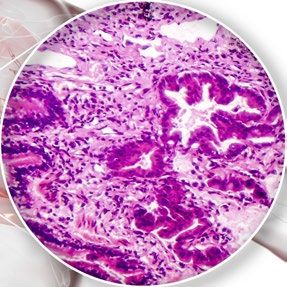
NSCLC is not the same in every person. “Histology” refers
to the structure of the cancer when viewed under a
microscope. There are many subtypes of NSCLC but the
most commonly diagnosed are:
ADENOCARCINOMA
Squamous cell carcinoma
LARGE CELL CARCINOMA
Knowing the subtype of NSCLC helps the healthcare team
identify the best treatment options.
When cancer has spread to other areas of the body, it is
still named after the type and histology of the primary
tumor. So lung cancer that has spread to the brain is
called lung cancer with brain metastasis, not brain cancer.
7staging
STAGE STAGE
I II
The tumor is only in The tumor is only in one
one lung and is no lung and may be larger
more than 5cm with than those in stage I.
no spread to nearby The cancer may have
lymph nodes. spread to nearby lymph
nodes but not beyond.
NSCLC is divided into four stages, based on the TNM System.
Stage is generally determined by the size of the cancer and
whether or not it has spread from the place it started (including
to lymph nodes).
77 T stands for tumor: Where the tumor is and how big it is.
77 N stands for lymph nodes: Whether the cancer has spread to
lymph nodes and where the affected lymph nodes are located.
77 M stands for metastasis: Whether the cancer has spread beyond
the lung to the other lung, the pleura or other parts of the body.
8STAGE STAGE
III IV
The tumor or tumors The tumor may be any
are only in one lung and size and the cancer
may have grown into has spread to the
other structures within other lung, the lining
the chest or spread to of the lung or organs
more lymph nodes. outside the lungs.
Your treatment team may tell you that your stage is an A or B,
for example, IIIA or IIIB. A is a lower stage (smaller or less spread) than B.
IT IS IMPORTANT TO KNOW THE STAGE OF THE CANCER.
Staging can help doctors create a treatment plan that is best
for you.
The terms “early stage” or “locally advanced” are sometimes
used to refer to stage I, stage II and some stage III tumors.
The term “advanced” may be used to describe some stage III
tumors and all stage IV tumors. Ask your healthcare team for
more details about tumor staging and how it may affect your
choices for treatment.
9The
Lymphatic
System
Lymph Node
The lymphatic system is a collection of organs, vessels and
nodes that are found throughout the body. It has two major
functions: to collect excess fluid and return it to the blood,
and to fight infection.
Lymph vessels are similar to blood vessels and help
to circulate lymph fluid throughout the body. Lymph fluid
contains white blood cells, which help to fight infection.
Lymph nodes are small, oval-shaped organs within the
lymphatic system.
The purpose of lymph nodes is to trap and collect invading
organisms that can be destroyed by white blood cells. Lymph
nodes are found throughout the body, but major clusters can be
found behind the knee and elbow joints and in the groin, armpits,
chest and neck. A large group is found in the center of the chest
(mediastinum) which drains lymph fluid from the lungs.
Cancer cells can break off from the main tumor and travel
through the lymphatic system. Some of these cells can become
trapped within a lymph node and start to grow. Determining
whether there are cancer cells in lymph nodes can help a doctor
estimate how far the cancer may have spread.
10Biomarker
TESTING
Tumor tissue removed during a biopsy can be tested for
biomarkers. Biomarkers are features of the cancer that give
the treatment team specific information about the cancer.
These features may be specific proteins on the surface of the
cell or genetic information inside the cell. Some biomarkers
can help predict how the cancer will act while others indicate
whether a specific treatment may be effective.
Ask your doctor if you have had biomarker testing. If not,
we can help provide it. Learn more at lungmatch.org or
call 1-800-298-2436.
making treatment decisions
Treatment for NSCLC has become much more personalized
and all lung cancers are not treated the same way. The
stage, subtype and biomarker testing results for your
cancer will direct your treatment team in determining what
treatment options are best for you. Discuss this information
with your treatment team and share your personal values
and goals to determine the options best for you.
11treatment
OPtions
treatment for nsclc include one or more of the
following:
77 Surgery
77 Radiation
77 Chemotherapy
77 Targeted Therapy
77 Immunotherapy
77 Clinical Trials
77 Palliative Care (Symptom Management)
Treatment depends on the following:
77 Subtype and stage of NSCLC
77 If certain biomarkers are present
77 How well your lungs are working
77 Other health problems which may increase the toxicity of
therapy
77 Your ability to perform activities of daily living without assistance
like eating, bathing and dressing
12surgery
Types of lung cancer surgery include:
77 SUB-LOBAR Resections: Removal of the tumor and
surrounding lung tissue without removing the whole lobe of the
lung. Types include wedge resection and segmentectomy.
77 Lobectomy: Removal of an entire lobe of the lung.
77 Bi-Lobectomy: Removal of two lobes of the lung.
77 Pneumonectomy: Removal of an entire lung.
77 Complex resections: Removal of part or all of a lung and
surrounding structures such as ribs, part of the chest wall or
windpipe (bronchus) when the cancer has spread to those areas.
All surgeries should include testing of the lymph nodes
associated with that part of the lung.
Types of surgical procedures:
77 Thoracotomy: An incision is made between the ribs to allow
removal of the cancer.
77 Minimally invasive surgery: A series of small incisions
allows insertion of a video camera along with small instruments
for removing cancerous tissue. Types include:
- Video assisted thoracic surgery (VATS)
- Robotic assisted thoracic surgery (RATS)
13radiation therapy
Radiation therapy is a treatment that uses high energy
x-rays or particles to kill or shrink cancer cells, to manage
pain or to prevent the cancer from spreading. It can be used
to get rid of tumors entirely, eradicate residual disease after
surgery. At times it might be helpful to reduce the size of
tumors before surgery.
Radiation therapy is also commonly used to treat brain
metastases and may be used for this purpose with other
treatments.
There are several types of radiation therapy:
77 External beam radiation: Use of carefully aimed beams of
radiation targeting the areas of cancer.
77 Intensity Modulated Radiation Therapy (IMRT): A type of
radiation treatment that carefully shapes the beams of radiation
around the areas of cancer.
77 Stereotactic Body Radiation Therapy (SBRT):
A type of treatment that can target small lung cancers that
cannot be removed by surgery. SBRT can be given either in a one-
day session with a single dose of radiation or on a “fractionated”
schedule in which smaller doses are given over time.
77 STEReOTACTIC RADIOSURGERY (SRS): Despite it’s name, this
type of radiation does not involve surgery. It delivers very high
doses of targeted radiation to tumors.
77 Brachytherapy (internal or implant radiation
therapy): Radioactive material is sealed in needles, seeds,
wires or catheters and placed directly into or near a tumor. This
technique allows very high doses of radiation to be more safely
delivered while helping to reduce side effects.
77 proton therapy: A type of radiation treatment that uses a
beam of protons or positively charged particles at high energy to
deliver radiation directly to cancer cells. This treatment is offered
only at select centers in the US.
77 CONCURRENT CHEMORADIATION: Sometimes chemotherapy
and radiation may be used at the same time to treat the cancer.
14chemotherapy
Chemotherapy is a treatment that kills cancer’s rapidly
growing and dividing cells. In NSCLC, chemotherapy may be
given as a single drug or as multiple drugs at the same time,
depending on the overall health of the patient as well as
stage and subtype of NSCLC.
Chemotherapy drugs most often used to treat NSCLC are:
77 Paraplatin (Carboplatin) 77 Alimta (Pemetrexed)
77 Platinol (Cisplatin) 77 Gemzar (Gemcitabine)
77 Taxol (Paclitaxel) 77 Navelbine (Vinorelbine)
77 Taxotere (Docetaxel) 77 Abraxane (Paclitaxel protein-
bound)
targeted therapy
Targeted therapies are aimed at a particular “target” in
a tumor cell with the goal of stopping the cancer from
continuing to grow. Your biomarker testing results will tell
the treatment team if you are a good candidate for targeted
therapy. The most common “targets” in NSCLC are gene
changes in EGFR, ALK, ROS1 and BRAF.
To learn more about Targeted Therapy, including the drugs
currently in use, please refer to our “Targeted Therapy for
Lung Cancer” brochure.
immunotherapy
Immunotherapy is a new type of treatment that helps the
body’s own immune system fight the cancer. A class of
immunotherapy drugs called “checkpoint inhibitors” are
approved for use in NSCLC .
To learn more about Immunotherapy, including the drugs
currently in use, please refer to our “Immunotherapy for
Lung Cancer” brochure.
15combination therapy
Sometimes using more than one type of treatment may
produce better results. For example, chemotherapy may be
combined with Avastin (a targeted therapy) or with radiation;
radiation and chemotherapy may be used before and/or after
surgery. Your treatment team will let you know if a single
treatment or combination therapy is best for your situation.
adjuvant therapy
A drug may be given as an “adjuvant” or additional therapy
along with surgery or radiation. Following surgical removal of
lung cancer, adjuvant therapy is sometimes recommended,
depending on the risk of recurrence. The goal is to increase
the cure rate compared to surgery alone.
neo-adjuvant therapy
Sometimes a drug may be given prior to surgery, often to
shrink the tumor. This is known as neo-adjuvant therapy.
maintenance therapy
Some drug therapies may be used after the initial therapy
regimen has ended. Maintenance therapy may be used as
long as it continues to work and is tolerable.
The two types of maintenance therapies are:
77 “Continuation”: When a drug that has been used before is continued
77 “Switch”: When a new drug is used under certain circumstances
16clinical trials
Clinical trials are research studies to determine whether
new approaches to therapy are safe and effective, as well
as to determine how they compare to existing treatments.
Clinical trials in lung cancer may involve new ways of giving
radiation, new chemotherapy drugs or new drugs which
target specific biomarker abnormalities in the cancer. Having
biomarker testing may help your treatment team identify
an appropriate clinical trial for you that is more precisely
targeted to your cancer.
There are also new types of clinical trials including LUNG-MAP
and NCI-MATCH (for all cancers) where you will undergo
biomarker testing as part of the clinical trial to put you in a
group testing a drug that is most likely to be effective for you.
Talk to your treatment team about whether a clinical trial is
right for you. To see if you may qualify for a research study, call
our HelpLine at 1-800-298-2436 or visit lungmatch.org.
17Side effects
radiation therapy
Common side effects of radiation therapy include:
77 Tiredness (fatigue)
77 Skin irritation
– Redness
– Itching
– Dryness
– Infection
77 Loss of appetite (anorexia)
77 Inflammation of the esophagus (esophagitis)
77 Inflammation of the lung(s) (pneumonitis)
Be sure to talk with your healthcare team about ways to
manage any side effects you may experience.
18drug therapy
The goal of treatment is to kill cancer cells, which are fast
growing. Chemotherapy can’t tell cancer cells from other fast
growing cells so it can damage them, too and cause side
effects. While side effects from targeted therapies and
immunotherapies tend to be milder than chemotherapy,
they can still result in challenging side effects.
Common side effects of many Treatments:
77 Tiredness (fatigue)
77 Diarrhea
77 Constipation
77 Loss of appetite (anorexia)
77 Nausea and vomiting
77 Shortness of breath (dyspnea)
Common side effects of chemotherapies:
77 Hair loss
77 Numbness or tingling in the hands or feet (neuropathy)
77 Low red/white blood cell counts
common side effects of targeted therapies:
(Note, these typically depend on the type of targeted therapy)
77 Rash
77 Eye irritation or vision problems
77 Swelling of hands and feet
77 Nosebleeds
77 High blood pressure
common side effects of IMMUnotherapies:
77 Pain in muscles and joints
77 Immune reactions which may lead to inflammation in various
organs. These immune-related side effects are uncommon
but can be serious and close monitoring is required.
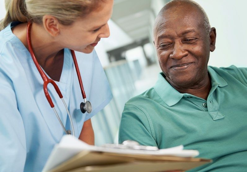
19A multidisciplinary team approach is when members of the
healthcare team discuss your situation and work together
to make treatment recommendations. It is thought that
this team approach improves coordination of care and
communication amongst the team.
20your treatment team may include
Thoracic Surgeon: A doctor who performs surgeries in
the chest region. Some thoracic surgeons specialize in lung
cancer.
Medical Oncologist: A doctor who specializes in
diagnosing and treating cancer. Medical oncologists may
use chemotherapy, hormonal, biologic or molecularly
targeted therapies as well as supportive therapies to treat
cancer.
Pathologist: A doctor who specializes in diagnosing and
classifying cancer by studying tissue, fluid or blood samples.
Radiation Oncologist: A doctor who specializes in
treating cancer using various forms of radiation by focusing
it on the tumor site in the body.
Pulmonologist: A doctor who specializes in treating
diseases and conditions involving the lungs.
Pulmonary Rehabilitation Specialist: A specialist
who works to reduce symptoms and side effects from
diseases of the lung—including lung cancer—and their
treatments.
Oncology Nurse: A nurse who specializes in helping
people with cancer and who may further specialize in the
surgical or medical management of a patient’s care.
Oncology Social Worker or Counselor: A social
worker or counselor who specializes in helping patients and
loved ones cope with the emotional impact of cancer and
who may help identify other needed resources.
Patient Navigator: A nurse, social worker or trained lay
person who assists patients and loved ones on their journey
through the health care system.
21Glossary
Biopsy: Removal of a small piece of tissue for examination
and analysis.
Cancer: A group of diseases in which cells grow and divide
uncontrollably, forming tumors. In some cases, the tumors
can invade nearby tissues. Tumor cells may also travel
through the bloodstream and lymphatic system to spread to
more distant parts of the body.
Carcinoma: Cancer that arises from epithelial cells, which
are cells that cover or line internal and external body surfaces.
Chemotherapy: Treatment with a chemical or a
combination of chemicals, to slow or kill rapidly dividing cells.
Clinical trial: A research study conducted to determine
whether investigational drugs or treatments are safe and
effective in humans.
Computed tomography (CT): An imaging technique that
uses a computer to create a series of precise X-ray images
of internal organs. CT scans show much more detail than
standard X-rays. Also known as “CAT” scanning.
Histology: The microscopic structure of tumor cells that
helps a doctor determine the subtype of a tumor.
Lobectomy: Surgery that removes the lobe (a portion) of
the lung that contains a tumor. The right lung is divided into
three lobes; the left lung has two lobes.
Lymph nodes: Small, oval structures located throughout the
body that together form part of the immune system.
Magnetic resonance imaging (MRI): The use of
magnetic fields to create images of internal organs.
22Metastasis: The spread of tumor cells to sites in the body
beyond the location in which the tumor began.
Pneumonectomy: Surgical removal of an entire lung.
Position emission tomography (PET) Scan: An
imaging technique that detects rapidly dividing cells. This
may help find cancers that are difficult to detect by other
means (e.g., X-ray, CT scan, MRI).
Radiation therapy: The use of focused beams of radiation
to kill cancer cells and reduce tumor size.
Resection: The surgical removal of part of a tissue or organ.
Side effects: Any undesired effects of a drug or treatment
on a patient.
Sputum: A phlegm-like substance brought up from the
lungs that contains mucus and cells and may contain
microorganisms, blood and/or pus.
Staging: Description of a tumor based on its size, location
and extent of spread to other organs.
TNM System: Staging of tumors according to three factors—
size and location of tumor (“T”), spread to lymph nodes (“N”),
and spread to other organs (also known as metastasis, “M”). In
lung cancer, a tumor is considered metastatic if it spreads to
the other lung or the pleura (the thin sac covering the lung).
Tumor: Abnormal tissue that results from uncontrolled cell
division. Tumors perform no useful bodily function and may
be either benign (not cancerous) or malignant (cancerous).
23Where can I go
for more
information?
For more information about lung cancer, treatments
and clinical trials, to discuss support options or for
referral to other resources, please contact us.
HELPLine | 1-800-298-2436 or support@go2foundation.org
Biomarker testing &
Clinical Trial Matching | lungmatch.org
website | go2foundation.org
2425
Founded by patients and survivors, GO2 Foundation
       for Lung Cancer transforms survivorship as the world’s
       leading organization dedicated to saving, extending,
       and improving the lives of those vulnerable, at risk, and
       diagnosed with lung cancer.
       GO2 Foundation works to change the reality of living with
       lung cancer by ending stigma, increasing public and
       private research funding, and ensuring access to care.

This publication is supported by our sponsors. They do not influence or provide input on content.
GO2 Foundation is a 501(c)(3) non-profit organization. Our programs are made possible by
generous support from people like you. All donations are tax deductible to the fullest extent
permitted by law.
The content of this brochure has been reviewed by members of our Medical and Professional
Advisory Board and our Patient & Caregiver Research Advocates.

          Copyright ©2021, GO2 Foundation. All rights reserved.

                 1-800-298-2436             |   go2foundation.org
You can also read